. 2023 Nov 23;20(1):274.
doi: 10.1186/s12985-023-02223-8.

Detection of monkeypox virus using helicase dependent amplification and recombinase polymerase amplification combined with lateral flow test

Affiliations

Detection of monkeypox virus using helicase dependent amplification and recombinase polymerase amplification combined with lateral flow test

Abudushalamu Gulinaizhaer et al. Virol J. .

Abstract

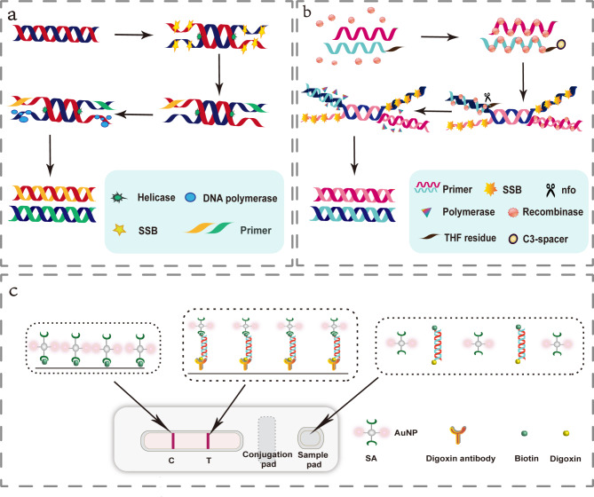
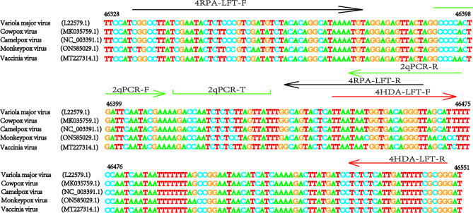
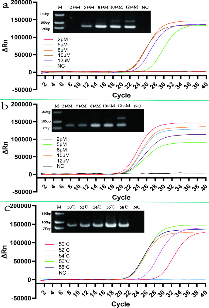
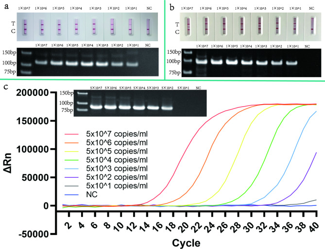
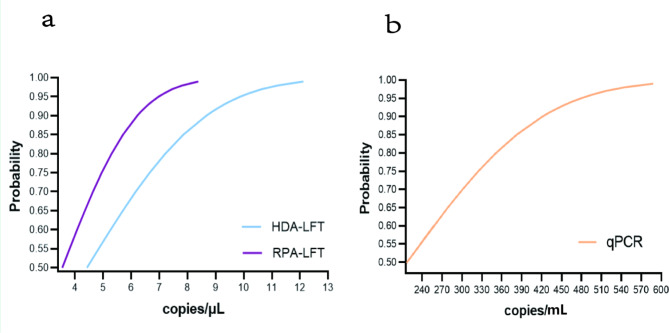
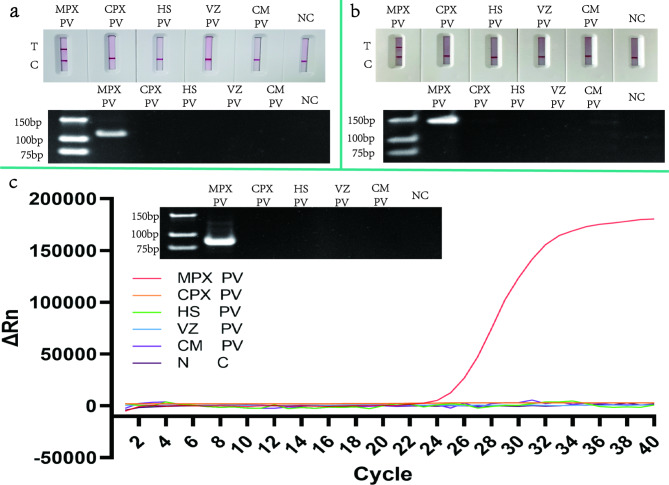
The monkeypox virus (MPXV) is a zoonotic DNA virus that belongs to the poxvirus family. Conventional laboratory methods for detecting MPXV are complex and expensive, making them unsuitable for detecting the virus in regions with limited resources. In this study, we using the Helicase dependent amplification (HDA) method and the Recombinase polymerase amplification (RPA) technique in combination with the lateral flow test (LFT), together with a self-designed qPCR technique for the detection of the MPXV specific conserved fragment F3L, to compare the sensitivity and specificity of the three assays. By analyzing the sensitivity detection results using Probit, it can be seen that the limit of detection (LOD) of the HDA-LFT detection target is 9.86 copies/µL (95% confidence interval, CI 7.52 copies/µL lower bound), the RPA-LFT detection target is 6.97 copies/µL (95% CI 3.90 copies/µL lower bound), and the qPCR detection target is 479.24 copies/mL (95% CI 273.81 copies/mL lower bound). The specificity test results showed that the specificity of the three methods mentioned above was higher than 90% in detecting pseudoviruses of the same genus of MPXV. The simple, highly sensitive, and specific MPXV assay developed in this study is anticipated to provide a solid foundation for future applications in the early screening, diagnosis, and evaluation of the efficacy of MPXV. This is the first time the HDA-LFT assay has been utilized to detect MPXV infection.

Keywords: HDA; INAAT; LFT; Monkeypox; RPA; qPCR.


Conflict of interest statement

The authors declare no conflict of interest.

Figures

Fig. 1
Fig. 1
Basic principles of detection technology. Figure 1a shows the basic principle of the HDA amplification technique: after the double-stranded DNA is deconvoluted using a helicase, a partial single-stranded sequence is created, and the single-stranded binding protein (SSB) binds to the newly formed single-stranded strand, and subsequent primers bind to the single-stranded strand and amplify it under the action of DNA polymerase. Figure 1b shows the basic principle of RPA amplification: Introduce tetrahydrofuran modification sites into the probe and modify the 3 ‘end to prevent elongation. When the probe is paired with the target DNA product amplified by the primer with a marker at the 5 ‘end, Nfo cuts the probe to form free 3’ - OH, which can be used as the primer to continue to extend, and the Synthon generation target DNA sequence; So the target DNA sequence of the offspring will also carry markers. SSB bind to the replaced DNA strand to prevent further replacement. Figure 1c shows the basic principle of the LFT technique: the digoxin antibody is fixed to the membrane in a strip (T-line) and the colloidal gold labelling reagent is adsorbed onto the gold pad. When the antigen to be tested is added to the sample pad at one end of the strip, the sample moves forward by capillary action, dissolves the colloidal gold labelling reagent on the binding pad and reacts with each other, and then moves to the area of the fixed antigen or antibody when the conjugate of the antigen to be tested and the gold labelling reagent When the sample is moved to the area of the fixed antigen or antibody, the conjugate of the test article and the gold standard reagent is specifically bound and retained on the test strip, which can be observed by the naked eye
Fig. 2
Fig. 2
Sequence alignment of MPXV and orthopoxvirus DNA. Compare F3L with DNA target sequences from several orthopox viruses. Virus strains: Monkeypox virus(ON585029.1) consulted on NCBI with Cowpox virus(MK035759.1), Camelpox virus(NC_003391.1), Vaccina virus(MT227314.1) and Variola major virus(L22579.1). F represents forward primer, R represents reverse primer, and T represents probe. Sequences were compared using Jalview and then drawn with Adobe illustrator
Fig. 3
Fig. 3
HDA-LFT reaction optimisation. The figure shows the optimisation of the HDA-LFT reaction, where graph figure a show the optimisation of the reaction time, from left to right, with a gradient of 35 − 75 min and the negative control (NC) results respectively. Figure b shows the optimisation of the reaction temperature, from left to right, with a gradient of 45-65 °C and the NC results respectively. Figure c shows the optimisation of primer concentrations, from left to right, with a gradient of 2µM (µmol/L) -12µM and the NC results respectively. Figure d shows the optimisation of dNTPs, from left to right, for 50µmol/L -130µmol/L and the NC results respectively
Fig. 4
Fig. 4
RPA-LFT reaction optimisation. The graph shows the optimisation of the RPA-LFT reaction, where graph a show the optimisation of the reaction time, from left to right, with a gradient of 10 − 30 min, and shows the NC results respectively. Figure b shows the optimisation of the reaction temperature, from left to right, with a gradient of 26-38 °C, and shows the NC results respectively. Figure c shows the optimisation of primer concentrations, from left to right, with a gradient of 2µM -12µM, and NC results respectively. Figure d shows the optimisation of MgOAc, from left to right, for 160 mM -280 mM and NC results respectively. Figure e shows the optimisation of Nfo, from left to right, for 0.04 mg/mL -0.12 mg/mL and NC results, respectively. PC stands for positive control
Fig. 5
Fig. 5
qPCR reaction optimization. The graph shows the optimisation of the qPCR reaction, where graph a show the optimisation of primer concentrations, from left to right, with a gradient of 2µM 12µM and the NC results. Figure b shows the optimisation of probe concentrations, from left to right, with a gradient of 2µM -12µM and the NC results. Figure c shows the optimisation of the annealing temperature, from left to right, with a gradient of 50-58 °C and the NC results
Fig. 6
Fig. 6
Sensitivity testing of HDA-LFT, RPA-LFT, qPCR. The graph shows the sensitivity detection results for the three methods mentioned above. Figure a shows the sensitivity of the HDA-LFT assay in gradients of 1.0 × 107 copy/µL to 1.0 × 101 copy/µL as well as the NC. Figure b shows the sensitivity of the RPA-LFT assay in gradients of 1.0 × 107 copy/µL to 1.0 × 101 copy/µL and a NC. Figure c shows the sensitivity of the qPCR assay in gradients of 5.0 × 107 copy/mL to 5.0 × 101 copy/mL and NC
Fig. 7
Fig. 7
Probit regression analysis for sensitivity analysis of the MPXV assay. Sensitivity analysis was conducted on the three nucleic acid detection methods mentioned above by using recombinant plasmids of different concentrations. The test results were analyzed using SPSS to obtain the above result graph. A is the probit analysis result of HDA-LFT and RPA-LFT technology detection sensitivity. B is the probit analysis result of qPCR technology detection sensitivity
Fig. 8
Fig. 8
Specificity testing of HDA-LFT, RPA-LFT, qPCR. This figure shows the specificity of the detection results of the three methods mentioned above. The specific detection sequence of the above three methods from left to right is Monkeypox pseudovirus (MPX PV), Cowpox pseudovirus (CPX PV), Herpes-simplex-Virus (HS PV), Varicella pseudovirus (VZ PV), Cytomegalo pseudovirus (CM PV) as well as the NC. Figure a shows the results of HDA-LFT specific detection. Figure b shows the specific results of RPA-LFT, while Figure c shows the specific results of qPCR

Similar articles

Cited by

References

    1. Parker S, Nuara A, Buller RM, Schultz DA. Human monkeypox: an emerging zoonotic Disease. Future Microbiol. 2007;2:17–34. doi: 10.2217/17460913.2.1.17. - DOI - PubMed
    1. Likos AM, Sammons SA, Olson VA, Frace AM, Li Y, Olsen-Rasmussen M, Davidson W, Galloway R, Khristova ML, Reynolds MG. A tale of two clades: monkeypox viruses. J Gen Virol. 2005;86:2661–72. doi: 10.1099/vir.0.81215-0. - DOI - PubMed
    1. Nolen LD, Osadebe L, Katomba J, Likofata J, Mukadi D, Monroe B, Doty J, Malekani J, Kabamba J, Bomponda PL. Introduction of monkeypox into a community and household: risk factors and zoonotic reservoirs in the Democratic Republic of the Congo. Am J Trop Med Hyg. 2015;93:410. doi: 10.4269/ajtmh.15-0168. - DOI - PMC - PubMed
    1. Beer EM, Rao VB. A systematic review of the epidemiology of human monkeypox outbreaks and implications for outbreak strategy. PloS Negl Trop D. 2019;13:e0007791. doi: 10.1371/journal.pntd.0007791. - DOI - PMC - PubMed
    1. Li Y, Olson VA, Laue T, Laker MT, Damon IK. Detection of monkeypox virus with real-time PCR assays. J Clin Virol. 2006;36:194–203. doi: 10.1016/j.jcv.2006.03.012. - DOI - PMC - PubMed

MeSH terms

LinkOut - more resources