

Molecular characterization of the CXCR4 / CXCR7 axis in germ cell tumors and its targetability using nanobody-drug-conjugates
- PMID: 37996954
- PMCID: PMC10668499
- DOI: 10.1186/s40164-023-00460-9
Molecular characterization of the CXCR4 / CXCR7 axis in germ cell tumors and its targetability using nanobody-drug-conjugates
Abstract
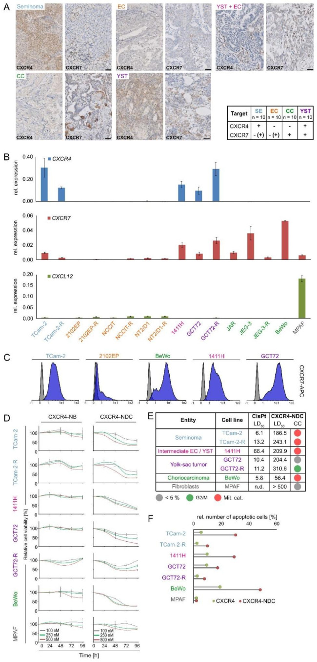
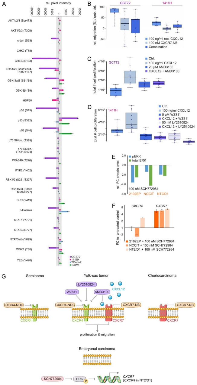
Being stimulated by the chemokine CXCL12, the CXCR4 / CXCR7 cascade is involved in tumor proliferation, migration, and metastasis. The interaction between CXCL12, secreted by cells from the microenvironment, and its receptors is complex and has been ascribed to promote chemotherapy resistance. However, the role of this signaling axis and its targetability in germ cell tumors (GCT) is not fully understood. Thus, this study investigated the therapeutic efficacy of a nanobody-drug-conjugate targeting CXCR4 (CXCR4-NDC) and functionally characterized this signaling pathway in GCT using small molecule inhibitors and nanobodies. As shown by diminished cell viability, enhanced apoptosis induction, and detection of mitotic catastrophes, we confirmed the cytotoxic efficacy of the CXCR4-NDC in CXCR4+-GCT cells (i.e. seminoma and yolk-sac tumor), while non-malignant CXCR4--fibroblasts, remained largely unaffected. Stimulation of CXCR4+ / CXCR7+-GCT cells with CXCL12 resulted in an enhanced proliferative and migratory capacity, while this effect could be reverted using CXCR4 inhibitors or a CXCR7-nanobody. Molecularly, the CXCR4 / CXCR7-signaling cascade could be activated independently of MAPK (ERK1 / 2)-phosphorylation. Although, in CXCR4- / CXCR7--embryonal carcinoma cells, CXCR7-expression was re-induced upon inhibition of ERK1 / 2-signaling. This study identified a nanobody-drug-conjugate targeting CXCR4 as a putative therapeutic option for GCT, i.e. seminoma and yolk-sac tumors. Furthermore, this study shed light on the functional role of the CXCR4 / CXCR7 / CXCL12-signaling cascade in GCT, demonstrating an important influence on proliferation and migration.
Keywords: CXCL12; CXCR4; CXCR7; Germ cell tumors; Nanobody-drug-conjugate; Resistance; Testis cancer; Therapy.
© 2023. The Author(s).
Conflict of interest statement
The authors declare no competing interests.
Figures
Similar articles
-
Inflammatory CXCL12-CXCR4/CXCR7 axis mediates G-protein signaling pathway to influence the invasion and migration of nasopharyngeal carcinoma cells.Tumour Biol. 2016 Jun;37(6):8169-79. doi: 10.1007/s13277-015-4686-2. Epub 2015 Dec 29. Tumour Biol. 2016. PMID: 26715277
-
The CXCL12-CXCR4/CXCR7 axis as a mechanism of immune resistance in gastrointestinal malignancies.Semin Cancer Biol. 2020 Oct;65:176-188. doi: 10.1016/j.semcancer.2019.12.007. Epub 2019 Dec 23. Semin Cancer Biol. 2020. PMID: 31874281 Review.
-
CXCR4, but not CXCR7, discriminates metastatic behavior in non-small cell lung cancer cells.Mol Cancer Res. 2014 Jan;12(1):38-47. doi: 10.1158/1541-7786.MCR-12-0334. Epub 2013 Sep 11. Mol Cancer Res. 2014. PMID: 24025971 Free PMC article.
-
Chemokine CXCL12 activates dual CXCR4 and CXCR7-mediated signaling pathways in pancreatic cancer cells.J Transl Med. 2012 Apr 2;10:68. doi: 10.1186/1479-5876-10-68. J Transl Med. 2012. PMID: 22472349 Free PMC article.
-
Drug Design Targeting the CXCR4/CXCR7/CXCL12 Pathway.Curr Top Med Chem. 2016;16(13):1441-51. doi: 10.2174/1568026615666150915120218. Curr Top Med Chem. 2016. PMID: 26369824 Review.
Cited by
-
Drug conjugates for the treatment of lung cancer: from drug discovery to clinical practice.Exp Hematol Oncol. 2024 Mar 1;13(1):26. doi: 10.1186/s40164-024-00493-8. Exp Hematol Oncol. 2024. PMID: 38429828 Free PMC article. Review.
-
Cisplatin‑resistant germ cell tumor models: An exploration of the epithelial‑mesenchymal transition regulator SLUG.Mol Med Rep. 2024 Dec;30(6):228. doi: 10.3892/mmr.2024.13352. Epub 2024 Oct 11. Mol Med Rep. 2024. PMID: 39392037 Free PMC article.
-
Modeling of cancer stem cells and the tumor microenvironment Via NT2/D1 cells to probe pathology and treatment for cancer and beyond.Discov Oncol. 2025 Apr 24;16(1):605. doi: 10.1007/s12672-025-02158-2. Discov Oncol. 2025. PMID: 40272656 Free PMC article. Review.
References
-
- Znaor A, Lortet-Tieulent J, Jemal A, Bray F. International variations and trends in testicular cancer incidence and mortality. Eur Urol. 2014;65(6):1095–106. - PubMed
Publication types
Grants and funding
LinkOut - more resources
Full Text Sources
Research Materials
Miscellaneous
