. 2024 Jan;78(1):e22284.
doi: 10.1002/syn.22284. Epub 2023 Nov 23.

Corticotropin-releasing factor-dopamine interactions in male and female macaque: Beyond the classic VTA

Affiliations

Corticotropin-releasing factor-dopamine interactions in male and female macaque: Beyond the classic VTA

E A Kelly et al. Synapse. 2024 Jan.

Abstract

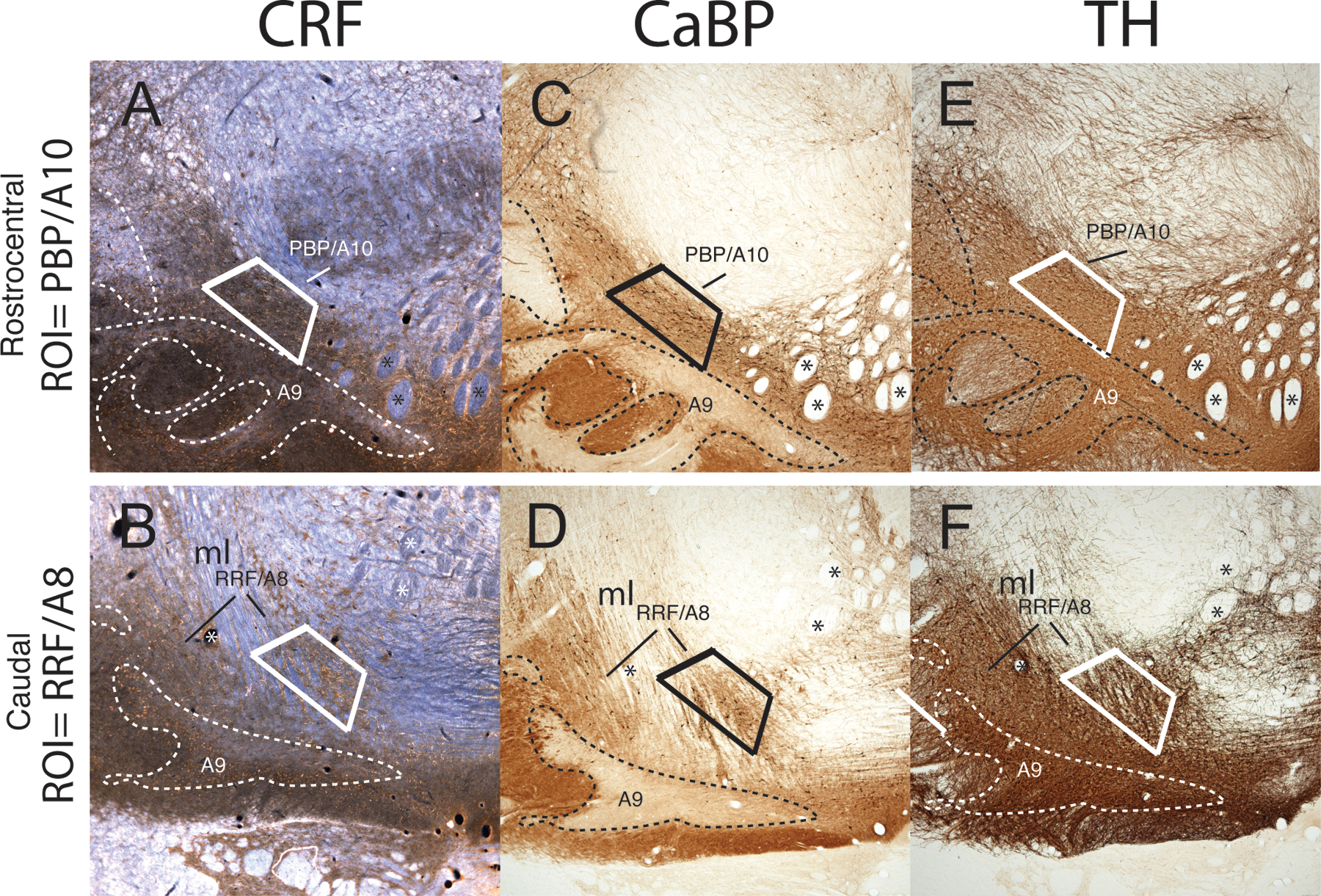
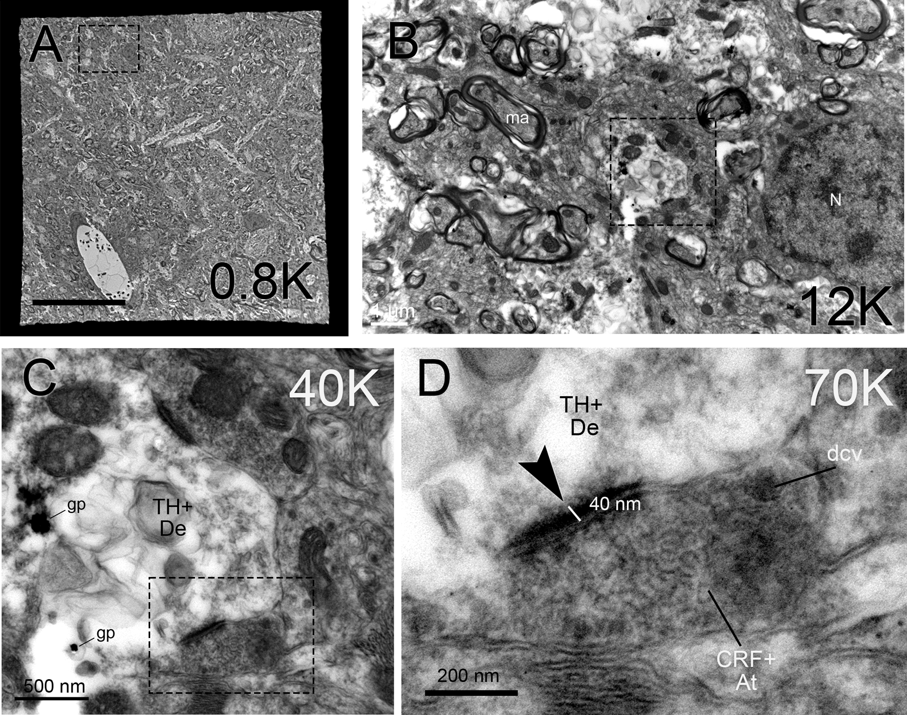
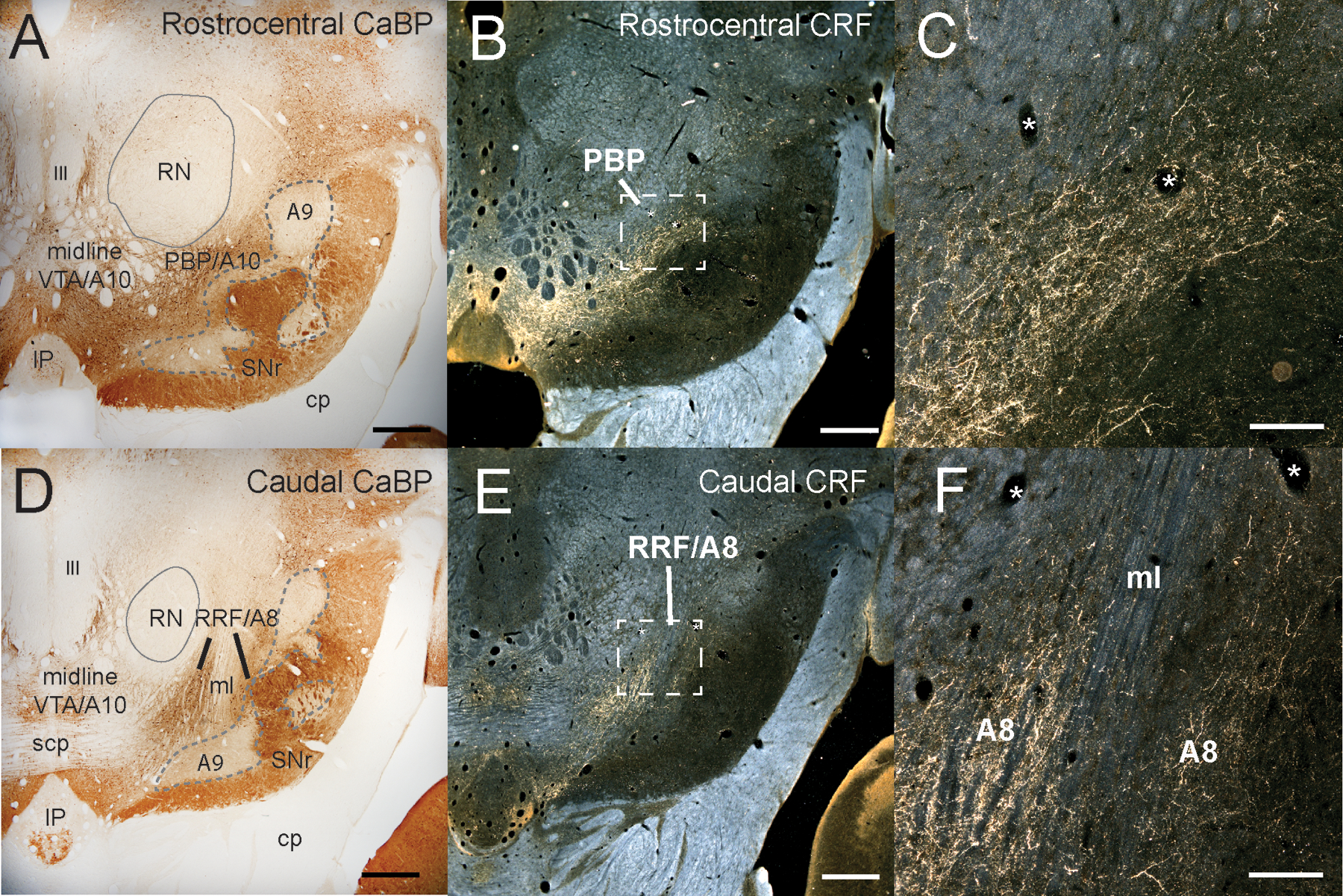
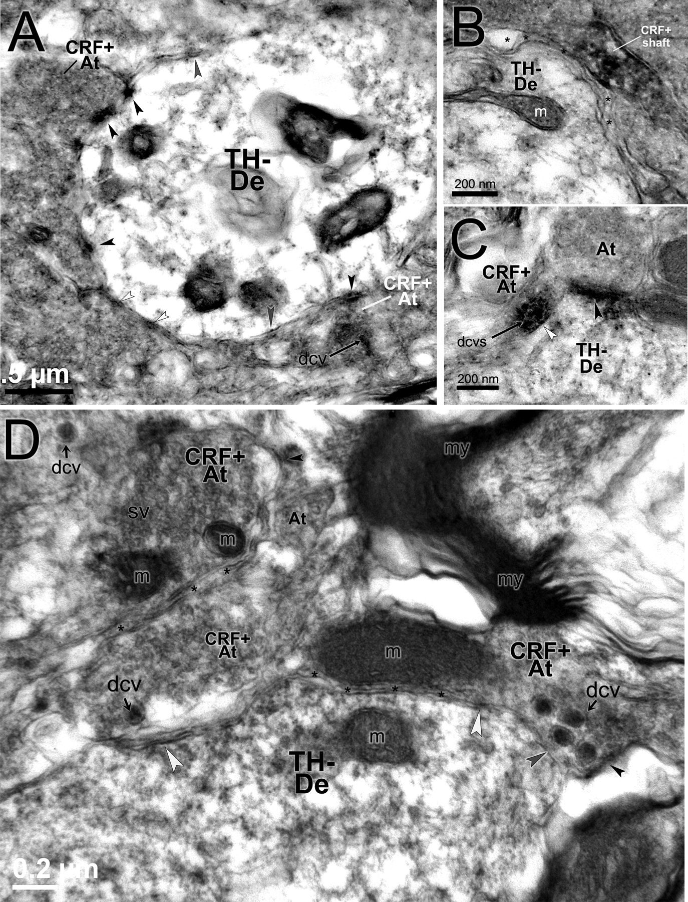
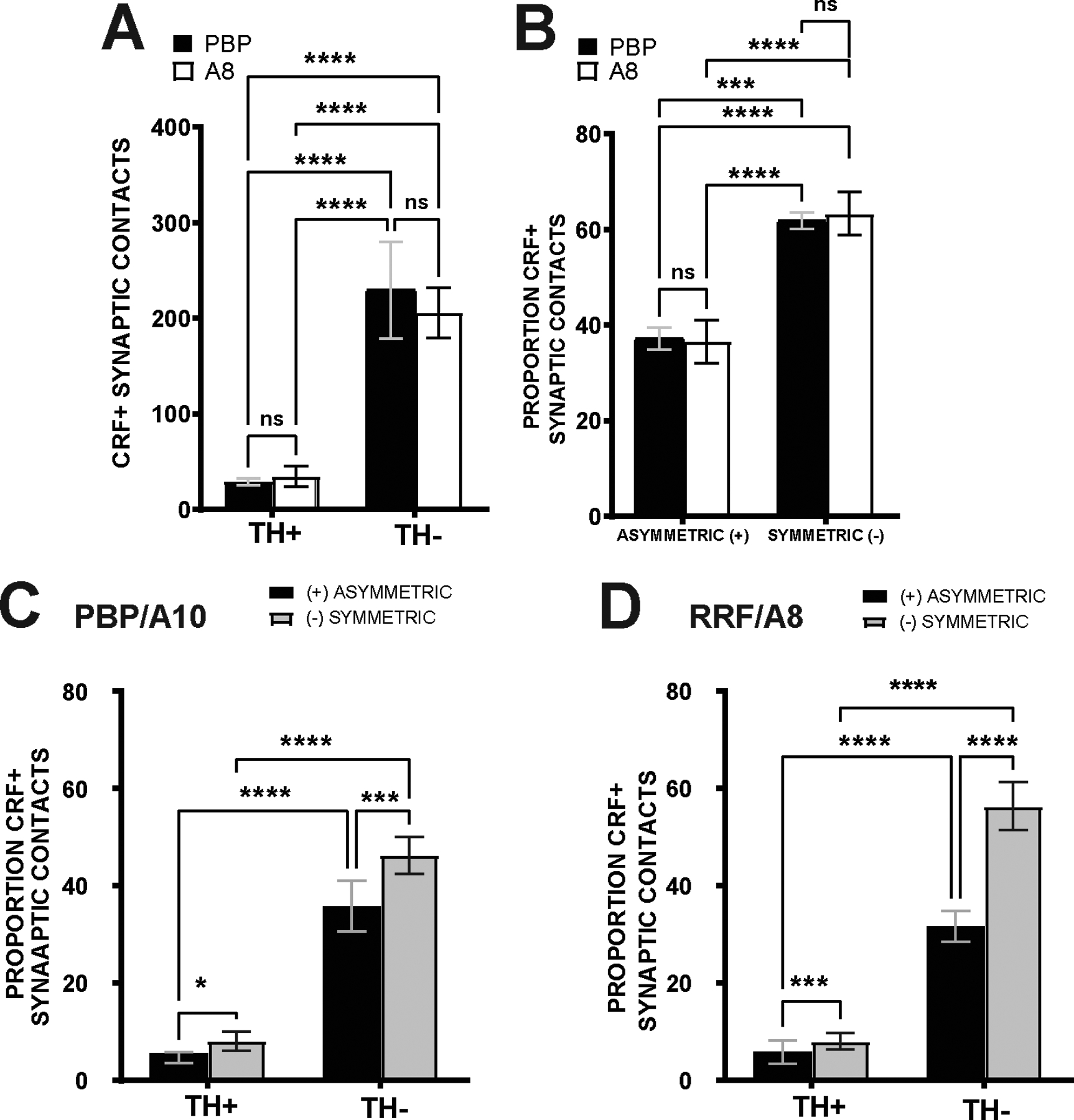
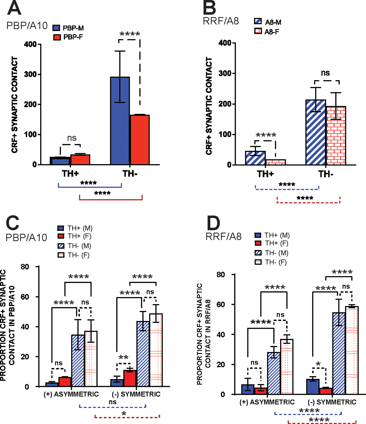
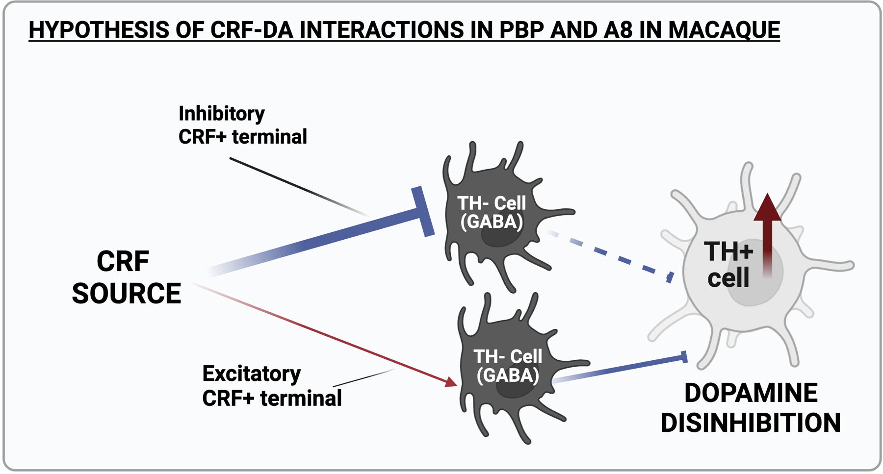
Dopamine (DA) is involved in stress and stress-related illnesses, including many psychiatric disorders. Corticotropin-releasing factor (CRF) plays a role in stress responses and targets the ventral midbrain DA system, which is composed of DA and non-DA cells, and divided into specific subregions. Although CRF inputs to the midline A10 nuclei ("classic VTA") are known, in monkeys, CRF-containing terminals are also highly enriched in the expanded A10 parabrachial pigmented nucleus (PBP) and in the A8 retrorubral field subregions. We characterized CRF-labeled synaptic terminals on DA (tyrosine hydroxylase, TH+) and non-DA (TH-) cell types in the PBP and A8 regions using immunoreactive electron microscopy (EM) in male and female macaques. CRF labeling was present mostly in axon terminals, which mainly contacted TH-negative dendrites in both subregions. Most CRF-positive terminals had symmetric profiles. In both PBP and A8, CRF symmetric (putative inhibitory) synapses onto TH-negative dendrites were significantly greater than asymmetric (putative excitatory) profiles. This overall pattern was similar in males and females, despite shifts in the size of these effects between regions depending on sex. Because stress and gonadal hormone shifts can influence CRF expression, we also did hormonal assays over a 6-month time period and found little variability in basal cortisol across similarly housed animals at the same age. Together our findings suggest that at baseline, CRF-positive synaptic terminals in the primate PBP and A8 are poised to regulate DA indirectly through synaptic contacts onto non-DA neurons.

Keywords: A8); corticotropin; dopamine; electron microscopy; nonhuman primate; parabrachial nucleus (PBP); retrorubral field (RRF; ventral tegmental area (VTA).


Conflict of interest statement

Competing Interests.

The authors have no financial or non-financial interests to disclose.

Figures

Figure 1.
Figure 1.. Localization of the PBP and A8 for EM.
Near adjacent compartments immuno-labeled for CRF, CaBP and TH to identify and block regions of interest (ROIs) for immuno-EM processing. Rostral adjacent compartments showing the PBP (A, C, E) and caudal adjacent compartments containing A8 (B, D, F) with CRF-, CaBP-, and TH-IR, respectively. Fiducial markers such as fiber bundles (e.g. fascicles of the 3rd nerve(asterisks) and medial lemniscus (ml)) and blood vessels were used to localize the region of interest in EM section.
Figure 2.
Figure 2.. EM micrograph acquisition.
(A) Example of a single electron microscopy grid opening at 0.8 K magnification. Scale bar= 20 um (B) 12K magnification of boxed region in (A) showing visualization of synaptic vesicle-rich presynaptic terminals and electron-lucent dendritic structures (with and without gold particle deposits). Scale bar= 1 um (C) Under 40K magnification, boxed region in (B) shows synaptic specialization and a CRF-positive dense core vesicle (dcv) is beginning to become visible in the axon terminal. At this magnification, pre- and postsynaptic membranes are discernible, and CRF+ immunoreactive accumulation is also seen in the terminal. Two gold particles identify the post-synaptic dendrite as TH-positive (TH+De) Scale bar= 500 nm (D) 70K magnification of boxed region in (C), to confirm structural identification. A CRF+ immunoreactive presynaptic terminal (CRF+At) abuts the TH-positive dendrite (TH+De). A dense core vesicle is apparent at the outer edge of the asymmetric synapse (black arrow), in addition to punctate reaction product. The asymmetric synapse is classified by the presence of a 40nm post-synaptic density, and little to no density on the presynaptic side. Scale bar=200 nm. Abbreviations: CRF+At, CRF-positive axon terminal; dcv, dense core vesicle; gp, gold particle; ma, myelinated axon; N, nucleus; TH+DE, TH-positive dendrite.
Figure 3.
Figure 3.. Localization of CRF immunoreactivity in ventral midbrain.
(A) Low magnification brightfield micrograph of the ventral midbrain rostrocentral level seen with CaBP-IR, a marker of the A10 and A8 neurons. CaBP-positive A10 neurons contrast with CaBP-negative SNc/A9 neurons, outlined with dotted line. The substantia nigra, pars reticulata (SNr) has dense CaBP-IR fibers. (B) CRF-IR in fibers in adjacent section, visualized with darkfield microscopy. (C) A higher magnification of boxed region in B showing patches of thin beaded CRF-positive fibers in a section of the PBP. (D) Low magnification brightfield micrograph of CaBP-IR in the caudal ventral midbrain. CaBP-IR neurons are found in the A10 and RRF/A8, and absent in SNc/A9 subregion (dotted line). (E) CRF-IR in a neighboring section to panel D, seen under dark-field microscopy. (F) CRF-labeled fibers course through the RRF/A8, which is bi-sected by the medial lemniscus (ml) seen under higher magnification (taken from boxed area in E). Scale bar in A,B,D,E= 1 mm; C,F= 250 um. Abbreviations: III, Third nerve; CRF, corticotropin releasing factor; CaBP, calbindin-28kD; IP, interpeduncular nucleus; cp, cerebral peduncle; scp, superior cerebellar peduncle; ml, medial lemniscus; SNr, substantia nigra reticulata; PBP, parabrachial pigmented nucleus; RN, red nucleus; VTA, ventral tegmental area.
Figure 4.
Figure 4.. CRF-IR axon terminals.
(A) Several CRF-IR axon terminals (CRF+At) with synaptic specializations on a TH- negative, electron-lucent dendrite. CRF-IR is seen as irregular peroxidase labeling surrounding clear vesicles throughout the presynaptic space, and in a dense core vesicle (dcv, arrow) in the lower right CRF+At, which lies away from the synaptic junction. The CRF+At in the lower right has both a symmetric synaptic profile (white arrowhead) and an apposition (gray arrowhead). Scale bar= 500nm (B) A CRF-labeled axonal shaft (CRF + shaft), enveloped by a clear astrocytic process (asterisks) neighboring a TH-negative dendrite. Scale bar= 200 nm. (C) CRF+ At, with CRF+ dcvs, making a symmetric synapse (white arrowhead) with a TH-negative dendrite. Adjacent is an At (CRF-negative) making an asymmetric contact (black arrowhead) with the same dendrite. Scale bar=200 nm. (D) CRF-positive axon terminals (CRF-At+); several are abutting a TH-negative dendrite (TH-De). A cluster of darkly labeled dense core vesicles (dcvs) are evident in the CRF-At+ structure on the right. Asterisks show astrocytic process interposed between the terminal and dendrite. Symmetric (white arrowheads), asymmetric (black arrowheads) synaptic profiles, and an apposition (gray arrowhead), are indicated. Scale bar=200 nm. Abbreviations: At, axon terminal (unlabeled) ; CRF+At, CRF-positive axon terminal; dcv, dense core vesicle; m, mitochondria; ma, myelinated axon; sv, small vesicles; TH+De, TH-positive dendrite; TH-dendrite, TH-negative dendrite.
Figure 5.
Figure 5.. CRF-positive terminals onto TH-positive and TH-negative dendrites.
(A) Lower magnification (30,0000X) electron micrograph detailing the specificity of TH-positive gold particles in several dendritic structures (TH+De). Gold particles are diffusely distributed throughout the cytoplasm of electron-lucent dendrites, allowing characterization of synaptic contacts by neighboring presynaptic terminals at higher power. Black arrowheads, asymmetric synapses; gray arrowhead, apposition; small arrow indicates appearance of a dense core vesicle (dcv) at low power. Scale bar= 1 micron. (B) A CRF-positive axon terminal (CRF+At) with dcv and punctate immunoreactive immunoreactivity making a symmetric contact (white arrowhead) with a TH-positive dendrite (TH+De). Several gold particles (gp) are seen in the dendrite. Other unlabeled axon terminals make contact with the same TH+De (black arrowheads, asymmetric synapse; white arrowheads, symmetric synapse; gray arrowhead, apposition). An unlabeled dense core vesicle (u-dcv) is also indicated. Scale bar= 200 nm. (C) A CRF-positive axon terminal (CRF+At) with several CRF-positive dcvs (arrow) making a symmetric contact with a TH-positive dendrite. Scale bar- 200 nm. (D) A large CRF-positive axon terminal (CRF+At) with dcv making an asymmetric (excitatory) synaptic contact onto a TH-negative dendrite (black arrowhead). Scale bar- 200 nm. Abbreviations: At, axon terminal (unlabeled) ; CRF+At, CRF-positive axon synaptic terminal; dcv, dense core vesicle; gp, gold particle; m, mitochondria; ma, myelinated axon; TH+De, TH-positive dendrite; TH-De, TH-negative dendrite, u-dcv, unlabeled dense core vesicle.
Figure 6.
Figure 6.. CRF positive axon contacts in PBP/A10 and RRF/A8.
(A) CRF- positive axon synapses across TH-positive versus TH-negative profiles in the PBP and A8. Significantly more synapses were found on TH-negative profiles in both PBP and A8. Between region comparisons were not significantly different. (B) Relative proportion of CRF synapse type (asymmetric,(+] excitatory; symmetric,[-] inhibitory) in PBP and A8. There are significantly more symmetric than asymmetric synapses in both PBP and A8, with no significant differences between regions. (C) Proportion of CRF-positive synaptic contacts by synapse type (asymmetric [+] vs symmetric [-]) onto TH-positive and TH-negative profiles in the PBP/A10. Significantly more CRF-positive synapses (both asymmetric and symmetric) were found on TH-negative profiles. The proportion of asymmetric versus symmetric synapses was significantly increased on both TH-positive versus TH-negative profiles. (D) Proportion of CRF-positive synaptic contacts across TH-positive and TH-negative profiles, and the relative proportion of synapse type onto each group (asymmetric [+] vs symmetric [-]) in RRF/A8. As in the PBP, significantly more synapses (both asymmetric and symmetric) were found in TH-negative profiles. Within each category of post-synaptic partners, the relative proportion of symmetric synapses was significantly higher than asymmetric synapses. *= p≤ 0.05. **= p<0.005, ***= p<0.001, ****= p<0.0001.
Figure 7.
Figure 7.. CRF-positive synaptic contacts in PBP/A10 and RRF/A8 in males vs females.
(A, B) Comparison of all CRF-positive synapses onto TH-positive versus TH-negative post-synaptic profiles in the PBP (A) and A8 (B) in males and females. (C-D) Proportion of CRF-IR asymmetric and symmetric contacts onto TH-positive and TH-negative dendrites in the PBP (C) and A8 (D), considered in males (blue) and females (red). There were no differences in the proportion of asymmetric (+) versus symmetric (−) type synapses between males and females. Proportions of both types of CRF-positive synapses were significantly higher on the TH-negative (compared to TH-positive) profiles, with no differences between in males versus females. In both PBP (C) and A8 (D), there was a significantly higher proportion of total asymmetric-type synapses versus symmetric synapses overall. Significantly more symmetric synapses were found on TH-negative profiles in both sexes (male: blue comparison bar below graph, female: red comparison bar below graph). See text for details. *= p≤ 0.05. **= p<0.005, ***= p<0.001, ****= p<0.0001.
Figure 8.
Figure 8.. Pubertal and stress hormone assays in adolescent male and female macaques.
Gender-specific developmental hormones and stress hormone concentrations from serum. Male hormones: androstenedione, testosterone, and DHEA (brown, orange, blue); Female hormones progesterone, estrone and estradiol (yellow, green, blue). Stress hormones: cortisone and cortisol (pink, purple). Asterisks show animals used in the present study. Descriptions of hormones can be found in Table 4.
Figure 9.
Figure 9.. Indirect CRF modulation of DA cells.
CRF-positive synaptic terminals predominantly make symmetric (inhibitory) synapses onto TH-negative profiles in both PBP and A8. We hypothesize that under stress, CRF may enhance inhibitory effects on TH-negative profiles (presumptive GABA interneurons), resulting in a disinhibition of DA neurons.

Similar articles

Cited by

References

    1. Arias-Carrion O, Stamelou M, Murillo-Rodriguez E, Menendez-Gonzalez M, Poppel E (2010) Dopaminergic reward system: a short integrative review. Int Arch Med 3:24. doi:10.1186/1755-7682-3-24 - DOI - PMC - PubMed
    1. Arsenault MY, Parent A, Seguela P, Descarries L (1988) Distribution and morphological characteristics of dopamine-immunoreactive neurons in the midbrain of the squirrel monkey (Saimiri sciureus). J Comp Neurol 267:489–506 - PubMed
    1. Balsters JH, Zerbi V, Sallet J, Wenderoth N, Mars RB (2020) Primate homologs of mouse cortico-striatal circuits. eLife 9. doi:10.7554/eLife.53680 - DOI - PMC - PubMed
    1. Bangasser DA, Reyes BA, Piel D, Garachh V, Zhang XY, Plona ZM, Van Bockstaele EJ, Beck SG, Valentino RJ (2013) Increased vulnerability of the brain norepinephrine system of females to corticotropin-releasing factor overexpression. Molecular psychiatry 18 (2):166–173. doi:10.1038/mp.2012.24 - DOI - PMC - PubMed
    1. Bangasser DA, Valentino RJ (2012) Sex differences in molecular and cellular substrates of stress. Cellular and molecular neurobiology 32 (5):709–723. doi:10.1007/s10571-012-9824-4 - DOI - PMC - PubMed