. 2023 Oct 27;12(11):1372.
doi: 10.3390/biology12111372.

Proton Microbeam Targeted Irradiation of the Gonad Primordium Region Induces Developmental Alterations Associated with Heat Shock Responses and Cuticle Defense in Caenorhabditis elegans

Affiliations

Proton Microbeam Targeted Irradiation of the Gonad Primordium Region Induces Developmental Alterations Associated with Heat Shock Responses and Cuticle Defense in Caenorhabditis elegans

Pierre Beaudier et al. Biology (Basel). .

Abstract

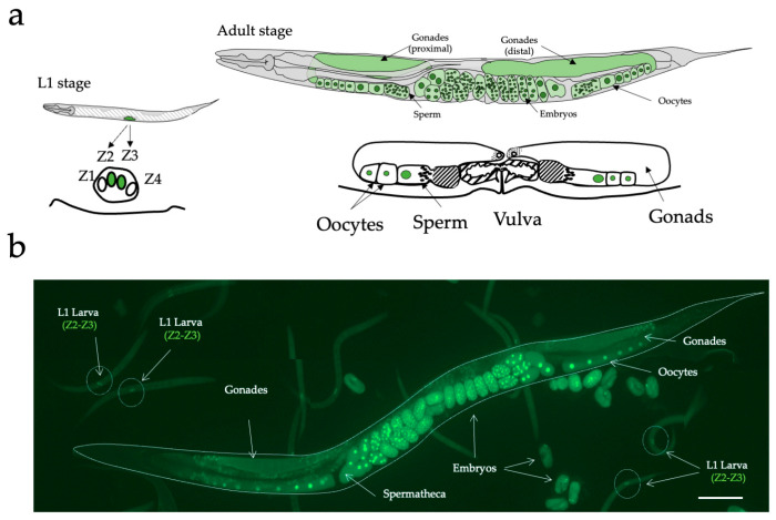
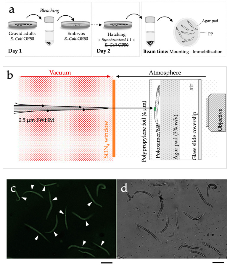
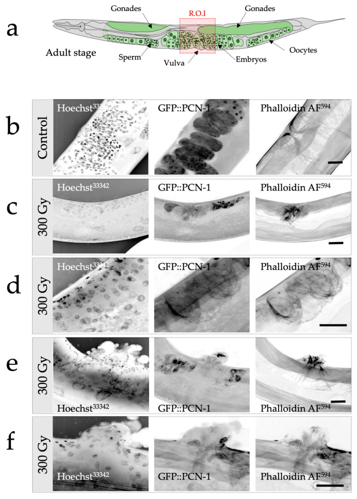
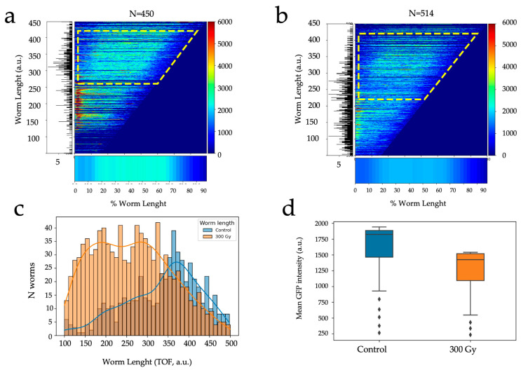
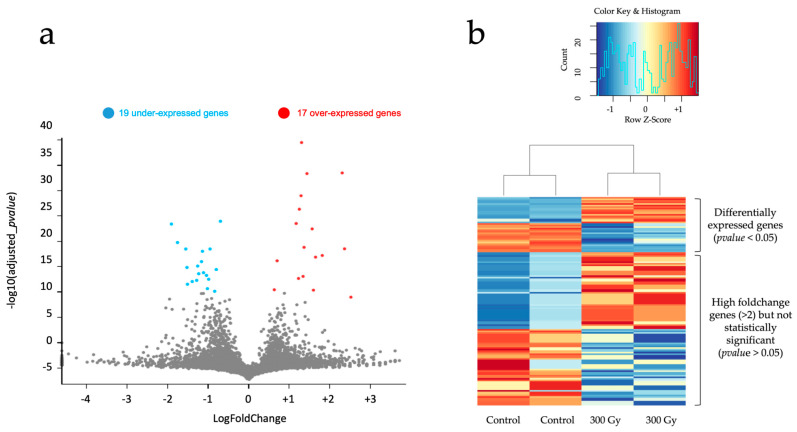
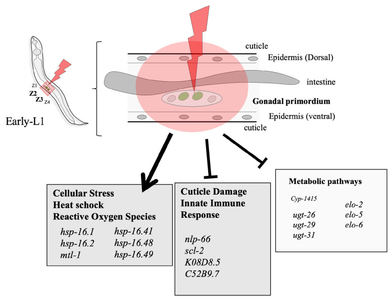
We describe a methodology to manipulate Caenorhabditis elegans (C. elegans) and irradiate the stem progenitor gonad region using three MeV protons at a specific developmental stage (L1). The consequences of the targeted irradiation were first investigated by considering the organogenesis of the vulva and gonad, two well-defined and characterized developmental systems in C. elegans. In addition, we adapted high-throughput analysis protocols, using cell-sorting assays (COPAS) and whole transcriptome analysis, to the limited number of worms (>300) imposed by the selective irradiation approach. Here, the presented status report validated protocols to (i) deliver a controlled dose in specific regions of the worms; (ii) immobilize synchronized worm populations (>300); (iii) specifically target dedicated cells; (iv) study the radiation-induced developmental alterations and gene induction involved in cellular stress (heat shock protein) and cuticle injury responses that were found.

Keywords: COPAS; Caenorhabditis elegans; gonadal–vulval development; microbeam; nanopore sequencing.


Conflict of interest statement

The authors declare no conflict of interest.

Figures

Figure 1
Figure 1
GZ264 transgenic C. elegans strain. (a) Schematic representation of post-embryonic hermaphrodite gonadal and vulval development in wild-type hermaphrodites. Comparative size of gonads at different stages is not to scale. Each diagram represents two stages of post-embryonic development of the gonad and the vulva. L1 stage: Two cells, Z1 and Z4, are the progenitors of the somatic tissues of gonad. Two other cells, Z2 and Z3, give rise to the germ line. The line below the gonad represents the underlying ventral hypodermis. Adult stage: somatic gonadal development is completed. The vulval invagination has everted to form the mature lips of the vulva. Oocytes and sperm are visible in the proximal part of the gonad. Description and scheme according to Seydoux et al., 1993 [30]. (b) Fluorescence imaging of different developmental stages expressing the GFP::PCN-1: adult, embryos, and L1 larvae. Scale bar: 100 µm.
Figure 2
Figure 2
Schematic representation of the different steps needed for micro-irradiation of C. elegans larvae (early L1 stage). (a) Preparation of large populations of early C. elegans L1 larvae by bleaching in the absence of E. coli OP50. (b) Detailed scheme of the microbeam end- station. 30 min before irradiation, an aliquot ~2 µL was directly deposited on a sterile 4 μm thick polypropylene (PP) foil and immediately covered with an afresh agar pad to maintain immobilized worms in a thin layer of medium. To prevent desiccation and contamination, the dish was closed with a glass side coverslip. Z2-Z3 nuclei were targeted using online fluorescence microscopy. The beam was positioned on the targeted cell. (c) PCN1::GFP detection in Z2-Z3 cells in synchronized early L1 using on-line fluorescence microscopy as obtained in real experimental conditions. White arrows indicate Z2-Z3 GFP-positive cells in synchronized early L1 larvae. (d) Synchronized early L1 larvae visualized using phase contrast imaging. scale bar: 100 µm.
Figure 3
Figure 3
Radiation-induced alterations of gonadal and vulval development in C. elegans following selective and targeted irradiation analyzed by confocal microscopy. (a) Schematic representation of the gonads and vulva in an adult worm (control). The region of interest (ROI) is depicted with the red square. (b) Confocal imaging of the ROI of an adult worm. Nuclei (DNA) are visualized using Hoechst33342 (left column), in utero fertilized embryos are revealed thanks to the GFP reporter (middle column), and the vulvar muscles with their typical cross-sectional profile are detected with the help of phalloidin (actin fibers; right column). (cf). Confocal imaging of the ROI of structural alterations detected in irradiated worms (300 Gy). Several configurations were observed. (c) Gonadal and vulval agenesis, absence of gonadal and vulval development with a limited actin fibrillar network, no embryos detected, absence of the typical cross-sectional profile of the vulval muscles. (d) Gonadal agenesis and vulvar developmental anomalies. Evidence of tissue disorganization illustrated by the actin fibrillar network and absence of gonads. (e,f) Gonadal agenesis and abnormal vulval eversion. Tissular disorganization, cell nuclei proliferation and disorganization, alteration of the actin fibrillar network, and evidence of abnormal vulvar eversion and agenesis. Scale bar: 10 µm.
Figure 4
Figure 4
Radiation-induced developmental alterations in C. elegans following selective irradiation, analyzed by COPAS. (a) Longitudinal fluorescence profiles (GFP) in control population as a function of development (worm length, TOF). The appearance of gonads at the L3/L4 stage and above (TOF > 250–300, dotted yellow region of interest) is converted into a bar where the calculated mean GFP longitudinal intensity profile is indicated by the length corresponding to the animal’s size and the color codes for the fluorescence intensity (bottom figure). The corresponding distribution of worm length is shown on the left panel. (b) Longitudinal profiles of GFP fluorescence in the population micro-irradiated at 300 Gy. The appearance of gonads at the L3/L4 stage and above (TOF > 250–300, dotted yellow region of interest) is visible in the calculated mean GFP longitudinal profile (bottom figure). (c) Length distribution of worms in control population (blue) and irradiated population (orange). (d) Average fluorescence intensity in worms above L3/L4 stage (control, N = 450; 300 Gy, N = 514).
Figure 5
Figure 5
Bulk transcriptomic analysis of cellular expression differences in two C. elegans samples containing multiple pooled replicates following selective irradiation. (a) Volcano plot of differentially expressed (DE, gray dots) genes in the 300 Gy condition compared to the control. DE genes were computed by fitting a linear model on each gene, extracting contrasts and performing an empirical Bayes test. The genes with a p-value < 0.05 (after Bonferroni correction) are considered as DE. (b) Heatmap of relative gene expression between control and 300 Gy samples on the 36 DE genes and 100 high foldchange genes (>2), 50 up-regulated and 50 down-regulated genes, not classified as DE. The 100 high foldchange genes selected were the top 50 highest foldchange in up-regulation and down-regulation.
Figure 6
Figure 6
Schematic representation of data obtained from differential expression analysis on C. elegans bulk transcriptomes, following selective irradiation performed on gonadal primordium cells at early L1 stage. Genes belonging to distinct pathways of interest were identified as differentially expressed 3 h post-irradiation.

References

    1. Sulston J.E. Neuronal Cell Lineages in the Nematode Caenorhabditis elegans. Pt 2Cold Spring Harb. Symp. Quant. Biol. 1983;48:443–452. doi: 10.1101/SQB.1983.048.01.049. - DOI - PubMed
    1. Sulston J.E., Horvitz H.R. Post-Embryonic Cell Lineages of the Nematode, Caenorhabditis elegans. Dev. Biol. 1977;56:110–156. doi: 10.1016/0012-1606(77)90158-0. - DOI - PubMed
    1. Kimble J., Hirsh D. The Postembryonic Cell Lineages of the Hermaphrodite and Male Gonads in Caenorhabditis elegans. Dev. Biol. 1979;70:396–417. doi: 10.1016/0012-1606(79)90035-6. - DOI - PubMed
    1. Sulston J.E., White J.G. Regulation and Cell Autonomy during Postembryonic Development of Caenorhabditis elegans. Dev. Biol. 1980;78:577–597. doi: 10.1016/0012-1606(80)90353-X. - DOI - PubMed
    1. Avery L., Horvitz H.R. A Cell That Dies during Wild-Type C. elegans Development Can Function as a Neuron in a Ced-3 Mutant. Cell. 1987;51:1071–1078. doi: 10.1016/0092-8674(87)90593-9. - DOI - PMC - PubMed

LinkOut - more resources