Review
. 2023 Nov 5;12(11):1402.
doi: 10.3390/biology12111402.

Caveolin-1: A Review of Intracellular Functions, Tissue-Specific Roles, and Epithelial Tight Junction Regulation

Affiliations
Review

Caveolin-1: A Review of Intracellular Functions, Tissue-Specific Roles, and Epithelial Tight Junction Regulation

Cody M Dalton et al. Biology (Basel). .

Abstract

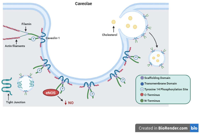
Caveolin-1 (Cav1) is a vital protein for many cellular processes and is involved in both the positive and negative regulation of these processes. Cav1 exists in multiple cellular compartments depending on its role. Of particular interest is its contribution to the formation of plasma membrane invaginations called caveolae and its involvement in cytoskeletal interactions, endocytosis, and cholesterol trafficking. Cav1 participates in stem cell differentiation as well as proliferation and cell death pathways, which is implicated in tumor growth and metastasis. Additionally, Cav1 has tissue-specific functions that are adapted to the requirements of the cells within those tissues. Its role has been described in adipose, lung, pancreatic, and vascular tissue and in epithelial barrier maintenance. In both the intestinal and the blood brain barriers, Cav1 has significant interactions with junctional complexes that manage barrier integrity. Tight junctions have a close relationship with Cav1 and this relationship affects both their level of expression and their location within the cell. The ubiquitous nature of Cav1 both within the cell and within specific tissues is what makes the protein important for ongoing research as it can assist in further understanding pathophysiologic processes and can potentially be a target for therapies.

Keywords: Caveolin-1; barrier function; caveolae; cellular processes; tight junction.


Conflict of interest statement

The authors declare no conflict of interest.

Figures

Figure 1
Figure 1
Caveolin-1 Structure and Function. Depiction of Caveolin-1 structure including its scaffolding domain, transmembrane domain, and Tyrosine 14 phosphorylation site. The structure of caveolae is represented with Caveolin-1 proteins forming the surface of the plasma membrane invagination. Important functions of Caveolin-1 are illustrated showing cytosolic interactions with actin filaments, endocytosis and cholesterol trafficking, tight junction shuttling, and intracellular signaling eNOS inactivation and decreased production of NO as an example.
Figure 2
Figure 2
Caveolin-1 Localization. Immunofluorescence staining of healthy small intestinal tissue showing a cross-sectional image of intestinal villi and polarized epithelial cells. Caveolin-1 (red) is seen localized to the apical and basolateral membranes rather than cytosolic compartments. Image taken at 20× magnification, the scale bar on the bottom right indicates 50 μm. Epithelial cell nuclei are stained blue using Fluroshield with DAPI (Sigma, St. Louis, MO, USA, CAT#F6057). Caveolin-1 is stained red using primary antibody (Novus, Centennial, CO, USA, CAT#NB100-615) and secondary antibody (Invitrogen, Waltham, MA, USA, CAT#A11032).

Similar articles

Cited by

References

    1. Kumar V. T cells and their immunometabolism: A novel way to understanding sepsis immunopathogenesis and future therapeutics. Eur. J. Cell Biol. 2018;97:379–392. doi: 10.1016/j.ejcb.2018.05.001. - DOI - PubMed
    1. Michell D.L., Shihata W.A., Andrews K.L., Abidin N.A.Z., Jefferis A.-M., Sampson A.K., Lumsden N.G., Huet O., Parat M.-O., Jennings G.L., et al. High intraluminal pressure promotes vascular inflammation via caveolin-1. Sci. Rep. 2021;11:5894. doi: 10.1038/s41598-021-85476-z. - DOI - PMC - PubMed
    1. Baker N., Zhang G., You Y., Tuan R.S. Caveolin-1 regulates proliferation and osteogenic differentiation of human mesenchymal stem cells. J. Cell. Biochem. 2012;113:3773–3787. doi: 10.1002/jcb.24252. - DOI - PubMed
    1. Park J.-S., Kim H.-Y., Kim H.-W., Chae G.-N., Oh H.-T., Park J.-Y., Shim H., Seo M., Shin E.Y., Kim E.G., et al. Increased caveolin-1, a cause for the declined adipogenic potential of senescent human mesenchymal stem cells. Mech. Ageing Dev. 2005;126:551–559. doi: 10.1016/j.mad.2004.11.014. - DOI - PubMed
    1. Volonte D., Zhang K., Lisanti M.P., Galbiati F. Expression of Caveolin-1 Induces Premature Cellular Senescence in Primary Cultures of Murine Fibroblasts. Mol. Biol. Cell. 2002;13:2502–2517. doi: 10.1091/mbc.01-11-0529. - DOI - PMC - PubMed

LinkOut - more resources