Review
. 2023 Nov 12;12(11):1423.
doi: 10.3390/biology12111423.

Iron Homeostasis in Azotobacter vinelandii

Affiliations
Review

Iron Homeostasis in Azotobacter vinelandii

Elena Rosa-Núñez et al. Biology (Basel). .

Abstract

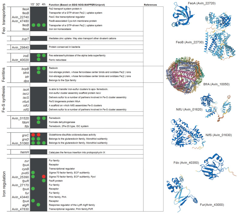
Iron is an essential nutrient for all life forms. Specialized mechanisms exist in bacteria to ensure iron uptake and its delivery to key enzymes within the cell, while preventing toxicity. Iron uptake and exchange networks must adapt to the different environmental conditions, particularly those that require the biosynthesis of multiple iron proteins, such as nitrogen fixation. In this review, we outline the mechanisms that the model diazotrophic bacterium Azotobacter vinelandii uses to ensure iron nutrition and how it adapts Fe metabolism to diazotrophic growth.

Keywords: biological nitrogen fixation; iron nutrition; iron transport; iron–sulfur cluster; nitrogenase.


Conflict of interest statement

The authors declare no conflict of interest.

Figures

Figure 1
Figure 1
Representative siderophores secreted by A. vinelandii cultures. (A) Catechol-based siderophores: 2,3-DHBA, aminochelin, azotochelin, and protochelin. (B) Mixed-type siderophores: azotobactin and vibrioferrin. (C) Chromosome position and organization of the A. vinelandii DJ (genome accession NC_012560) gene clusters involved in siderophore production. OR indicates the origin of replication.
Figure 2
Figure 2
Current understanding of iron acquisition and trafficking in A. vinelandii cells. Siderophores secreted into the environment bind Fe3+. The resulting complex is introduced in the periplasm through TonB transporters. In the periplasm, different pathways can be followed: (i) the Fe3+–siderophore complex is transported through an ABC system into the cytosol, (ii) Fe3+ dissociates or is released from the siderophore and then it is transported into the cytosol by a different ABC transporter, or (iii) dissociated Fe3+ is reduced by an unknown ferroreductase (1) into Fe2+, which is transported by a Feo iron import system into the cytosol. Cytosolic Fe2+ or Fe3+ can be delivered directly to Fe-S scaffold proteins or to other iron-using enzymes (dotted lines). Alternatively, the excess iron can be stored within (bacterio) ferritins, and may later be mobilized after reducing Fe3+ to Fe2+ including siderophore biosynthesis. Iron trafficking in the cytosol is facilitated by yet-to-be-identified iron chaperones (2). As in the periplasm, iron is also recovered from the internalized Fe3+–siderophore complex, and Fe3+ may be reduced to Fe2+. Finally, an unknown protein (3) will be responsible for iron delivery to the ferrochelatase for heme synthesis. Siderophores are shown in green. Bacterioferritin (BfrA) protein subunits are shown as blue circles. TonB stands for (phage) T-one resistance B; ExbB/D, for Excretion of colicin B inhibitor B/D; MFS is Major Facilitator Superfamily; RND, Resistance-Nodulation-Division; Feo, ferrous iron transport; IscU, Iron Sulfur-Cluster assembly U; NifU, Nitrogen Fixation U; VnfU, Vanadium nitrogen fixation U; and AnfU, Alternative nitrogen fixation U (iron-only nitrogenase). O.m: outer membrane; i.m. cytosolic membrane; and ?: proteins that have not been identified to date. * indicates that VnfU or AnfU are used instead of NifU for alternative nitrogenases.
Figure 3
Figure 3
List of A. vinelandii genes involved or putatively involved in iron homeostasis and their regulation under diazotrophic conditions. Genes are organized by function and named according to their accession numbers in the published genome [93,103]. Green and red dots indicate the up-regulation or down-regulation, respectively, of the expression of each gene in nitrogenase de-repressing conditions compared to nitrogen-sufficient conditions (NH3) at three time points after the removal of NH3 from the medium (15 min, 30 min, and 4 h). No dot means that no change in expression was observed. This comparison was made using the transcriptomic data deposited in Gene Expression Ommibus Accesion GSE244772. Functional annotations were obtained from EGG NOG-MAPPER and Uniprot. Structural models were generated using AlphaFold [109] and visualized with PyMOL (Schörindger, Inc, New York, NY, USA). The references indicated are Baars et al., 2016 [31], Yoneyama et al., 2011 [110], and Tindale et al., 2021 [111].
Figure 3
Figure 3
List of A. vinelandii genes involved or putatively involved in iron homeostasis and their regulation under diazotrophic conditions. Genes are organized by function and named according to their accession numbers in the published genome [93,103]. Green and red dots indicate the up-regulation or down-regulation, respectively, of the expression of each gene in nitrogenase de-repressing conditions compared to nitrogen-sufficient conditions (NH3) at three time points after the removal of NH3 from the medium (15 min, 30 min, and 4 h). No dot means that no change in expression was observed. This comparison was made using the transcriptomic data deposited in Gene Expression Ommibus Accesion GSE244772. Functional annotations were obtained from EGG NOG-MAPPER and Uniprot. Structural models were generated using AlphaFold [109] and visualized with PyMOL (Schörindger, Inc, New York, NY, USA). The references indicated are Baars et al., 2016 [31], Yoneyama et al., 2011 [110], and Tindale et al., 2021 [111].

References

    1. Bandyopadhyay S., Chandramouli K., Johnson M.K. Iron-sulfur cluster biosynthesis. Biochem. Soc. Trans. 2008;36:1112–1119. doi: 10.1042/BST0361112. - DOI - PMC - PubMed
    1. Layer G. Heme biosynthesis in prokaryotes. Biochim. Biophys. Acta Mol. Cell Res. 2021;1868:118861. doi: 10.1016/j.bbamcr.2020.118861. - DOI - PubMed
    1. Winterbourn C.C. Toxicity of iron and hydrogen peroxide: The Fenton reaction. Toxicol. Lett. 1995;82:969–974. doi: 10.1016/0378-4274(95)03532-X. - DOI - PubMed
    1. Stanbury D.M. The principle of detailed balancing, the iron-catalyzed disproportionation of hydrogen peroxide, and the fenton reaction. Dalton Trans. 2022;51:2135–2157. doi: 10.1039/D1DT03645A. - DOI - PubMed
    1. Tang D., Chen X., Kang R., Kroemer G. Ferroptosis: Molecular mechanisms and health implications. Cell Res. 2021;31:107–125. doi: 10.1038/s41422-020-00441-1. - DOI - PMC - PubMed