Review
. 2023 Nov 9;13(22):3410.
doi: 10.3390/diagnostics13223410.

Adhesive Capsulitis of the Shoulder: Current Concepts on the Diagnostic Work-Up and Evidence-Based Protocol for Radiological Evaluation

Affiliations
Review

Adhesive Capsulitis of the Shoulder: Current Concepts on the Diagnostic Work-Up and Evidence-Based Protocol for Radiological Evaluation

Riccardo Picasso et al. Diagnostics (Basel). .

Abstract

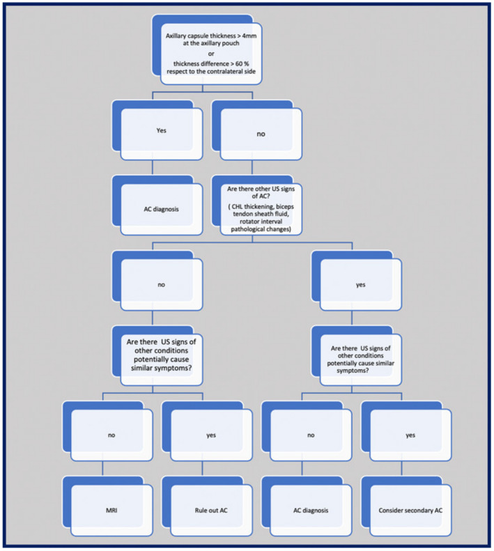
Adhesive capsulitis is an idiopathic and disabling disorder characterized by intense shoulder pain and progressive limitation of active and passive glenohumeral joint range of motion. Although adhesive capsulitis has been traditionally considered a diagnosis of exclusion that can be established based on a suggestive medical history and the detection of supporting findings at the physical exam, imaging studies are commonly requested to confirm the diagnostic suspicion and to exclude other causes of shoulder pain. Indeed, clinical findings may be rather unspecific, and may overlap with diseases like calcific tendinitis, rotator cuff pathology, acromioclavicular or glenohumeral arthropathy, autoimmune disorders, and subacromial/subdeltoid bursitis. Magnetic resonance imaging, magnetic resonance arthrography, and high-resolution ultrasound have shown high sensitivity and accuracy in diagnosing adhesive capsulitis through the demonstration of specific pathological findings, including thickening of the joint capsule and of the coracohumeral ligament, fibrosis of the subcoracoid fat triangle, and extravasation of gadolinium outside the joint recesses. This narrative review provides an updated analysis of the current concepts on the role of imaging modalities in patients with adhesive capsulitis, with the final aim of proposing an evidence-based imaging protocol for the radiological evaluation of this condition.

Keywords: MRA; MRI; adhesive capsulitis; frozen shoulder; ultrasound.


Conflict of interest statement

The authors declare that they have no competing interests.

Figures

Figure 1
Figure 1
Glenohumeral joint capsule. (A) Schematic drawing and (B) anatomic dissection show the glenohumeral joint capsule (arrows) and its relationship with the overlaying rotator cuff tendons. The superior glenohumeral ligament (SGHL), the middle glenohumeral ligament (MGHL), and the anterior (IGHLa) and the posterior (IGHLp) bands of the inferior glenohumeral ligament are demonstrated as focal thickening and folding of the anteroinferior capsule. SS, supraspinatus tendon; IS, infraspinatus tendon; Tm, teres minor tendon; Sub, subscapularis tendon; LhB, long head of the biceps tendon. Co, coracoid process; AR, axillary recess; asterisk, subscapularis recess.
Figure 2
Figure 2
Glenohumeral joint capsule, normal MRI findings. (A) Sagittal tSE T1-weighted MRI image shows the joint capsule (arrow) as a structure of intermediate signal located deep to the rotator cuff tendons. Note the intra-articular part of the long head of the biceps tendon (outlined arrowhead) running on the inner surface of the capsule. The coracohumeral ligament (white arrowhead) is demonstrated as a thin and low signal fibrillar structure running from the coracoid (Co) to the humeral head (HH). The subcoracoid fat triangle (asterisk) is a fat-filled space delimited by the coracohumeral ligament, the joint capsule, and the subscapularis (Sub) muscle. SS, supraspinatus muscle; black arrowhead, coracoacromial ligament. (B) Coronal tSE fat-suppressed T2-weighted MRI scan shows the inferior part of the joint capsule (arrow) as a low signal folding that delimits the axillary recess. Note the thin superior capsule (outlined arrow) located underneath the supraspinatus muscle and tendon (arrowhead). Gl, scapular glenoid; HH, humeral head; star, greater tuberosity.
Figure 3
Figure 3
Adhesive capsulitis, spectrum of MRI findings. (A) Sagittal tSE T1-weighted MRI scan from a 42 year old woman with a three-month history of shoulder pain demonstrates mild thickening of the coracohumeral ligament (white arrowhead) and initial effacement of the subcoracoid fat triangle (asterisk) by hypointense synovium. Black arrowhead, coracoacromial ligament. (B) Coronal tSE Proton Density MRI scan from a 68 year old woman with recent onset of pain and progressive limitation of glenohumeral ROM shows a marked thickening of the inferior capsule (arrows), which appears edematous and demonstrates increased signal intensity in fluid-sensitive sequences. (C) Sagittal tSE T1-weighted and (D) Sagittal tSE fat-suppressed T2-weighted MRI images from a 70 year old male with a one-year history of severe limitation of active and passive shoulder motion demonstrate complete obliteration of the subcoracoid fat triangle by synovial tissue (arrows), which is also extended underneath the long head of the biceps tendon (arrowhead) in the area of the pulley (asterisk). The coracohumeral ligament appears embedded by the synovium. Note severe thickening and hyperintensity of the anteroinferior capsule (outlined arrowhead). Ac, acromion; Cl, clavicle; Co, coracoid; SS, supraspinatus; Sub, subscapularis; GL, scapular glenoid; HH, humeral head.
Figure 4
Figure 4
Adhesive capsulitis in a 45 year old man with severe limitation of the glenohumeral ROM after a trauma, who was submitted for an MRA for a suspected labral tear. (A) Axial and (B) Coronal tSE fat-suppressed T1-weighted MR images obtained after intraarticular injection of gadolinium demonstrate anterior extravasation of the contrast medium (outlined arrows) into and underneath the subscapularis muscle (Sub) as a consequence of capsular stiffness and fissuration. Note the abnormally low distension of the axillary recess (arrows) and its markedly thickened walls. In (B) a partial thickness tear (white outlined arrowhead) of the articular side of the supraspinatus tendon (black arrowhead) is also evident. In effect, the tear involves both the inner fibers of the supraspinatus and the joint capsule, which are merged at this level to form the superior complex. As a consequence, note the superior migration of the contrast outside the joint cavity (black arrow). Black outlined arrowhead, long head of the bicep tendon; white arrowhead, subscapularis tendon; asterisk greater tuberosity; star, lesser tuberosity; HH, humeral head; Gl, glenoid; Ac, acromion.
Figure 5
Figure 5
Glenohumeral joint capsule and pericapsular ligaments, normal US findings. (A) Oblique transverse 18–5 MHz US image shows the normal thin and fibrillar appearance of the coracohumeral ligament (outlined arrowheads), which is demonstrated connecting the coracoid (Co) and humeral head (HH) in a deeper position respective to the coracoacromial ligament (arrowheads). Note the homogeneous and hyperechoic appearance of the subcoracoid fat (asterisk). (B) Short-axis 18–5 MHz US image shows the distal part of the coracohumeral ligament (outlined arrowheads) in the area of the rotator interval and the biceps pulley (asterisk). Bt, long head of the biceps tendon. (C) Longitudinal 18–5 MHz US obtained orienting the probe parallel to the humerus in the axillary region shows the inferior capsule (arrowheads) overlying the humeral head (HH) and folding over the humeral neck (HN).
Figure 6
Figure 6
US findings in a 53 year old woman with adhesive capsulitis. (A) Oblique transverse 18–5 MHz US image demonstrates the markedly thickened coracohumeral ligament (outlined arrowheads), which has lost the normal fibrillar echotexture and appears homogeneously hypoechoic due to fibrotic changes and degeneration of the fibers. Note the presence of hypoechoic synovial tissue in the subcoracoid triangle (asterisk). Arrowheads, coracoacromial ligament. (B) Short-axis 18–5 Mhz US evidences the thickening and fibrotization of the coracohumeral ligament (arrowheads) and the biceps pulley (asterisks) in the rotator interval. (C) Longitudinal 18–5 MHz US image shows a significant thickening of the inferior capsule (arrowheads). HH, humeral head; Co, coracoid; Bt, long head of the biceps tendon; HN, humeral neck.
Figure 7
Figure 7
Protocol for imaging evaluation of patients with suspected AC.

References

    1. Zappia M., Di Pietto F., Aliprandi A., Pozza S., De Petro P., Muda A., Sconfienza L.M. Multi-modal imaging of adhesive capsulitis of the shoulder. Insights Imaging. 2016;7:365–371. doi: 10.1007/s13244-016-0491-8. - DOI - PMC - PubMed
    1. Codman E.A. The Shoulder: Rupture of the Supraspinatus Tendon and Other Lesions in or about the Subacromial Bursa. Thomas Todd Co.; Boston, MA, USA: 1934. pp. 216–222.
    1. Neviaser J.S. Adhesive capsulitis of the shoulder (the frozen shoulder) Med. Times. 1962;90:783–807. - PubMed
    1. Papalexis N., Parmeggiani A., Facchini G., Miceli M., Carbone G., Cavallo M., Spinnato P. Current concepts in the diagnosis and treatment of adhesive capsulitis: Role of diagnostic imaging and ultrasound-guided interventional procedures. Radiol. Med. 2022;127:1390–1399. doi: 10.1007/s11547-022-01566-6. - DOI - PubMed
    1. Fields B.K.K., Skalski M.R., Patel D.B., White E.A., Tomasian A., Gross J.S., Matcuk G.R. Adhesive capsulitis: Review of imaging findings, pathophysiology, clinical presentation, and treatment options. Skelet. Radiol. 2019;48:1171–1184. doi: 10.1007/s00256-018-3139-6. - DOI - PubMed