. 2023 Nov 10;9(11):1097.
doi: 10.3390/jof9111097.

The Early Endocytosis Gene PAL1 Contributes to Stress Tolerance and Hyphal Formation in Candida albicans

Affiliations

The Early Endocytosis Gene PAL1 Contributes to Stress Tolerance and Hyphal Formation in Candida albicans

Miranda Yu et al. J Fungi (Basel). .

Abstract

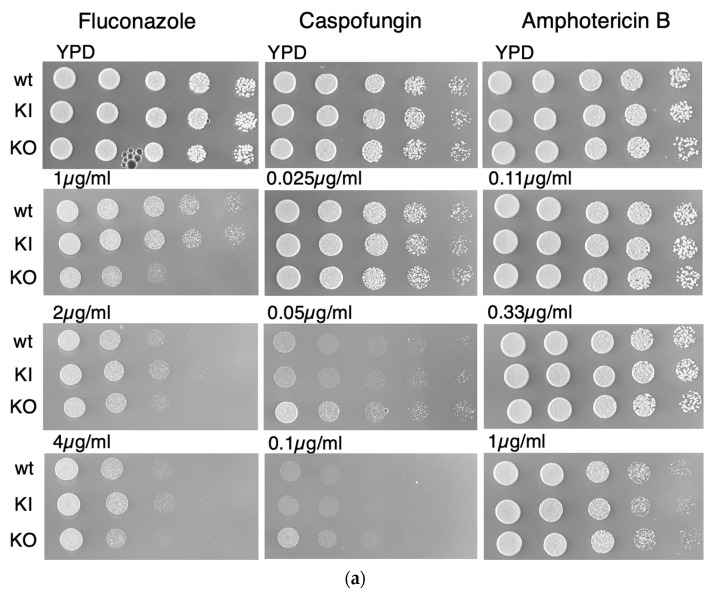
The endocytic and secretory pathways of the fungal pathogen Candida albicans are fundamental to various key cellular processes such as cell growth, cell wall integrity, protein secretion, hyphal formation, and pathogenesis. Our previous studies focused on several candidate genes involved in early endocytosis, including ENT2 and END3, that play crucial roles in such processes. However, much remains to be discovered about other endocytosis-related genes and their contributions toward Candida albicans secretion and virulence. In this study, we examined the functions of the early endocytosis gene PAL1 using a reverse genetics approach based on CRISPR-Cas9-mediated gene deletion. Saccharomyces cerevisiae Pal1 is a protein in the early coat complex involved in clathrin-mediated endocytosis that is later internalized with the coat. The C. albicans pal1Δ/Δ null mutant demonstrated increased resistance to the antifungal agent caspofungin and the cell wall stressor Congo Red. In contrast, the null mutant was more sensitive to the antifungal drug fluconazole and low concentrations of SDS than the wild type (WT) and the re-integrant (KI). While pal1Δ/Δ can form hyphae and a biofilm, under some hyphal-inducing conditions, it was less able to demonstrate filamentous growth when compared to the WT and KI. The pal1Δ/Δ null mutant had no defect in clathrin-mediated endocytosis, and there were no changes in virulence-related processes compared to controls. Our results suggest that PAL1 has a role in susceptibility to antifungal agents, cell wall integrity, and membrane stability related to early endocytosis.

Keywords: Candida albicans; clathrin-mediated endocytosis; hyphal formation; stress tolerance.


Conflict of interest statement

The authors declare no conflict of interest.

Figures

Figure 1
Figure 1
Protein sequence alignments between C. albicans C3_01890C, S. cerevisiae YDR348C (PAL1), and orthologs in three other species of Candida and one in the Debaryomyces species. The alignment was produced using data from the Candida Genome Database. Regions of amino acid conservation at >80% are highlighted in green. Among the highly conserved sequences are NPF motifs that bind EH domains (boxed in blue), and many proline residues. The sequences in the orange box are a conserved region that are weakly alpha-helical. The red V marks the splice junction in C. albicans that is conserved in many but not all the orthologs examined.
Figure 2
Figure 2
The C. albicans pal1∆/∆ null mutant (KO) does not demonstrate impaired growth nor altered cell morphology. (a) Growth on yeast extract peptone dextrose (YPD) plates at 30 °C, 37 °C, and 42 °C. The KO does not display any significant reduction in growth compared to the wild-type (WT) and re-integrant (KI) strains. All three temperatures showed similar levels of growth, indicating an absence of temperature sensitivity. (b) Growth curve in liquid yeast nitrogen base (YNB) at 30 °C. OD600 values were recorded every 30 min for 16 h. Experiments were conducted in triplicate, with three replicates per strain. Error bars indicate the 95% confidence interval of OD600 values at each time point for each strain. The doubling times for the WT, KI, and KO strains were calculated as 3.28 ± 0.27 h, 3.16 ± 0.08 h, and 3.37 ± 0.29 h, respectively.
Figure 2
Figure 2
The C. albicans pal1∆/∆ null mutant (KO) does not demonstrate impaired growth nor altered cell morphology. (a) Growth on yeast extract peptone dextrose (YPD) plates at 30 °C, 37 °C, and 42 °C. The KO does not display any significant reduction in growth compared to the wild-type (WT) and re-integrant (KI) strains. All three temperatures showed similar levels of growth, indicating an absence of temperature sensitivity. (b) Growth curve in liquid yeast nitrogen base (YNB) at 30 °C. OD600 values were recorded every 30 min for 16 h. Experiments were conducted in triplicate, with three replicates per strain. Error bars indicate the 95% confidence interval of OD600 values at each time point for each strain. The doubling times for the WT, KI, and KO strains were calculated as 3.28 ± 0.27 h, 3.16 ± 0.08 h, and 3.37 ± 0.29 h, respectively.
Figure 3
Figure 3
The C. albicans pal1Δ/Δ mutant strain (KO) altered stress tolerance. As shown by the plate assays, the null mutant demonstrated increased sensitivity to SDS, which permeabilizes cell membranes. Reduced sensitivity to Congo Red, which disrupts fungal cell walls, was observed in the KO compared to the wild-type (WT) and re-integrant (KI) strains. The KO grew comparably to the WT and KI under Calcofluor White conditions.
Figure 4
Figure 4
The C. albicans pal1Δ/Δ mutant strain (KO) shows reduced sensitivity to the antifungal drug caspofungin compared to the wild-type (WT) and re-integrant (KI) strains. (a) As demonstrated in the plate assays, the KO strain exhibits better growth than the WT and KI strains in media containing caspofungin. However, the KO strain grew comparably to the WT and KI in all concentrations of antifungal drug fluconazole and amphotericin B. (b) The KO strain also exhibits reduced susceptibility compared with the WT and KI strains in a liquid YPD medium assay with caspofungin at a concentration of 0.05 µg/mL.
Figure 4
Figure 4
The C. albicans pal1Δ/Δ mutant strain (KO) shows reduced sensitivity to the antifungal drug caspofungin compared to the wild-type (WT) and re-integrant (KI) strains. (a) As demonstrated in the plate assays, the KO strain exhibits better growth than the WT and KI strains in media containing caspofungin. However, the KO strain grew comparably to the WT and KI in all concentrations of antifungal drug fluconazole and amphotericin B. (b) The KO strain also exhibits reduced susceptibility compared with the WT and KI strains in a liquid YPD medium assay with caspofungin at a concentration of 0.05 µg/mL.
Figure 5
Figure 5
Membrane-related endocytosis is unaffected in the C. albicans pa11Δ/Δ mutant (KO). Membrane-related endocytosis was observed over time using lipophilic dye FM4-64 (red), then visualized using DIC and fluorescence microscopy. At 5 and 15 min after incubation at room temperature, the dye was observed to move from the cell periphery towards the vacuole. At 30 min after incubation at room temperature, the dye was accumulated in the vacuolar membrane in all three strains, as evidenced by the presence of a fluorescent vacuolar “ring”, indicating a lack of active endocytosis delay in the KO mutant. The scale bar is 10 μm.
Figure 6
Figure 6
The C. albicans pal1Δ/Δ mutant strain (KO) is defective in filamentation but not biofilm formation. (a) Filaments visualized under differential interference contrast (DIC) and fluorescence microscopy with Calcofluor White (CW) staining after 2 and 6 h of liquid RPMI-1640 incubation after overnight growth in YPD. While wild-type (WT) and re-integrant (KI) filaments demonstrated hyphal morphology, the null mutant formed pseudohyphae with abnormal septal junctions. The scale bar is 10 μm. (b) Proportions of yeast, hyphae, and pseudohyphae present in each of the WT, KI, and KO strains assessed for cells incubated for 6 h in RPMI-1640. Statistical significance was determined using a generalized linear model (GLM) with a least-square means estimation of difference. Compared to the WT and KI strains, the KO strain contained a significantly greater proportion of pseudohyphae and a significantly lower proportion of hyphae (p < 0.0001 for all). Significant differences between the WT and KO strains are marked with *. Significant differences between the KI and KO strains are denoted by **. (c) Agar plate assays of C. albicans hyphal formation. WT, KI, and KO strains were spotted on filamentation-inducing media (RPMI-1640, M199, FCS, and Spider) and incubated at 37 °C for 72 h, then photographed. Under RPMI-1640 and M199 conditions, the KO exhibited a substantial reduction in filamentous growth around the spotted colony, suggesting impaired filamentation. (d) Biofilm metabolic activity measured using an XTT reduction assay. Error bars indicate standard deviation. Statistical significance was determined with Student’s t test (WT/KO p < 0.0001; KI/KO p < 0.0001; WT/KI p = 0.07). Relative to the WT and KI strains, the biofilm activity in the KO was not significantly lower, indicating a lack of defect in biofilm formation in the C. albicans pal1∆/∆ null mutant.
Figure 6
Figure 6
The C. albicans pal1Δ/Δ mutant strain (KO) is defective in filamentation but not biofilm formation. (a) Filaments visualized under differential interference contrast (DIC) and fluorescence microscopy with Calcofluor White (CW) staining after 2 and 6 h of liquid RPMI-1640 incubation after overnight growth in YPD. While wild-type (WT) and re-integrant (KI) filaments demonstrated hyphal morphology, the null mutant formed pseudohyphae with abnormal septal junctions. The scale bar is 10 μm. (b) Proportions of yeast, hyphae, and pseudohyphae present in each of the WT, KI, and KO strains assessed for cells incubated for 6 h in RPMI-1640. Statistical significance was determined using a generalized linear model (GLM) with a least-square means estimation of difference. Compared to the WT and KI strains, the KO strain contained a significantly greater proportion of pseudohyphae and a significantly lower proportion of hyphae (p < 0.0001 for all). Significant differences between the WT and KO strains are marked with *. Significant differences between the KI and KO strains are denoted by **. (c) Agar plate assays of C. albicans hyphal formation. WT, KI, and KO strains were spotted on filamentation-inducing media (RPMI-1640, M199, FCS, and Spider) and incubated at 37 °C for 72 h, then photographed. Under RPMI-1640 and M199 conditions, the KO exhibited a substantial reduction in filamentous growth around the spotted colony, suggesting impaired filamentation. (d) Biofilm metabolic activity measured using an XTT reduction assay. Error bars indicate standard deviation. Statistical significance was determined with Student’s t test (WT/KO p < 0.0001; KI/KO p < 0.0001; WT/KI p = 0.07). Relative to the WT and KI strains, the biofilm activity in the KO was not significantly lower, indicating a lack of defect in biofilm formation in the C. albicans pal1∆/∆ null mutant.
Figure 7
Figure 7
The ability to dissolve cell–cell adhesions in human VK-2 cells remains unaffected in the presence of C. albicans pal1Δ/Δ null mutant (KO). (a) Human VK-2 cells were infected with wild-type (WT), re-integrant (KI), and KO strains then incubated for 6 and 24 h. E-cadherin was fluorescently labeled using a GFP-tagged antibody and observed as punctate structures present at epithelial cell junctions. DAPI dye was used to label the nucleus, and the merged E-cadherin and DAPI images are displayed in the top row. E-cadherin-labeled junctions were degraded at comparable levels after 24 h by all Candida strains. The scale bar is 10 μm. (b) Western blot of E-cadherin in C. albicans-infected VK-2 cells. E-cadherin is absent in the WT-, KI-, and KO-infected VK-2 cells 24 h post infection, indicating a lack of defect in the ability to disrupt host cell–cell junctions in the C. albicans pal1Δ/Δ null mutant. Tubulin was used as a loading control.
Figure 8
Figure 8
The C. albicans pal1Δ/Δ mutant demonstrates a similar level of ability to kill human VK-2 cells in vitro. Proportion of live (white bars) and dead (black bars) VK-2 cells 4 h and 24 h after C. albicans infection. Error bars represent the standard deviation. While 50% of cells infected with the wild-type (WT) strain and 55% of cells infected with the re-integrant (KI) strain were dead after 24 h, nearly 60% of the cells infected with the KO strain were dead as well, suggesting that the capacity to kill host cells is unaffected in the KO strain.

References

    1. Wächtler B., Citiulo F., Jablonowski N., Förster S., Dalle F., Schaller M., Wilson D., Hube B. Candida albicans-Epithelial Interactions: Dissecting the Roles of Active Penetration, Induced Endocytosis and Host Factors on the Infection Process. PLoS ONE. 2012;7:e36952. doi: 10.1371/journal.pone.0036952. - DOI - PMC - PubMed
    1. Rollenhagen C., Mamtani S., Ma D., Dixit R., Eszterhas S., Lee S.A. The Role of Secretory Pathways in Candida albicans Pathogenesis. J. Fungi. 2020;6:26. doi: 10.3390/jof6010026. - DOI - PMC - PubMed
    1. Goode B.L., Eskin J.A., Wendland B. Actin and Endocytosis in Budding Yeast. Genetics. 2015;199:315–358. doi: 10.1534/genetics.112.145540. - DOI - PMC - PubMed
    1. Tsui C., Kong E.F., Jabra-Rizk M.A. Pathogenesis of Candida albicans Biofilm. Pathog. Dis. 2016;74:ftw018. doi: 10.1093/femspd/ftw018. - DOI - PMC - PubMed
    1. Schaller M., Korting H.C., Schäfer W., Bastert J., Chen W., Hube B. Secreted Aspartic Proteinase (Sap) Activity Contributes to Tissue Damage in a Model of Human Oral Candidosis. Mol. Microbiol. 1999;34:169–180. doi: 10.1046/j.1365-2958.1999.01590.x. - DOI - PubMed

LinkOut - more resources