. 2023 Nov 18;14(11):893.
doi: 10.3390/insects14110893.

The Morphological Transformation of the Thorax during the Eclosion of Drosophila melanogaster (Diptera: Drosophilidae)

Affiliations

The Morphological Transformation of the Thorax during the Eclosion of Drosophila melanogaster (Diptera: Drosophilidae)

Si-Pei Liu et al. Insects. .

Abstract

The model organism Drosophila melanogaster, as a species of Holometabola, undergoes a series of transformations during metamorphosis. To deeply understand its development, it is crucial to study its anatomy during the key developmental stages. We describe the anatomical systems of the thorax, including the endoskeleton, musculature, nervous ganglion, and digestive system, from the late pupal stage to the adult stage, based on micro-CT and 3D visualizations. The development of the endoskeleton causes original and insertional changes in muscles. Several muscles change their shape during development in a non-uniform manner with respect to both absolute and relative size; some become longer and broader, while others shorten and become narrower. Muscular shape may vary during development. The number of muscular bundles also increases or decreases. Growing muscles are probably anchored by the tissues in the stroma. Some muscles and tendons are absent in the adult stage, possibly due to the hardened sclerites. Nearly all flight muscles are present by the third day of the pupal stage, which may be due to the presence of more myofibers with enough mitochondria to support flight power. There are sexual differences in the same developmental period. In contrast to the endodermal digestive system, the functions of most thoracic muscles change in the development from the larva to the adult in order to support more complex locomotion under the control of a more structured ventral nerve cord based on the serial homology proposed herein.

Keywords: Drosophila melanogaster; anatomical morphology; eclosion; metamorphosis; thorax.


Conflict of interest statement

The authors declare no conflict of interest. The funders had no role in the design of the study; in the collection, analyses, or interpretation of data; in the writing of the manuscript; or in the decision to publish the results.

Figures

Figure 1
Figure 1
Drosophila melanogaster, micro-CT scans. Female: (A): PD3; (C): PD4; (E): AD1; (G): AD3. Male: (B): PD3; (D): PD4; (F): AD1; (H): AD3. The scale bar above refers to (AD); the scale bar below refers to (EH). Abbreviations: bm—basal membrane; mu—muscles; pup—puparium; str—stroma; ths—thoracic skeleton; vnc–ventral nerve cord.
Figure 2
Figure 2
Whole bodies of Drosophila melanogaster from the late pupal stage to after emergence, represented using a 3D visualization. The exoskeletons are colored light grey. The wings are colored light yellow. The halteres are colored blue. Parts of the wings are removed to show the halteres surrounded by green lines. (A): PD3; (B): PD4; (C): AD1; (D): AD3. The scale bar above refers to (A,B); the scale bar below refers to (C,D). Abbreviations: abd—abdomen; bm—basal membrane; hd—head; ht—halter; L1/2/3—foreleg/midleg/hindleg; pup—puparium; thx—thorax; wg—wing.
Figure 3
Figure 3
Hand-drawings of endoskeletal and muscular structures. (A): female endoskeleton on PD3, sagittal section; (B): the central area of the probasisternum, of Iscm1 origin; (C): the discrimen of the probasisternum, of Iscm1 origin; (D): male IItpm8 on PD3; (E): male IItpm8 on PD4; (F): male IItpm9 on PD3; (G): male IItpm9 on PD4. The scale bar on the upper left refers to (A); the scale bar on the upper right refers to (B,C); the scale bar below refers to (DG). Abbreviations: bs1—probarsisternum; cx1/2/3—pro-/meso-/metacoxa; dc—discrimen; fs1/2/3—pro-/meso-/metafurca; n1—pronotum; pa1/2—pro-/mesopleural apophysis; pl1/2/3—pro-/meso-/metapleuron; pn2—mesopostnotum; scl2—mesoscutellum; sct2—mesoscutum.
Figure 4
Figure 4
The female endoskeletons of Drosophila melanogaster, 3D visualization. (A): PD4; (B): AD1; (C): AD3. Abbreviations: aes2—mesanepisternum; apr—anapleural ridge; ba—mesobasalare; bs1—probasisternum; cx1/2/3—pro-/meso-/metacoxa; fs1/2/3—pro-/meso-/metafurca; n1—pronotum; npr—notopleural ridge; pa1/2—pro-/mesopleural apophysis; pes2—mesopreepisternum; pl1/2/3—pro-/meso-/metapleuron; plscr—posterolateral scutal ridge; pn2—mesopostnotum; ppt—postpronotal trabecle; pr3—metapleural ridge; scl2—mesoscutellum; scr—scutal ridge; sct2—mesoscutum; tr—transverse ridge.
Figure 5
Figure 5
Measurement rules for muscles with different shapes. Lengths are denoted with red dotted lines. Widths are denoted with blue dotted lines. (A): broad medially and narrowing towards both ends; (B): parallelogram; (C): approximate parallelogram; (D): isosceles long triangle; (E): approximate long isosceles triangle; (F): long triangle; (G): approximate long triangle; (H): flat triangle; (I): approximate flat triangle; (J): trapezoid.
Figure 6
Figure 6
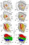
Musculature of Drosophila melanogaster in the PD3 sagittal section. Female: (A,C,E,G); Male: (B,D,F,H). Abbreviations: ANm–abdominal neuromere; car—cardia; cr—crop; MesoNm–mesothoracic neuromere; MetaNm–metathoracic neuromere; mg—midgut; nc—neck connective.
Figure 7
Figure 7
Musculature of Drosophila melanogaster in the PD4 sagittal section. Female: (A,C,E,G); Male: (B,D,F,H). The female IIdlm3 and the male Itpm2, Iscm4, and Ivlm1 are hidden by the skeleton. Abbreviations: ANm–abdominal neuromere; car—cardia; cr—crop; MesoNm–mesothoracic neuromere; MetaNm–metathoracic neuromere; mg—midgut; nc—neck connective.
Figure 8
Figure 8
Musculature of Drosophila melanogaster in the AD1 sagittal section. Female: (A,C,E,G); Male: (B,D,F,H). Abbreviations: ANm–abdominal neuromere; car—cardia; cr—crop; MesoNm–mesothoracic neuromere; MetaNm–metathoracic neuromere; mg—midgut; nc—neck connective; slgl—salivary gland.
Figure 9
Figure 9
Musculature of Drosophila melanogaster in the AD3 sagittal section. Female: (A,C,E,G); Male: (B,D,F,H). The male IIscm1 is hidden by the skeleton. Abbreviations: ANm–abdominal neuromere; car—cardia; cr—crop; MesoNm–mesothoracic neuromere; MetaNm–metathoracic neuromere; mg—midgut; nc—neck connective; slgl—salivary gland.

References

    1. Engel M.S. Insect evolution. Curr. Biol. 2015;25:R868–R872. doi: 10.1016/j.cub.2015.07.059. - DOI - PubMed
    1. Hall M.J.R., Martín-Vega D. Visualization of insect metamorphosis. Philos. Trans. R. Soc. B. 2019;374:20190071. doi: 10.1098/rstb.2019.0071. - DOI - PMC - PubMed
    1. Rolff J., Johnston P.R., Reynolds S. Complete metamorphosis of insects. Philos. Trans. R. Soc. B. 2019;374:20190063. doi: 10.1098/rstb.2019.0063. - DOI - PMC - PubMed
    1. Jindra M., Palli S.R., Riddiford L.M. The juvenile hormone signaling pathway in insect development. Annu. Rev. Entomol. 2012;58:181–204. doi: 10.1146/annurev-ento-120811-153700. - DOI - PubMed
    1. Lee G., Park J.H. Programmed cell death reshapes the central nervous system during metamorphosis in insects. Curr. Opin. Insect Sci. 2021;43:39–45. doi: 10.1016/j.cois.2020.09.015. - DOI - PMC - PubMed

LinkOut - more resources