. 2023 Nov 15;10(11):657.
doi: 10.3390/vetsci10110657.

Intestine Health and Barrier Function in Fattening Rabbits Fed Bovine Colostrum

Affiliations

Intestine Health and Barrier Function in Fattening Rabbits Fed Bovine Colostrum

Lucia Aidos et al. Vet Sci. .

Abstract

The permeability of the immature intestine is higher in newborns than in adults; a damaged gut barrier in young animals increases the susceptibility to digestive and infectious diseases later in life. It is therefore of major importance to avoid impairment of the intestinal barrier, specifically in a delicate phase of development, such as weaning. This study aimed to evaluate the effects of bovine colostrum supplementation on the intestinal barrier, such as the intestinal morphology and proliferation level and tight junctions expression (zonulin) and enteric nervous system (ENS) inflammation status (through the expression of PGP9.5 and GFAP) in fattening rabbits. Rabbits of 35 days of age were randomly divided into three groups (n = 13) based on the dietary administration: commercial feed (control group, CTR) and commercial feed supplemented with 2.5% and 5% bovine colostrum (BC1 and BC2 groups, respectively). Rabbits receiving the BC1 diet showed a tendency to have better duodenum morphology and higher proliferation rates (p < 0.001) than the control group. An evaluation of the zonulin expression showed that it was higher in the BC2 group, suggesting increased permeability, which was partially confirmed by the expression of GFAP. Our results suggest that adding 2.5% BC into the diet could be a good compromise between intestinal morphology and permeability, since rabbits fed the highest inclusion level of BC showed signs of higher intestinal permeability.

Keywords: bovine colostrum; enteric nervous system; intestinal barrier; intestinal health; rabbits; zonulin.


Conflict of interest statement

The authors declare no conflict of interest.

Figures

Figure 1
Figure 1
Body weight (BW) expressed in kilograms (kg) in rabbits aged 42, 49, 56, 63, and 70. Values are expressed as the mean ± S.E.M. * p < 0.05; ** p < 0.01. Group: p < 0.001; time: p < 0.001; group × time: p = 0.088; BW at weaning: p < 0.001; sex: p = 0.047.
Figure 2
Figure 2
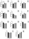
Histometrical analyses: (AC) duodenum; (DF) jejunum; (GI) ileum; (J) cecum; (K) colon. Villus height, expressed in μm: (A) duodenum; (D) jejunum; (G) ileum. Crypts depth, expressed in μm: (B) crypt depth of the duodenum; (E) jejunum; (H) ileum; (J) cecum; (K) colon. villus-crypt ratio: (C) duodenum; (F) jejunum; (I) ileum. One-way ANOVA was performed. Values are expressed as the mean ± S.E.M; t, tendency (p = 0.07).
Figure 3
Figure 3
Representative images of the AB-PAS-stained (A) duodenum; (B) jejunum; (C) ileum; (D) cecum; (E) colon. Goblet cells, arrows; acidic, blue; neutral, magenta; mixed, purple. Scale bars: 50 μm.
Figure 4
Figure 4
(A) Representative image of an enlargement of the tip of a villus in which the mucus layer is visible (bold, black arrows). Scale bar: 20 μm. Quantitative representation of the thickness of the mucus, expressed in μm: (B) duodenum; (C) jejunum; (D) ileum; (E) cecum; (F) colon. One-way ANOVA was performed. Values are expressed as the mean ± S.E.M.
Figure 5
Figure 5
(A) Representative image of the PCNA-immunostained duodenum crypts. Positive nuclei, arrows. Scale bar: 20 µm. (B) Quantitative representation of PCNA counts in the duodenum, expressed as the number of immunopositive cells over the total number of cells per crypt. One-way ANOVA was performed. Values are expressed as the mean ± S.E.M. ** p < 0.01; *** p < 0.001.
Figure 6
Figure 6
(A) Representative image of a ZO-1 immunofluorescence-stained duodenum villus (white arrows). Scale bar: 20 µm. (B) Quantitative representation of the expression of ZO-1 in the duodenum, expressed in intensity per µm2. One-way ANOVA was performed. Values are expressed as the mean ± S.E.M. * p < 0.05.
Figure 7
Figure 7
Representative image of (A) submucosal and (B) myenteric neuronal plexuses of the duodenum stained with PGP9.5 (green color)—GFAP (red color) double immunofluorescence. Ganglions, asterisk; longitudinal muscular layer, LM; circular muscular layer, CM; submucosa, SM. Scale bar: 50 µm. Quantitative representation of the expression of PGP9.5 in the (C) submucosal and (D) myenteric neuronal ganglia and GFAP in the (E) submucosal and (F) myenteric neuronal ganglia in the duodenum, expressed in intensity per µm2. One-way ANOVA was performed. Values are expressed as the mean ± S.E.M. * = p < 0.05; t, tendency (p = 0.06).
Figure 8
Figure 8
Number of ganglia per section area of the (A) submucosal and (B) myenteric plexuses of the duodenum. One-way ANOVA was performed. Values are expressed as the mean ± S.E.M.

References

    1. Gidenne T., Fortun-Lamothe L. Feeding Strategy for Young Rabbits around Weaning: A Review of Digestive Capacity and Nutritional Needs. Anim. Sci. 2002;75:169–184. doi: 10.1017/S1357729800052942. - DOI
    1. Modina S.C., Polito U., Rossi R., Corino C., Di Giancamillo A. Nutritional Regulation of Gut Barrier Integrity in Weaning Piglets. Animals. 2019;9:1045. doi: 10.3390/ani9121045. - DOI - PMC - PubMed
    1. Oglesbee B.L., Lord B. Gastrointestinal Diseases of Rabbits. Ferrets Rabbit. Rodents. 2020:174–187. doi: 10.1016/B978-0-323-48435-0.00014-9. - DOI
    1. Bivolarski B.L., Vachkova E.G. Morphological and Functional Events Associated to Weaning in Rabbits. J. Anim. Physiol. Anim. Nutr. 2014;98:9–18. doi: 10.1111/jpn.12058. - DOI - PubMed
    1. Martens E.C., Neumann M., Desai M.S. Interactions of Commensal and Pathogenic Microorganisms with the Intestinal Mucosal Barrier. Nat. Rev. Microbiol. 2018;16:457–470. doi: 10.1038/s41579-018-0036-x. - DOI - PubMed

Grants and funding