. 2024 May;20(5):1134-1146.
doi: 10.1080/15548627.2023.2287932. Epub 2023 Nov 28.

Deacetylation of ATG7 drives the induction of macroautophagy and LC3-associated microautophagy

Affiliations

Deacetylation of ATG7 drives the induction of macroautophagy and LC3-associated microautophagy

Yinfeng Xu et al. Autophagy. 2024 May.

Abstract

LC3 lipidation plays an important role in the regulation of macroautophagy and LC3-associated microautophagy. The E1-like enzyme ATG7 is one of the core components that are directly involved in LC3 lipidation reaction. Here, we provide evidence showing that acetylation of ATG7 tightly controls its enzyme activity to regulate the induction of macroautophagy and LC3-associated microautophagy. Mechanistically, acetylation of ATG7 disrupts its interaction with the E2-like enzyme ATG3, leading to an inhibition of LC3 lipidation in vitro and in vivo. Functionally, in response to various different stimuli, cellular ATG7 undergoes deacetylation to induce macroautophagy and LC3-associated microautophagy, which are necessary for cells to eliminate cytoplasmic DNA and degrade lysosome membrane proteins, respectively. Taken together, these findings reveal that ATG7 acetylation acts as a critical rheostat in controlling LC3 lipidation and related cellular processes.Abbreviations: AMPK: AMP-activated protein kinase; ATG: autophagy-related; cGAMP: cyclic GMP-AMP; CGAS: cyclic GMP-AMP synthase; CREBBP/CBP: CREB binding protein; EGF: epidermal growth factor; EGFR: epidermal growth factor receptor; EP300/p300: E1A binding protein p300; IFNB1: interferon beta 1; ISD: interferon stimulatory DNA; MAP1LC3/LC3: microtubule associated protein 1 light chain 3; MCOLN1/TRPML1: mucolipin TRP cation channel 1; MEF: mouse embryonic fibroblast; MTOR: mechanistic target of rapamycin kinase; NAM: nicotinamide; PE: phosphatidylethanolamine; PTM: post-translational modification; RB1CC1/FIP200: RB1 inducible coiled-coil 1; SIRT: sirtuin; SQSTM1/p62: sequestosome 1; STING1: stimulator of interferon response cGAMP interactor 1; TSA: trichostatin A; ULK1: unc-51 like autophagy activating kinase 1; WIPI2: WD repeat domain, phosphoinositide interacting 2; WT: wild-type.

Keywords: ATG7; Acetylation; LC3 lipidation; macroautophagy; microautophagy.


Conflict of interest statement

No potential conflict of interest was reported by the author(s).

Figures
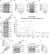
Figure 1.
Figure 1.
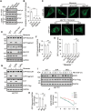
Identification of the acetylation sites of ATG7. (A) acetylation of ATG7 in HEK293 cells treated with or without nicotinamide (NAM), an inhibitor of SIRT family deacetylases, and/or trichostatin a (TSA), an inhibitor of HDAC family deacetylases. ATG7 was immunoprecipitated from the cell lysate with an anti-ATG7 antibody, and the immunoprecipitates were analyzed by western blot using an anti-acetyl-lysine antibody (Ace-Lys). (B) statistical analysis of (A). (C) acetylation of ATG7 in HEK293 cells treated with or without C646, an inhibitor of acetyltransferases EP300-CREBBP, or EX-527, an inhibitor of deacetylase SIRT1. (D) statistical analysis of (C). (E) acetylation of ATG7 in HEK293 cells treated with either nontargeting siRNA or siRNAs for EP300-CREBBP or SIRT1. (F) statistical analysis of (E). (G) in vitro acetylation assays of ATG7. Purified recombinant GST-ATG7 from Escherichia coli BL21 cells was incubated with immunoprecipitated Flag-tagged wild-type (WT) EP300 or EP300S1396R,Y1397R, an inactive mutant, from 293T cells, with or without acetyl-CoA and C646. (H) MS/MS spectrum of the tryptic peptide (inset) from recombinant ATG7 protein underwent in vitro acetylation assay with a mass shift of 42.01057 Da at the lysine residue. (I) in vitro acetylation assays of WT ATG7 or ATG7-2KR (K284R,K296R) with or without acetyl-CoA in the presence of EP300-Flag. (J) statistical analysis of (I). All statistical data are presented as mean ± SEM of three independent experiments. **p < 0.01, ***p < 0.001 (Student’s t-test).
Figure 2.
Figure 2.
Acetylation of ATG7 inhibits LC3 lipidation. (A) acetylation of ATG7 in HEK293 cells. The cells were treated with starvation medium (ST) or torin1, an inhibitor of MTORC1, or followed by fresh culture medium treatment. (B) statistical analysis of (A). (C) acetylation of Flag-tagged WT ATG7 or ATG7 mutants in HEK293 cells. The cells were starved or re-stimulated with fresh culture medium. (D) statistical analysis of (C). (E) schematic of the workflow for in vitro reconstitution of LC3 lipidation. Purified GST-LC3 (LC3-G120, the processed form of LC3) and Flag-tagged WT ATG7 or ATG7 mutants were incubated with the membranes from atg5 knockout mouse embryonic fibroblasts (MEFs) and the cytosol from atg7 knockout MEFs plus GTP and an ATP regeneration system (ATPR) for the indicated times. Then the reaction was terminated and the generated LC3–PE was subjected to western blot analysis. (F) the in vitro LC3 lipidation assays were carried out using Flag-tagged WT ATG7 or ATG7C567S, an inactive mutant, from 293T cells treated with or without CTB, an EP300-CREBBP activator. (G) statistical analysis of LC3–PE production in (F). (H) the in vitro LC3 lipidation assays were carried out using Flag-tagged WT ATG7, ATG7-2KR or ATG7-2KQ from 293T cells. (I) statistical analysis of LC3–PE production in (H). All statistical data are presented as mean ± SEM of three independent experiments. *p < 0.05, ***p < 0.001 (Student’s t-test).
Figure 3.
Figure 3.
Acetylation of ATG7 inhibits its binding to ATG3. (A) interaction between LC3 and ATG7. GST-tagged WT LC3 or LC3-2KQ (K49Q,K51Q) was incubated with WT ATG7, ATG7-2KR or ATG7-2KQ, and GST-tagged WT LC3 or LC3-2KQ was then pulled down using glutathione sepharose beads, and the bound ATG7 was analyzed by western blot. (B) statistical analysis of (A). (C) interaction between ATG7 and ATG3. GST-tagged WT ATG7, ATG7-2KR or ATG7-2KQ was incubated with ATG3 and the GST affinity-isolation assays were carried out using glutathione sepharose beads. (D) statistical analysis of (C). (E) interaction between ATG7 and ATG3. GST-tagged WT ATG7, ATG7-2KR or ATG7-2KQ was incubated with or without acetyl-CoA in the presence of EP300-Flag, and further incubated with ATG3 and followed by GST affinity-isolation assays. (F) statistical analysis of (E). (G) co-immunoprecipitation of Flag-tagged WT ATG7 with ATG3 in 293T cells treated with C646, an inhibitor of acetyltransferases EP300-CREBBP, or EX-527, an inhibitor of deacetylase SIRT1. The cells were lysed and incubated with anti-Flag magnetic beads, and the immunoprecipitates were subjected to western blot analysis using anti-ATG3. (H) statistical analysis of (G). (I) co-immunoprecipitation of Flag-tagged WT ATG7, ATG7-2KR or ATG7-2KQ with ATG3 in 293T cells treated with or without torin1. (J) statistical analysis of (I). All statistical data are presented as mean ± SEM of three independent experiments. **p < 0.01, ***p < 0.001 (Student’s t-test).
Figure 4.
Figure 4.
Deacetylation of ATG7 is essential for macroautophagy. (A) formation of GFP-LC3 puncta in MEFs stably expressing GFP-LC3. The cells with or without atg7 knockout were re-introduced with WT ATG7, ATG7-2KR or ATG7-2KQ and treated with or without starvation medium (ST) for 1 h. (B) statistical analysis of the number of GFP-LC3 puncta in cells treated as in (A). n = 30. (C) the production of LC3–PE and the degradation of SQSTM1 in MEFs treated with or without torin1 or chloroquine (CQ) for 3 h. The cells with or without atg7 knockout were re-introduced with WT ATG7, ATG7-2KR or ATG7-2KQ. (D and E) statistical analysis of the protein levels of SQSTM1 and LC3–PE in (C). (F) LC3 conversion in atg7 knockout MEFs with re-introduction of WT ATG7, ATG7-2KR or ATG7-2KQ. The cells were treated with or without cGAMP. (G) statistical analysis of LC3–PE production in (F). (H) the clearance of cytoplasmic DNA in WT or atg7 knockout MEFs re-introduced with WT ATG7, ATG7-2KR or ATG7-2KQ. The cells were transfected with Cy3 labeled-ISD (Cy3-ISD) (4 μg/ml) for 6 h and then stimulated by cGAMP for another 12 h. (I) statistical analysis of the fluorescence intensity of Cy3-ISD in cells treated as in (H). All the statistical data are presented as mean ± SEM of three independent experiments. **p < 0.01, ***p < 0.001 (Student’s t-test). Scale bars: 10 µm.
Figure 5.
Figure 5.
Deacetylation of ATG7 is required for LC3-associated microautophagy. (A) acetylation of ATG7 in MEFs treated with ammonium chloride, monensin or nigericin. (B) statistical analysis of (A). (C) formation of GFP-LC3 puncta in MEFs stably expressing GFP-LC3. The cells with or without rb1cc1 or atg7 knockout were re-introduced with or without WT ATG7, ATG7-2KR or ATG7-2KQ, and treated with or without monensin for 2 h. Scale bars: 10 µm. (D) statistical analysis of the number of GFP-LC3 puncta in cells treated as in (C). n = 30. (E) GFP-MCOLN1 turnover in MEFs. The cells with or without knockout of rb1cc1 or atg7 were treated with or without monensin for 8 h. GFP-tagged full-length MCOLN1 and free GFP fragments are indicated. (F) statistical analysis of the production of free GFP fragments in (E). (G) GFP-MCOLN1 turnover in atg7 knockout MEFs with re-introduction of WT ATG7, ATG7-2KR or ATG7-2KQ. The cells were treated with or without monensin for 8 h. (H) statistical analysis of the production of free GFP fragments in (G). (I) EGFR degradation in atg7 knockout MEFs with re-introduction of WT ATG7, ATG7-2KR or ATG7-2KQ. The cells were subjected to 2-h monensin treatment and 4-h monensin washout, and then serum starved and stimulated by EGF with the indicated times. (J) statistical analysis of (I). All statistical data are presented as mean ± SEM of three independent experiments. **p < 0.01, ***p < 0.001 (Student’s t-test).
Figure 6.
Figure 6.
Schematic model for the role of ATG7 acetylation in the induction of macroautophagy and LC3-associated microautophagy. In response to various intracellular or environmental stimuli, ATG7 undergoes deacetylation, which promotes the interaction between ATG7 and ATG3, leading to the activation of LC3 lipidation and the induction of macroautophagy or LC3-associated microautophagy.

References

    1. Xu Y, Wan W.. Acetylation in the regulation of autophagy. Autophagy. 2023;19(2):379–387. doi: 10.1080/15548627.2022.2062112 - DOI - PMC - PubMed
    1. Yim WW, Mizushima N. Lysosome biology in autophagy. Cell Discov. 2020;6:6. doi: 10.1038/s41421-020-0141-7 - DOI - PMC - PubMed
    1. Xu Y, Wan W. Emerging roles of p300/CBP in autophagy and autophagy-related human disorders. J Cell Sci. 2023;136(12):jcs261028. doi: 10.1242/jcs.261028 - DOI - PubMed
    1. Yamamoto H, Matsui T. Molecular mechanisms of macroautophagy, microautophagy, and chaperone-mediated autophagy. J Nippon Med Sch. 2023. doi: 10.1272/jnms.JNMS.2024_91-102 - DOI - PubMed
    1. Feng Y, He D, Yao Z, et al. The machinery of macroautophagy. Cell Res. 2014;24(1):24–41. doi: 10.1038/cr.2013.168 - DOI - PMC - PubMed

Publication types