. 2023 Nov 24;9(47):eadi8454.
doi: 10.1126/sciadv.adi8454. Epub 2023 Nov 24.

Partial in vivo reprogramming enables injury-free intestinal regeneration via autonomous Ptgs1 induction

Affiliations

Partial in vivo reprogramming enables injury-free intestinal regeneration via autonomous Ptgs1 induction

Jumee Kim et al. Sci Adv. .

Abstract

Tissue regeneration after injury involves the dedifferentiation of somatic cells, a natural adaptive reprogramming that leads to the emergence of injury-responsive cells with fetal-like characteristics. However, there is no direct evidence that adaptive reprogramming involves a shared molecular mechanism with direct cellular reprogramming. Here, we induced dedifferentiation of intestinal epithelial cells using OSKM (Oct4, Sox2, Klf4, and c-Myc) in vivo. The OSKM-induced forced dedifferentiation showed similar molecular features of intestinal regeneration, including a transition from homeostatic cell types to injury-responsive-like cell types. These injury-responsive-like cells, sharing gene signatures of revival stem cells and atrophy-induced villus epithelial cells, actively assisted tissue regeneration following damage. In contrast to normal intestinal regeneration involving Ptgs2 induction, the OSKM promotes autonomous production of prostaglandin E2 via epithelial Ptgs1 expression. These results indicate prostaglandin synthesis is a common mechanism for intestinal regeneration but involves a different enzyme when partial reprogramming is applied to the intestinal epithelium.


Figures
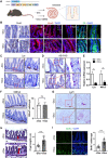
Fig. 1.
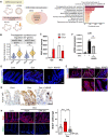
Fig. 1.. Dedifferentiation of intestinal epithelium by partial reprogramming.
(A) Experimental scheme on the intestine of Dox inducible OSKM (iOSKM) mouse model. (B) Immunohistochemistry (IHC) of Oct4 and immunofluorescence (IF) of Sox2 and 2A peptide in the intestine of iOSKM mice. Samples were analyzed 4 days after Dox administration. Hematoxylin or DAPI for nuclear staining. (C) IHC of Lysozyme for PC in the intestine of iOSKM mice. (D) AB-PAS staining for GC in the intestine of iOSKM mice. (E) Relative mRNA expressions of Lysozyme (Lyz for PC) and Mucin 2 (Muc2 for GC) in intestinal epithelial cells of iOSKM mice. n = 6; 3 mice × 2 technical replicates. (F) IHC of Olfm4 in the intestine of iOSKM mice (left) and the quantification of Olfm4+ cells per crypt (−Dox, n = 30; +Dox, n = 28 in total two mice of each condition). (G) RNA scope of Lgr5 in the intestine of iOSKM mice (H) H&E histology in the intestine of iOSKM mice. Red line indicates the length of crypt (left). Quantification of crypt length (−Dox, n = 29; +Dox, n = 46 in total three mice of each condition) (right). (I) IF of BrdU in the intestine (left) and quantification of BrdU-positive cells per crypt (−Dox, n = 25; +Dox, n = 39 in total three mice of each condition) (right). BrdU was administered 3 hours before the euthanasia. Data represent the mean with SD. Student’s t test: **P < 0.01 and ****P < 0.0001; ns, not significant. Scale bar, 50 μm.
Fig. 2.
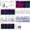
Fig. 2.. Cellular and molecular alterations induced by partial reprogramming.
(A) Experimental scheme for scRNA-seq using intestine of iOSKM mouse in Dox condition. (B) UMAP plots of scRNA-seq from OSKM-induced mouse intestinal epithelium in −Dox and + Dox conditions (Total, left), −Dox (control, middle), and + Dox (Dox-treated, right). (C) Bar plot indicating differentiation potency inferred by CytoTRACE per condition in different cell types. (D and E) UMAP plot (D) and violin plot (E) showing gene module score of 1275 fetal signature genes. (F and G) UMAP plot (F) and violin plot (G) showing gene module score of 394 yap signature genes. (H) IF of Sca1 in the intestine. DAPI for nuclear staining (left). Relative mRNA expression of Ly6a (encoding Sca1) in intestinal epithelial cells of iOSKM mice after Dox treatment (right). (I) IHC of Active YAP in the intestine of iOSKM mice 4 days after Dox administration. Red arrowheads indicate active YAP. (J) Microscopic images of Dox-treated intestinal organoids from rtTA mouse or iOSKM mouse. (K) IF of fetal markers, Trop2 and Sca1, in iOSKM intestinal organoids with Dox treatment for 3 days. DAPI for nuclear staining. (L) IF of Active YAP and 2A peptide in iOSKM intestinal organoids. DAPI for nuclear staining. (M) Microscopic images (top) and IF of Active YAP (bottom) in XMU-MP-1 (Mst1/2 inhibitor)–treated intestinal organoids. (N) Gene enrichment of YAP or fetal signatures in control and iOSKM intestinal organoids. Data represent the mean with SD. Student’s t test: *P < 0.05 and **P < 0.01. Scale bar, 50 μm [(H), (I), (K), (L), and (M)], 500 μm (J).
Fig. 3.
Fig. 3.. Generation of two distinct injury-responsive–like cells by partial reprogramming.
(A) Projection of revSC (left) and aVEC (right) cells onto our scRNA-seq data. Cells in our study are indicated in gray. (B) Volcano plot showing differentially expressed genes (DEGs) between DC1 (right) and DC2 (left). (C) Dot plot for relative expression of marker genes of revSC, aVEC, top-villus, EC, and fetal signature genes in DC1 and DC2. (D) UMAP plots showing gene expression of Clu and Msln. (E) In situ hybridization (ISH) of Clu and Msln in the intestine of iOSKM mice. White arrowhead indicates Clu+ revSC-like cells, while black arrowhead indicates Msln+ aVEC-like cells. (F) IF of Sca1 and Avil in the intestine of iOSKM mice. White arrowhead indicates Sca1Avil+ aVEC-like cells, while yellow arrow indicates Sca1+Avli+ revSC-like cells. (G) Graphical presentation of spatial presence of revSC (DC1) or aVEC (DC2)- like cells. (H) PAGA graphs showing velocity-directed arrows from cluster to cluster in −Dox (left) and +Dox (right). Scale bar, 50 μm.
Fig. 4.
Fig. 4.. Promotion of intestinal repair by partial reprogramming.
(A) Experimental scheme for 10-Gy IR and Dox treatment in iOSKM mice. The day after IR was indicated. Dox was treated 2 days before IR for 4 days and then removed to produce differentiated cells from OSKM-induced dedifferentiated cells for regeneration. Analysis at 2 and 4 dpi were showed in (B) and (C), respectively. (B to D) H&E histology and IHC of Olfm4 and Ki67 in the intestine of iOSKM mice after IR at 2 dpi (B) and at 4 dpi (C) with the quantification of Olfm4+ cells per crypt (n = 42 in total 3 mice of each condition) (D). (E) IF of phospho-histone H3(pHH3) and BrdU in the intestine of iOSKM mice after IR at 4 dpi (left) and quantification of BrdU+ cells per crypt (−Dox and non-IR, n = 20; −Dox and IR, n = 19; +Dox and IR, n = 22 in total 3 mice of each condition) (right). (F) IF of Sca1 and BrdU in the intestine of iOSKM mice after IR at 2 and 4 dpi. Top of the villi were magnified. (G) IHC of Active YAP in the intestine of iOSKM mice after IR at 4 dpi. Red arrowheads indicate active YAP. Data represent the mean with SD. Student’s t test: *P < 0.05, ****P < 0.0001. Scale bar, 50 μm.
Fig. 5.
Fig. 5.. Enhanced epithelial PGE2 synthesis.
(A) Experimental scheme for RNA-seq for OSKM- or IR-induced regenerating intestinal organoids (left), and a bar plot showing enrichR combined scores of top 10 commonly up-regulated pathways in OSKM-, IR-induced regenerating intestinal organoids compared to their control intestinal organoids (right). (B) A violin plot showing “Prostaglandin synthesis and Regulation” signature score in cell types (C) PGE2 level (pg/mg) of mouse intestine in epithelium (Epi) and mesenchyme (Mes) with or without Dox treatment. (D) IF of BrdU in the intestine of iOSKM mice, Cont (left), Dox (Dox-treated, middle), and Dox + NSAID (Dox- and dexibuprofen-treated, right). (E) IF of fetal markers, Sca1, in iOSKM mice intestine treated with Dox and NSAID for 4 days. DAPI for nuclear staining. (F) mRNA expressions of Ly6a in intestinal epithelial cells of iOSKM mice. n = 6; 2 mice × 3 technical replicates. (G) IHC of Active YAP in the intestine of iOSKM mice after Dox and NSAID administration for 4 days. (H) IF of Sca1 and BrdU in the intestine of iOSKM mice, Cont (left), Dox (Dox-treated, middle), and Dox + Ep4 ant (Dox and GW627368 treated, right) and quantification of BrdU+ cells per crypt. Data represent the mean with SD. Student’s t test: *P < 0.05, **P < 0.01, and ****P < 0.0001. Scale bar, 50 μm.
Fig. 6.
Fig. 6.. Cox1 activity for partial reprogramming-induced regeneration.
(A) mRNA expressions of Ptgs1 and Ptgs2 in the intestinal epithelial cells of iOSKM mice. n = 18; 6 mice × 3 technical replicates (left). UMAP plots showing gene expression of Ptgs1 and Ptgs2 (right). (B) IF of Cox1 (left) and Cox2 (right) in the intestine of iOSKM mice 4 days after Dox administration. DAPI for nuclear staining. (C) IF of Sca1 and BrdU in the intestine of iOSKM mice. Dox was treated for 4 days with Cox1 inhibitor (iCox1: SC-560) or Cox2 inhibitor (iCox2: celecoxib) treatment for 2 days. DAPI for nuclear staining. (D) In situ hybridization of Clu in the intestine of iOSKM mice. (E) IHC of Active YAP in the intestine of iOSKM mice. (F) H&E histology and IHC of Olfm4 in the intestine of iOSKM mice at 4 dpi. Dox for 4 days and Cox1 inhibitor for 2 days were treated. (G) IF of Sca1 and BrdU in the intestine of iOSKM mice (left) and quantification of BrdU-positive cells per crypt (n = 20 in total three mice for each condition) (right). Data represent the mean with SD. Student’s t test: *P < 0.05 and ****P < 0.0001. Scale bar, 50 μm.

References

    1. S. H. Tan, P. Phuah, L. T. Tan, S. Yada, J. Goh, L. B. Tomaz, M. Chua, E. Wong, B. Lee, N. Barker, A constant pool of Lgr5(+) intestinal stem cells is required for intestinal homeostasis. Cell Rep. 34, 108633 (2021). - PubMed
    1. N. Barker, Adult intestinal stem cells: critical drivers of epithelial homeostasis and regeneration. Nat. Rev. Mol. Cell Biol. 15, 19–33 (2014). - PubMed
    1. H. Tian, B. Biehs, S. Warming, K. G. Leong, L. Rangell, O. D. Klein, F. J. de Sauvage, A reserve stem cell population in small intestine renders Lgr5-positive cells dispensable. Nature 478, 255–259 (2011). - PMC - PubMed
    1. U. Jadhav, M. Saxena, N. K. O’Neill, A. Saadatpour, G. C. Yuan, Z. Herbert, K. Murata, R. A. Shivdasani, Dynamic Reorganization of Chromatin Accessibility Signatures during Dedifferentiation of Secretory Precursors into Lgr5+ Intestinal Stem Cells. Cell Stem Cell 21, 65–77.e5 (2017). - PMC - PubMed
    1. J. H. van Es, Dll1+ secretory progenitor cells revert to stem cells upon crypt damage. Nat. Cell Biol. 14, 1099–1104 (2012). - PMC - PubMed

MeSH terms