. 2023 Nov 24;9(47):eadh9673.
doi: 10.1126/sciadv.adh9673. Epub 2023 Nov 24.

Intestinal Paneth cell differentiation relies on asymmetric regulation of Wnt signaling by Daam1/2

Affiliations

Intestinal Paneth cell differentiation relies on asymmetric regulation of Wnt signaling by Daam1/2

Gabriele Colozza et al. Sci Adv. .

Abstract

The mammalian intestine is one of the most rapidly self-renewing tissues, driven by stem cells residing at the crypt bottom. Paneth cells form a major element of the niche microenvironment providing various growth factors to orchestrate intestinal stem cell homeostasis, such as Wnt3. Different Wnt ligands can selectively activate β-catenin-dependent (canonical) or -independent (noncanonical) signaling. Here, we report that the Dishevelled-associated activator of morphogenesis 1 (Daam1) and its paralogue Daam2 asymmetrically regulate canonical and noncanonical Wnt (Wnt/PCP) signaling. Daam1/2 interacts with the Wnt inhibitor RNF43, and Daam1/2 double knockout stimulates canonical Wnt signaling by preventing RNF43-dependent degradation of the Wnt receptor, Frizzled (Fzd). Single-cell RNA sequencing analysis revealed that Paneth cell differentiation is impaired by Daam1/2 depletion because of defective Wnt/PCP signaling. Together, we identified Daam1/2 as an unexpected hub molecule coordinating both canonical and noncanonical Wnt, which is fundamental for specifying an adequate number of Paneth cells.


Figures
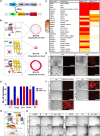
Fig. 1.
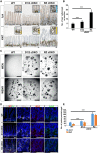
Fig. 1.. Functional CRISPR screening of RNF43-interacting proteins.
(A) Schematic of the DNA constructs used to generate small intestinal organoids expressing doxycycline-inducible RNF43-IRES-mCherry. mCherry is used as a proxy for RNF43 expression. (B) Schematic showing the CRISPR-based screening of RNF43-expressing organoids. Doxycyclin addition turns on RNF43 expression and organoids die unless downstream components necessary for RNF43 activity are eliminated via CRISPR-Cas9 knockout (KO). (C) List of RNF43-interacting proteins identified via mass spectrometry. The abundance of each protein is represented as the number of peptides detected, according to the color scale on the left. Pull-down results from full-length (RNF43_Full) and truncated RNF43 (RNF43_Del) are shown. See Material and Methods for details. (D) Organoid assay showing robust RNF43 expression upon doxycycline treatment. Note that, at day 7, all organoids expressing RNF43 (here visualized through mCherry fluorescence) are dead. (E) Bar plot quantification of organoid survival after indicated passages. Only Axin and Daam CRISPR-KO organoids grow in the presence of RNF43 overexpression. (F) Survival assay of wild-type (WT) and indicated CRISPR KO mutant organoids after RNF43 induction. (G) Schematic illustrating the growth factor withdrawal assay used in (H), to determine RNF43 interactor epistasis in the Wnt pathway. (H) Growth factor withdrawal assay on WT and indicated CRISPR mutant organoids. All scale bars represent 1000 μm. CMV, cytomegalovirus; IRES, internal ribosomal entry site. BF, brightfield; TCF/LEF, T-cell factor/lymphoid enhancer factor; WENR, Wnt EGF Noggin R-spondin1-containing medium.
Fig. 2.
Fig. 2.. Daam is required for RNF43-dependent Frizzled endocytosis.
(A) Subcellular localization of SNAP-Fzd5 in WT or Daam1/2 DKO HEK293T cells cotransfected with control empty vector or RNF43. Surface SNAP-Fzd5 was labeled with SNAP-surface Alexa 549 for 15 min and chased for 30 min. Scale bars, 20 μm. (B) Fluorescence-activated cell sorting (FACS) analysis of plasma membrane levels of Fzd receptors in WT and D1/2 DKO HEK293T cells, with or without RNF43 overexpression. (C) Schematic of Nano-Glo/HiBiT assay. The N terminus of Fzd5 contains a HibiT tag, to which LgBiT binds, restoring nano-luciferase activity. In the presence of RNF43, less HibiT-Fzd5 is exposed on the plasma membrane, preventing the reconstitution of luciferase activity. (D) Nano-Glo–based quantification of cell surface Fzd5. Levels are expressed as percentages of relative light unit fractions. Data from three independent biological experiments.
Fig. 3.
Fig. 3.. Daam interacts with RNF43 through its N-terminal domain.
(A) Western blot of representative cell surface protein biotinylation and pull-down assay on HEK293T cells transfected with indicated constructs, showing that Daam1/2 KO also prevents RNF43-dependent internalization of the Wnt co-receptor Lrp6. Note that N-Daam1 cooperates with RNF43 in reducing Lrp6 surface levels. Transferrin receptor (TfR) was used as a negative control. Colored arrowheads correspond to the different Daam1 constructs, as shown in (C). (B) Immunoprecipitation (IP) assay used to show the interaction between hemagglutinin (HA)-tagged RNF43 and Myc-tagged Daam1. (C) Schematic of the Daam1 full-length and deletion constructs used in this study to map the RNF43-interacting domain of Daam1. Daam1 architecture domain and relative amino acid position are indicated. GBD, Rho GTPase binding domain; FH3, formin homology 3 domain; DD, dimerization domain; CC, coiled-coil domain; FH1, formin homology 1 domain; FH2, formin homology 2 domain; Helix, an amphipathic helix involved in interaction with FH3 domain; DAD, diaphanous autoregulatory domain. (D) IP experiment showing that the N-terminal domain of Daam1 is required for RNF43 interaction. Colored arrowheads correspond to the different Daam1 constructs, as shown in (C), and indicate their migration position on the blot. (E) Western blots showing Frizzled degradation by RNF43 in WT, D1/2 DKO, and Dvl TKO HEK293T cells. (F) Western blot showing ubiquitin levels of Fzd5 in WT, D1/2, and Dvl mutant cells. α-Tubulin was used as a loading control in (A) and (F). Gapdh was used as a loading control in (B), (D), and (E).
Fig. 4.
Fig. 4.. Daam1/2 cDKO mice show a milder phenotype than RZ cDKO, despite maintaining Rspo1 independence.
(A) Small intestine histological sections from WT, D1/2, and RZ conditional double KO mice stained for lysozyme (Lyz), a PC marker, and Ki67, used as a proliferation marker. Insets show magnification of dash-boxed areas. The extent of Lyz-positive Paneth zones and Ki67-positive proliferative zones are indicated by blue and yellow sidebars, respectively. (B) Bar plot representing experiment quantification, showing mean values and SD. P values (D1/2: 0.0009; RZ: 5.42 × 10⁻¹⁰) were calculated using an unpaired two-tailed t test. (C) Organoids derived from WT, D1/2, and RZ cDKO mice, showing that D1/2 and RZ mutant organoids can survive in the absence of R-spondin, unlike WT organoids. (D) RNAscope in situ hybridization analysis with probes targeting Olfm4 (O4, stem cell marker), Wnt3 (W3, PC marker), and Axin2 (A2, canonical Wnt target gene). White dashed boxed areas shown in the left “merge” panels are enlarged and shown on the right as single probe staining. Scale bars, 50 μm [(A) and (D)] and 1000 μm (C). (E) Bar plot representing experiment quantification, showing mean values and SD. P values were calculated using an unpaired two-tailed t test. P values: (D1/2 Axin2: 2.03 × 10⁻¹⁰; Wnt3: 1.93 × 10−08); (RZ Axin2: 2.97 × 10⁻¹⁸; Wnt3: 6.25 × 10⁻²⁴).
Fig. 5.
Fig. 5.. Daam knockout impairs noncanonical Wnt while enhancing canonical Wnt signaling.
(A) Active Rho pull-down assay. WT and D1/2 KO cells were treated with recombinant Wnt5a (0, 200, or 400 ng/ml) for 30 min at 37°C before Western blot analysis. White and black arrowheads point to phosphorylated and unphosphorylated Dvl2, respectively, which served to monitor for Wnt5a activity. Daam1 immunoblot was used to confirm its absence in D1/2 KO cells. (B) WT and D1/2 KO HEK293T cells transfected with LifeAct-GFP were treated with Wnt5a CM for 2 hours at 37°C, before immunofluorescence analysis. Insets show magnification of the dash-boxed area. White lines indicate transfected cell location among nontransfected cells. Scale bars, 20 μm. (C) Western blot comparing levels of active β-catenin in WT and D1/2 KO HEK293T cells. Cells were treated with Wnt3a CM for the indicated time, before analysis. α-Tubulin was used as a loading control in (A) and (C). (D) Reverse transcription quantitative polymerase chain reaction (RT-qPCR) analysis of canonical Wnt target genes Sp5 and Axin2 on WT and D1/2 KO cells treated overnight with Wnt3a CM. Expression levels are normalized to Actin mRNA. Error bars represent SD across three biological replicates.
Fig. 6.
Fig. 6.. Daam knockout in small intestinal organoids prevents the efficient generation of Paneth cells.
(A) RT-qPCR analysis of canonical Wnt target genes Lgr5 and Axin2 on WT, D1/2, and RZ cDKO mouse small intestinal organoids. Expression levels are normalized to Gapdh mRNA. Error bars represent SD across three biological replicates. (B) Organoids derived from WT, RZ, and D1/2 cDKO mice cultured in ENR medium and stained for lysozyme and Ki67. Arrowheads point at PCs (Lyz+, in red). (C) FACS analysis of organoids from indicated mouse genotypes, stained with fluorescein isothiocyanate (FITC)–labeled Ulex europaeus agglutinin 1 (UEA1). (D) Confocal imaging of lysozyme::mRuby WT SI organoid reporter line, expressing mRuby red fluorescent protein in PCs. Organoids were cultured in ENR, and WENR on Wnt5a-containing ENR. Scale bars, 50 μm [(B) and (D)]. (E) Schematic showing PC ISC double-positive feedback in normal homeostatic and Wnt high conditions (such as in RZ cDKO intestine). a.u., arbitrary units.
Fig. 7.
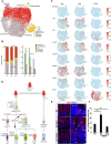
Fig. 7.. scRNA-seq analysis on WT, RZ, and D1/2 cDKO organoids shows that Daam1/2 is required for Paneth cell differentiation.
(A) Integrated Uniform Manifold Approximation and Projection (UMAP) cluster map including WT, RZ, and D1/2 samples. (B) Cell-type composition of WT, RZ, and D1/2 organoids. P values were calculated by Fisher’s exact tests. Corrected P values were described as *P < 0.01 and ***P < 0.001. (C) Expression pattern of selected cell type markers on UMAP clusters from individual samples. The red color indicates the maximum expression level, while the blue color indicates the minimum expression level for each gene. (D) Cellular flow chart showing the stepwise commitment from Lgr5+ stem cells. Dllhigh progenitors can generate secretory cells including tuft, Paneth, enteroendocrine (EE), and goblet cells, while Dlllow will only generate enterocytes. Among Dllhigh progenitors, only cells with active Wnt/PCP signaling can mature into Paneth, EE, and goblet cells. Note that Daam1/2 activity is positioned after the first secretory progenitor specification, where it regulates Wnt/PCP and consequently PC differentiation (arrows). (E) In situ hybridization using an RNAscope probe specific for Cfap126 (Fltp) in the small intestinal crypts of WT, RZ, and D1/2 cDKO mice. The right panels are the enlargement of areas included in the dash boxes on the left. White arrowheads indicate the fluorescent signal from single mRNA transcripts (red puncta). Note the decrease in Cfap126/Fltp expression in D1/2 KO mice. Scale bars, 20 μm. (F) Bar plot showing quantification of Cfap126/Fltp probe puncta shown in (E). Mean values and SD are shown, and P values were calculated with unpaired two-tailed t test. P values: (RZ: 1.57 × 10−08); (D1/2: 0.00046).
Fig. 8.
Fig. 8.. Model summarizing the role of Daam1/2 in the context of Wnt signaling and Paneth cell differentiation.
(A) Diagram showing the effects of RNF43, Dvl1/2/3, and Daam1/2 KOs on canonical and noncanonical Wnt signaling. RNF43 KO increases both pathways, due to the increase of Frizzled receptors. Conversely, triple KO of Dvl1/2/3, a common effector of canonical and noncanonical Wnt, leads to a decrease in both. Unlike RZ and Dvl1/2/3, Daam1/2 shows an asymmetric regulation of the two Wnt pathways as the KO of Daam1/2 triggers canonical Wnt activation while inactivating noncanonical Wnt signaling. (B) Diagram showing Daam1/2 within the different Wnt pathways. Left: Daam1/2 interacts with RNF43, allowing endocytosis of Fzd receptors ubiquitinated by RNF43. This terminates the Wnt/β-catenin signal. Right: Daam1/2 interacts with Dvl, regulating positively downstream events of the Wnt/PCP pathway. (C) Comparison between the effects of RZ and D1/2 KO in PC differentiation. RZ mutant mice undergo rapid intestinal adenoma formation caused by ISCs and Wnt-secreting PC co-hyperplasia. The absence of RZ boosts both canonical and noncanonical Wnt, increasing the number of PCs. In turn, this leads to more Wnt3 production, sustaining high stem cell proliferation, which is stimulated to differentiate into more PCs (rather than other secretory cells) because of the high levels of Wnt/PCP. On the other hand, Daam1/2 mutants show a completely alleviated phenotype, thanks to the noncanonical Wnt defect that prevents efficient PC differentiation, thereby reducing Wnt3 ligands in the microenvironment.

References

    1. Beumer J., Clevers H., Cell fate specification and differentiation in the adult mammalian intestine. Nat. Rev. Mol. Cell Biol. 22, 39–53 (2021). - PubMed
    1. Colozza G., Park S. Y., Koo B. K., Clone wars: From molecules to cell competition in intestinal stem cell homeostasis and disease. Exp. Mol. Med. 54, 1367–1378 (2022). - PMC - PubMed
    1. Nusse R., Clevers H., Wnt/β-catenin signaling, disease, and emerging therapeutic modalities. Cell 169, 985–999 (2017). - PubMed
    1. Koo B. K., Spit M., Jordens I., Low T. Y., Stange D. E., van de Wetering M., van Es J. H., Mohammed S., Heck A. J., Maurice M. M., Clevers H., Tumour suppressor RNF43 is a stem-cell E3 ligase that induces endocytosis of Wnt receptors. Nature 488, 665–669 (2012). - PubMed
    1. Hao H. X., Xie Y., Zhang Y., Charlat O., Oster E., Avello M., Lei H., Mickanin C., Liu D., Ruffner H., Mao X., Ma Q., Zamponi R., Bouwmeester T., Finan P. M., Kirschner M. W., Porter J. A., Serluca F. C., Cong F., ZNRF3 promotes Wnt receptor turnover in an R-spondin-sensitive manner. Nature 485, 195–200 (2012). - PubMed