. 2024 Jan 15:341:122997.
doi: 10.1016/j.envpol.2023.122997. Epub 2023 Nov 22.

Molecular mechanisms underlying NLRP3 inflammasome activation and IL-1β production in air pollution fine particulate matter (PM2.5)-primed macrophages

Affiliations

Molecular mechanisms underlying NLRP3 inflammasome activation and IL-1β production in air pollution fine particulate matter (PM2.5)-primed macrophages

Lourdes Caceres et al. Environ Pollut. .

Abstract

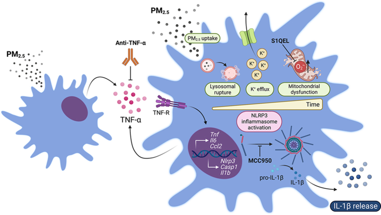
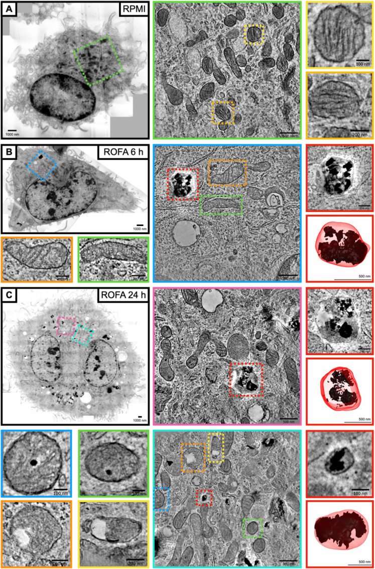
Exposure to air pollution fine particulate matter (PM2.5) aggravates respiratory and cardiovascular diseases. It has been proposed that PM2.5 uptake by alveolar macrophages promotes local inflammation that ignites a systemic response, but precise underlying mechanisms remain unclear. Here, we demonstrate that PM2.5 phagocytosis leads to NLRP3 inflammasome activation and subsequent release of the pro-inflammatory master cytokine IL-1β. Inflammasome priming and assembly was time- and dose-dependent in inflammasome-reporter THP-1-ASC-GFP cells, and consistent across PM2.5 samples of variable chemical composition. While inflammasome activation was promoted by different PM2.5 surrogates, significant IL-1β release could only be observed after stimulation with transition-metal rich Residual Oil Fly Ash (ROFA) particles. This effect was confirmed in primary human monocyte-derived macrophages and murine bone marrow-derived macrophages (BMDMs), and by confocal imaging of inflammasome-reporter ASC-Citrine BMDMs. IL-1β release by ROFA was dependent on the NLRP3 inflammasome, as indicated by lack of IL-1β production in ROFA-exposed NLRP3-deficient (Nlrp3-/-) BMDMs, and by specific NLRP3 inhibition with the pharmacological compound MCC950. In addition, while ROFA promoted the upregulation of pro-inflammatory gene expression and cytokines release, MCC950 reduced TNF-α, IL-6, and CCL2 production. Furthermore, inhibition of TNF-α with a neutralizing antibody decreased IL-1β release in ROFA-exposed BMDMs. Using electron tomography, ROFA particles were observed inside intracellular vesicles and mitochondria, which showed signs of ultrastructural damage. Mechanistically, we identified lysosomal rupture, K+ efflux, and impaired mitochondrial function as important prerequisites for ROFA-mediated IL-1β release. Interestingly, specific inhibition of superoxide anion production (O2•-) from mitochondrial respiratory Complex I, but not III, blunted IL-1β release in ROFA-exposed BMDMs. Our findings unravel the mechanism by which PM2.5 promotes IL-1β release in macrophages and provide a novel link between innate immune response and exposure to air pollution PM2.5.

Keywords: Inflammation; K(+) efflux; Lysosomal disruption; Macrophages; Mitochondria; Particulate matter.


Conflict of interest statement

Declaration of competing interest The authors declare that they have no known competing financial interests or personal relationships that could have appeared to influence the work reported in this paper.

Figures

Image 1
Graphical abstract
Fig. 1
Fig. 1
ROFA induces inflammasome priming, ASC-specks formation, and IL-1β secretion in human monocytes and macrophages. (A) Representative dot plots of inflammasome-reporter THP-1-ASC-GFP cells incubated with different PM2.5 surrogates at 100 μg/mL for 6 or 24 h. (B) Quantification of ASC-GFP fluorescence indicative of inflammasome priming and specks formation in THP-1-ASC-GFP cells incubated with increasing PM2.5 concentrations for 6 or 24 h. (C) IL-1β levels in cell culture supernatants from THP-1-ASC-GFP cells after PM2.5 incubation at 100 μg/mL for 6 or 24 h. (D) IL-1β release in cell culture supernatants from monocyte-derived macrophages obtained from differentiated PBMCs from healthy donors and incubated with ROFA at 100 μg/mL for 24 h. Data are presented as mean ± SEM from at least three independent experiments. *p < 0.05, **p < 0.01, and ***p < 0.001 versus RPMI (priming). §p < 0.05 and §§§p < 0.001 versus RPMI (specks).
Fig. 2
Fig. 2
ROFA promotes the NLRP3 inflammasome-dependent release of IL-1β in murine BMDMs. (A) Confocal microscopy of BMDMs from inflammasome-reporter ASC-Citrine mice incubated with ROFA at 100 μg/mL for 6 or 24 h. White arrows indicate ASC-specks formation. LPS stimulation at 20 ng/mL for 4 h followed by the addition of 5 μM Nigericin for 2 h was used as a positive control. Time course analysis of (B) Nlrp3, Casp1, and Il1b mRNA expression (C) pro-IL-1β protein levels, and (D) Caspase-1 activity and IL-1β release in BMDMs from wild type (wt) mice incubated with ROFA at 100 μg/mL. (E) IL-1β release in cell culture supernatants from wt, Nlrp3−/−, and Casp1−/− BMDMs incubated with ROFA at 100 μg/mL for 6 or 24 h. (F) Representative dot-plots of ASC-Citrine BMDMs incubated with ROFA at 100 μg/mL for 6 or 24 h, with or without pre-incubation with MCC950. (G) Quantification of ASC-Citrine fluorescence in ASC-Citrine BMDMs incubated with ROFA at 100 μg/mL for 6 or 24 h. (H) IL-1β levels in cell culture supernatants from wt BMDMs incubated with ROFA at 100 μg/mL for 6 or 24 h, with or without pre-incubation with MCC950. Data are presented as mean ± SEM from at least three independent experiments.
Fig. 3
Fig. 3
The pro-inflammatory phenotype induced by ROFA incubation in BMDMs partially depends on the NLRP3 inflammasome. (A) Gene expression in wild type BMDMs incubated with ROFA at 100 μg/mL for 24 h. (B) Cytokine levels in cell culture supernatants from BMDMs incubated with ROFA at 100 μg/mL for 6 or 24 h, and (C) with or without pre-incubation with MCC950. (D) IL-1β levels in cell culture supernatants from BMDMs incubated with ROFA at 100 μg/mL for 6 or 24 h, and with or without pre-incubation with a blocking anti-TNF-α antibody. Data are presented as mean ± SEM from at least three independent experiments.
Fig. 4
Fig. 4
ROFA uptake by BMDMs accumulates in vesicles and reaches mitochondria leading to structural damage. Representative electron tomography overview of BMDMs after incubation with (A) RPMI for 24 h, or ROFA at 100 μg/mL for (B) 6 or (C) 24 h. (A) Control BMDMs show preserved mitochondrial integrity and absence of particle-containing vesicles (green frame close-up). Higher magnification images show preserved mitochondrial structure (yellow and orange frames). (B) BMDMs incubated with ROFA for 6 h show particle accumulation in vesicles and signs of early structural damage in mitochondria (blue frame close-up). Higher magnification images show mild mitochondrial structure alterations, i.e. local absence of cristae (orange and green frames). (C) BMDMs incubated with ROFA for 24 h show a higher density of particle-containing vesicles and severe structural alterations in mitochondria (magenta and cyan frame close-ups). Higher magnification images show ROFA inside mitochondria (blue and green frames) and prominently altered mitochondrial structure (yellow and orange frames). (B and C) Vesicles containing ROFA particles after 6 or 24 h of incubation, and 3D reconstruction modelling, are shown in red frames. (For interpretation of the references to colour in this figure legend, the reader is referred to the Web version of this article.)
Fig. 5
Fig. 5
Lysosomal disruption, K+ efflux, and mitochondrial dysfunction drive NLRP3 activation and IL-1β release following ROFA incubation. (A) Confocal microscopy of wild type BMDMs incubated with ROFA at 100 μg/mL for 6 or 24 h were stained with Acridine Orange. LLO-Me was used as positive control for lysosomal destabilization. (B) Representative histograms for Acridine Orange staining assessed by flow cytometry in BMDMs incubated with ROFA at 100 μg/mL. Quantification of PerCP-Cy5.5low events was indicative of lysosomal disruption after 6 or 24 h. (C) IL-1β levels in cell culture supernatants from BMDMs pre-incubated with increasing [K+]ex, followed by incubation with ROFA at a concentration of 100 μg/mL for 6 or 24 h. (D) Mitochondrial oxygen consumption rate (OCR) was assessed by the Seahorse MitoStress Test in BMDMs incubated with ROFA at 100 μg/mL for 6 or 24 h. (E) Representative histograms of MitoSOX staining assessed by flow cytometry in BMDMs incubated with ROFA at 100 μg/mL for 6 or 24 h. Quantification of MitoSOX+ cells indicated mitochondrial O2•- production after 6 or 24 h of ROFA incubation. (F and G) IL-1β levels in cell culture supernatants from BMDMs with or without pre-incubation with (F) a selective O2•- production inhibitor targeting mitochondrial Complex I (S1QEL 1.1) or (G) Complex III (S3QEL 2), followed by incubation with ROFA at 100 μg/mL for 6 or 24 h. Data are presented as mean ± SEM from at least three independent experiments. *p < 0.05, **p < 0.01, ***p < 0.001 versus ROFA at 5 mM [K+]ex.
Fig. 6
Fig. 6
Mechanistic insights into NLRP3 inflammasome activation and IL-1β release by PM2.5. PM2.5 uptake by macrophages promotes gene expression of pro-inflammatory cytokines and inflammasome priming (i.e. upregulation of Nlrp3, Casp1, and Il1b). Lysosomal rupture, K+ efflux, and O2•- production from mitochondrial Complex I sequentially promote NLRP3 inflammasome oligomerization, ASC-specks formation, pro-IL-1β cleavage by activated Caspase-1, and IL-1β release. PM2.5-induced inflammation involves cytokine production, such as of TNF-α, which further contributes to NLRP3 inflammasome activation and IL-1β production. TNF-α blockade, O2•- scavenging by S1QEL 1.1, and inhibition of NLRP3 oligomerization by MCC950 reduces PM2.5-induced IL-1β release in macrophages.

References

    1. Afonina I.S., Zhong Z., Karin M., Beyaert R. Limiting inflammation-the negative regulation of NF-kappaB and the NLRP3 inflammasome. Nat. Immunol. 2017;18:861–869. - PubMed
    1. Al-Kindi S.G., Brook R.D., Biswal S., Rajagopalan S. Environmental determinants of cardiovascular disease: lessons learned from air pollution. Nat. Rev. Cardiol. 2020;17:656–672. - PMC - PubMed
    1. Antunes F., Cadenas E., Brunk U.T. Apoptosis induced by exposure to a low steady-state concentration of H2O2 is a consequence of lysosomal rupture. Biochem. J. 2001;356:549–555. - PMC - PubMed
    1. Bai L., Wang J., Ma X., Lu H. Air pollution forecasts: an overview. Int. J. Environ. Res. Publ. Health. 2018;15 - PMC - PubMed
    1. Brand M.D. Mitochondrial generation of superoxide and hydrogen peroxide as the source of mitochondrial redox signaling. Free Radic. Biol. Med. 2016;100:14–31. - PubMed