. 2024 Jan 3;226(1):iyad183.
doi: 10.1093/genetics/iyad183.

Finishing the egg

Affiliations

Finishing the egg

Celeste Berg et al. Genetics. .

Abstract

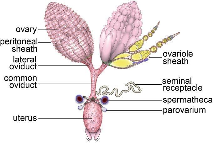
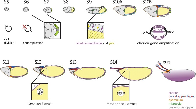
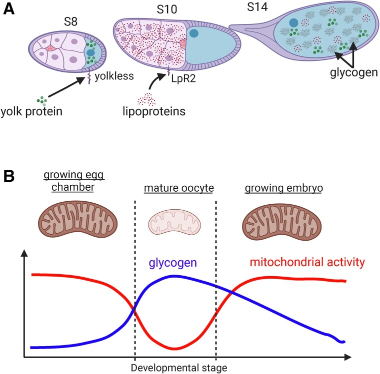
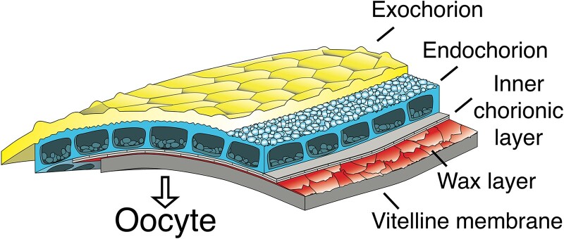
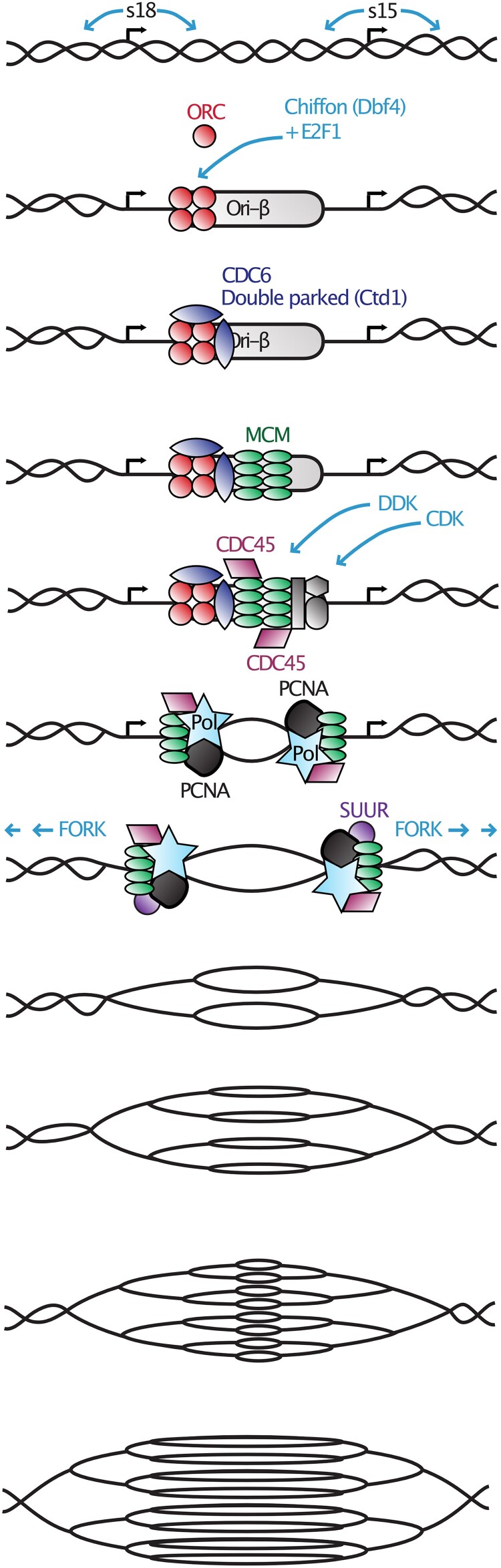
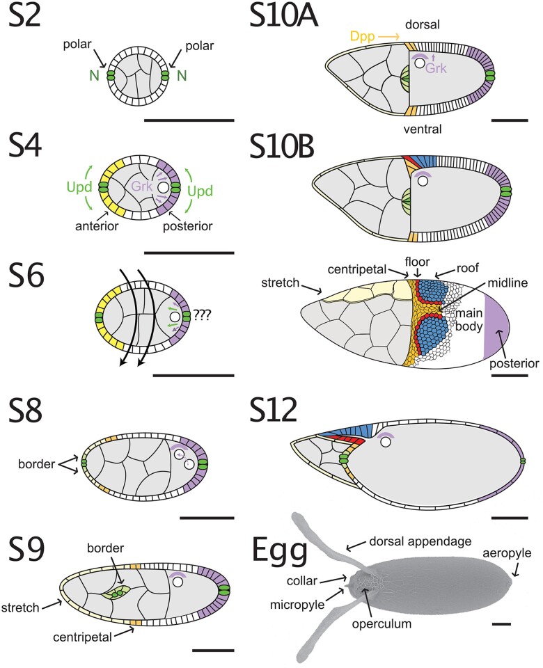
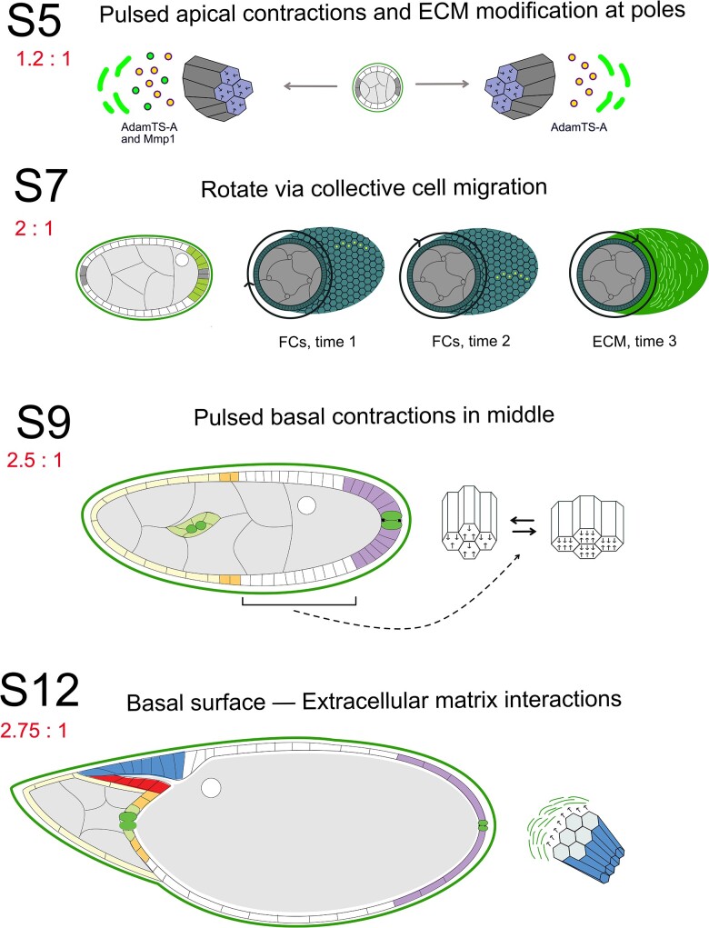
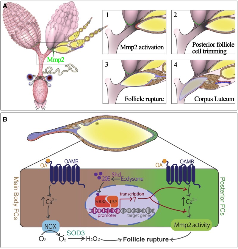
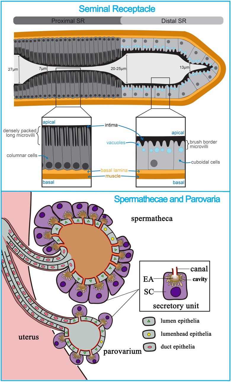
Gamete development is a fundamental process that is highly conserved from early eukaryotes to mammals. As germ cells develop, they must coordinate a dynamic series of cellular processes that support growth, cell specification, patterning, the loading of maternal factors (RNAs, proteins, and nutrients), differentiation of structures to enable fertilization and ensure embryonic survival, and other processes that make a functional oocyte. To achieve these goals, germ cells integrate a complex milieu of environmental and developmental signals to produce fertilizable eggs. Over the past 50 years, Drosophila oogenesis has risen to the forefront as a system to interrogate the sophisticated mechanisms that drive oocyte development. Studies in Drosophila have defined mechanisms in germ cells that control meiosis, protect genome integrity, facilitate mRNA trafficking, and support the maternal loading of nutrients. Work in this system has provided key insights into the mechanisms that establish egg chamber polarity and patterning as well as the mechanisms that drive ovulation and egg activation. Using the power of Drosophila genetics, the field has begun to define the molecular mechanisms that coordinate environmental stresses and nutrient availability with oocyte development. Importantly, the majority of these reproductive mechanisms are highly conserved throughout evolution, and many play critical roles in the development of somatic tissues as well. In this chapter, we summarize the recent progress in several key areas that impact egg chamber development and ovulation. First, we discuss the mechanisms that drive nutrient storage and trafficking during oocyte maturation and vitellogenesis. Second, we examine the processes that regulate follicle cell patterning and how that patterning impacts the construction of the egg shell and the establishment of embryonic polarity. Finally, we examine regulatory factors that control ovulation, egg activation, and successful fertilization.

Keywords: FlyBook; chorion gene amplification; egg activation; egg shape; eggshell; female reproductive tract; follicle cell differentiation; nutrient storage; oocyte maturation; ovulation.


Conflict of interest statement

Conflicts of interest statement The author(s) declare no conflict of interest.

Figures

Fig. 1.
Fig. 1.
Overview of the Drosophila female reproductive system. A schematic drawing (adapted from Deady et al. 2017) of the Drosophila female reproductive system shows the germline-containing ovary, which is comprised of 16 ovarioles that house the germline stem cells and developing egg chambers. The remainder of the reproductive tract (also named the lower reproductive tract) is comprised of somatic tissues such as the lateral oviducts, common oviduct, and the uterus, through which the egg passes during ovulation. The lower reproductive tract has a layer of epithelium separating the lumen and an outer muscle layer. This diagram also shows somatic structures such as the seminal receptacle, spermatheca, and parovarium, which house sperm or produce reproductive secretions that play key roles in ensuring fertilization success.
Fig. 2.
Fig. 2.
Stages of oogenesis. Germline cysts enveloped by a follicular epithelium emerge from the germarium as S1 egg chambers. This chapter focuses on the events from S5 onward that are needed to finish the egg. At S5, the follicle cells complete their last mitotic cycle and enter an endoreplication cycle at S6. They shut down endocycling at the transition from S10A to 10B, but they continue to amplify regions encoding the chorion genes. At S6, a signal from posterior follicle cells induces a reorganization of the oocyte cytoskeleton, and the oocyte nucleus moves to the anterior. From S8 to S10, the follicle cells synthesize and secrete yolk proteins and vitelline membrane proteins, and from S10 to S14, they synthesize and secrete the layers and specializations of the eggshell. At S9, the border cells delaminate and migrate between the nurse cells while the stretch cells flatten. At S10B, the centripetal cells begin to ingress. At S11, the nurse cells transfer their contents into the oocyte and begin to break down; at the same time, the dorsal appendage cells wrap to make 2 tubes. From S1 to S12, the oocyte chromosomes are held in a prophase I arrest. At S13, the oocyte nuclear envelope breaks down and the chromosomes line up on the metaphase plate. When the egg chamber moves into the oviduct, the follicle cells and nurse cell remnants slough off, revealing the eggshell.
Fig. 3.
Fig. 3.
Metabolic transitions drive stepwise nutrient storage during oogenesis. a) Nutrient storage occurs in a stepwise fashion during S8–S14, beginning with Yolk protein uptake during S8–S10, followed by lipoprotein uptake from S9 to S10A, and glycogen storage beginning at S10B. b) A working model of metabolic transitions shows the suppression of mitochondrial respiration that signifies the onset of MRQ (mitochondrial respiratory quiescence) as egg chambers transition from growth in earlier stages to quiescence at S10B. This suppression of mitochondrial metabolism during MRQ helps drive glycogen storage in mature oocytes for use by the developing embryo. Created with BioRender.com.
Fig. 4.
Fig. 4.
Composition of the Eggshell. Moving from oocyte proximal (bottom) to external surfaces (top), the eggshell consists of a vitelline membrane (thick, solid), wax layer (thin, scalloped), inner chorionic layer (thin, brick-like), endochorion with its floor, roof, and pillars (pebbly windows), and the exochorion (surface craters). (Redrawn with permission from Margaritis et al. 1980.) See also Turner and Mahowald (1976) and Margaritis (1986) for more detail.
Fig. 5.
Fig. 5.
Chorion gene amplification. Six locations in the genome amplify the DNA encoding major and minor chorion proteins. Shown here is the portion of DAFC-66D that contains s18 and s15, genes encoding the late chorion proteins Cp18 and Cp15. DNA replication initiation occurs at discrete origins associated with open areas of chromatin, which are indicated by curved arrows bracketing each gene. The winged helix-turn-helix E2F transcription factor and the Dbf4-like zinc finger protein encoded by chiffon (so named for the translucent eggshells of mutants; Landis and Tower 1999) facilitate the binding of origin recognition complex (ORC), which in turn recruits several additional winged helix-turn-helix proteins (Cdc6, double-parked/Cdt1) and the mini-chromosome maintenance (MCM) DNA helicase complex, allowing association of the helicase CDC45. Two kinases, Cyclin-dependent kinase (Cdk2), with its partner Cyclin E, and Dbf4-dependent kinase (Cdc7; Stephenson et al. 2015), activate this prereplication complex. Activation facilitates binding of proliferating cell nuclear antigen (PCNA) and the DNA polymerase complex (Pol), thereby initiating bidirectional fork movement. SUUR limits fork movement to ∼50 kb in each direction. Several rounds of reinitiation create branched duplexes such that DNA levels are highest at the origins and gradually decrease on each side (reviewed by Tower 2004; Claycomb and Orr-Weaver 2005; Nordman and Orr-Weaver 2012).
Fig. 6.
Fig. 6.
Patterning and morphogenesis. Representative stages showing progressive patterning and movements of follicle cells. All drawings are lateral cross sections, except the lower S10B example, which is a dorsolateral surface view. By S2, Notch signaling has defined 2 polar cells (dark green) at the anterior and posterior of the egg chamber. During S1–S5, the polar follicle cells secrete the JAK/STAT ligand Upd to pattern terminal regions. At the anterior (yellow), the gradient will specify border cells (light green), squamous stretch cells (yellow), and centripetal cells (orange) (see S8 and S9), while at the posterior, Gurken (Grk, EGF) signaling converts these cells to a posterior fate (purple). At S6, an unknown signal from posterior cells to the oocyte induces a microtubule rearrangement that moves the oocyte nucleus to the anterior. grk RNA and protein (crescent) move with the oocyte nucleus. Rotation of the egg chamber by migration of follicle cells on the ECM, which begins slowly at S1 but speeds up at S6, alters egg chamber shape from round to elongated, particularly during S6–S8. At S9, the border cells move between nurse cells toward the oocyte, carrying the polar cells, while the stretch cells flatten. At the transition from S10A to S10B, dorsal anterior follicle cells begin to express markers responding to Grk (EGF) and Dpp (BMP) signals (red, floor cells; blue, roof cells; orange, midline cells). At S10B, the centripetal and midline cells move inward. At S11, the dorsal appendage-forming cells wrap to make 2 tubes while the nurse cells dump their contents into the oocyte, and at S12–S13 the dorsal appendage-forming cells move out over the stretch cells, which envelope the degenerating nurse cells. Scanning electron microscope image of a laid egg (wild-type, Oregon R) reveals structures synthesized by the anterior cells (operculum, micropyle, collar), roof and floor cells (dorsal appendages), main body cells (majority of eggshell), and posterior cells (aeropyle). Image courtesy of Dr. Miriam Osterfield. All black bars = 50 µ.
Fig. 7.
Fig. 7.
Mechanisms of egg elongation. The round egg chamber elongates through distinct follicle cell behaviors. During S3–S7 (represented by the S5 egg chamber), a gradient of JAK/STAT signaling at the poles induces apical constriction of the follicle cells, pulling the germ cells outward. JAK/STAT signaling also induces expression of proteases (AdamTS-A and MMP1) that modify the ECM (outer, thick, light green lines). As shown by the S7 egg chamber, the main force driving elongation is the rotation of the egg chamber along its long axis. The follicle cells (middle two egg chambers, dark green) walk in the same direction; yellow dots mark the position of a row of cells at different times during development. The cells move slowly at first (S1–S5), but the speed picks up during S6–S8. During their migration, the follicle cells secrete ECM proteins into the basement membrane (far right egg chamber, light green) in a graded fashion such that the polar regions exert less tension on the egg chamber than the middle. During S9–S10A, pulsed basal contractions in the middle of the egg chamber squeeze the tissue outward. During the last stages of oogenesis (represented by S12), basal cell surfaces maintain contact with the ECM and exert tension on the tissue. The aspect ratio (length to width) of each stage is shown at the far left.
Fig. 8.
Fig. 8.
Process of ovulation and intrinsic factors in ovulation. a) A model shows the key steps in ovulation (adapted from Deady et al. 2017). Ovulation begins with activation of Matrix metalloprotease 2 (MMP2) in posterior follicle cells. Activation is followed by follicle cell trimming and follicle rupture. Once the oocyte is ovulated, the follicle cell sheath is maintained as a corpus luteum whose function is completely unknown. b) Describes the intrinsic roles for ecdysone signaling, calcium signaling, reactive oxygen species, and octopamine in main body or posterior follicle cells to control ovulation.
Fig. 9.
Fig. 9.
Anatomical structure of seminal receptacle, spermathecae, and parovaria. The top panel shows the anatomy of the proximal and distal seminal receptacle. The bottom panel shows the anatomy of the spermathecae and parovarian structures of the reproductive tract. The secretory unit consists of a secretory cell (SC), acellular end apparatus (EA), secretory cavity (space between the apical surface of SC and EA), and a canal that connects the secretory cell to the lumen. The cuticular intima lining the lumen and canal is highlighted in red.

References

    1. Aggarwal BD, Calvi BR. 2004. Chromatin regulates origin activity in Drosophila follicle cells. Nature. 430(6997):372–376. doi:10.1038/nature02694. - DOI - PubMed
    1. Aigaki T, Fleischmann I, Chen P-S, Kubli E. 1991. Ectopic expression of sex peptide alters reproductive behavior of female D. melanogaster. Neuron. 7(4):557–563. doi:10.1016/0896-6273(91)90368-A. - DOI - PubMed
    1. Airoldi SJ, McLean PF, Shimada Y, Cooley L. 2011. Intercellular protein movement in syncytial Drosophila follicle cells. J Cell Sci. 124(23):4077–4086. doi:10.1242/jcs.090456. - DOI - PMC - PubMed
    1. Akey CW, Edelstein SJ. 1987. The innermost chorionic layer of Drosophila. I. The role of chorin octamers in the formation of a family of interdigitating crystalline plates. J Mol Biol. 193(4):673–683. doi:10.1016/0022-2836(87)90350-0. - DOI - PubMed
    1. Alégot H, Pouchin P, Bardot O, Mirouse V. 2018. Jak-Stat pathway induces Drosophila follicle elongation by a gradient of apical contractility. Elife. 7:773. doi:10.7554/eLife.32943. - DOI - PMC - PubMed

Publication types

LinkOut - more resources