Clinical Trial
. 2023 Nov 25;14(1):7725.
doi: 10.1038/s41467-023-43175-5.

Tamoxifen for the treatment of myeloproliferative neoplasms: A Phase II clinical trial and exploratory analysis

Affiliations
Clinical Trial

Tamoxifen for the treatment of myeloproliferative neoplasms: A Phase II clinical trial and exploratory analysis

Zijian Fang et al. Nat Commun. .

Abstract

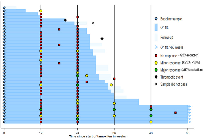
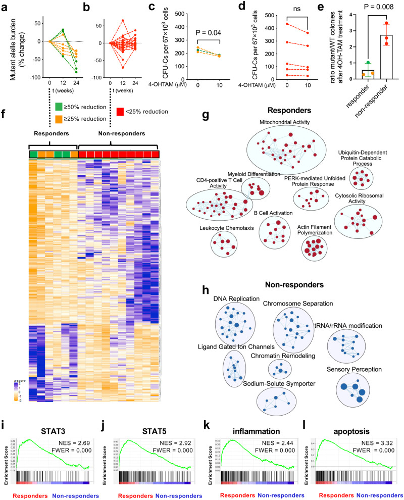
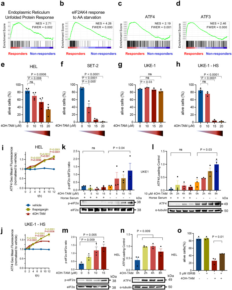
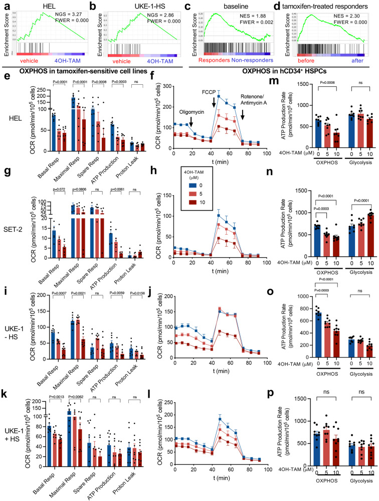
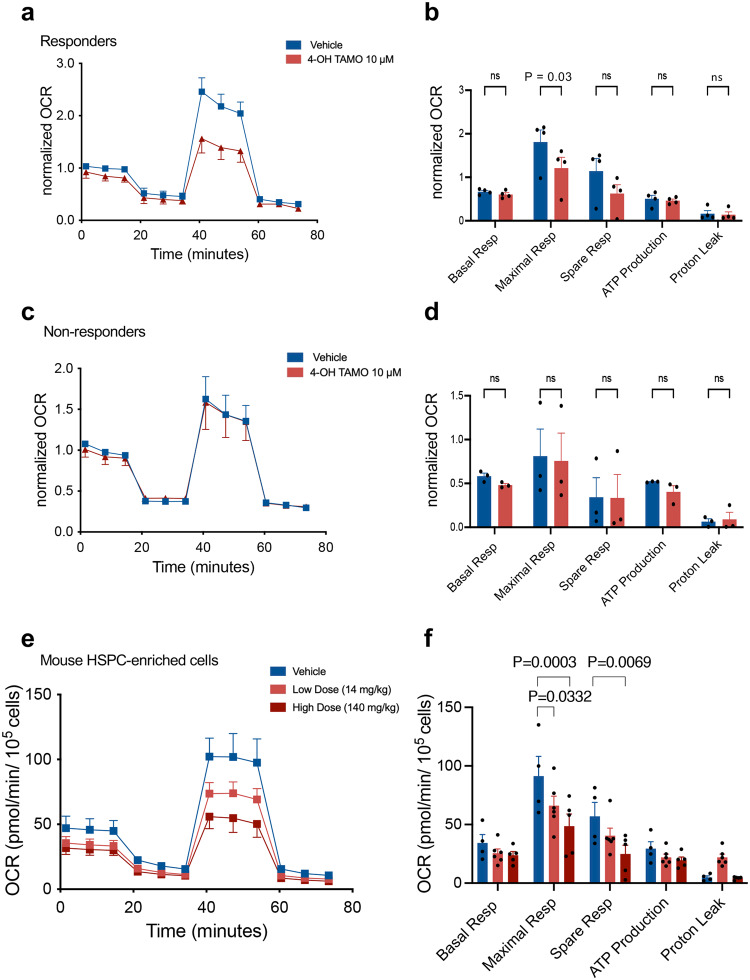
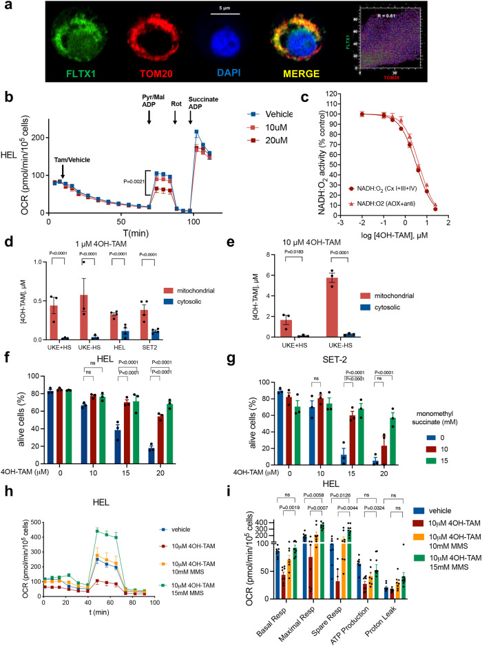
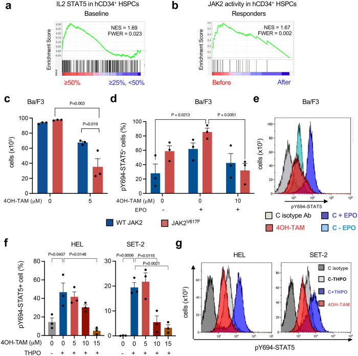
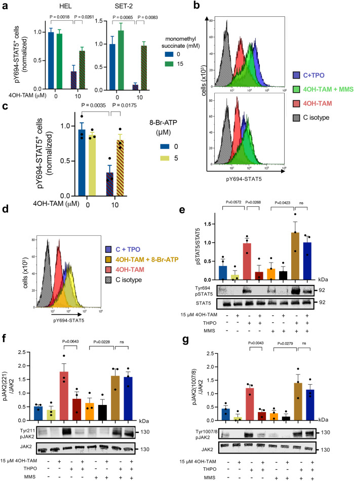
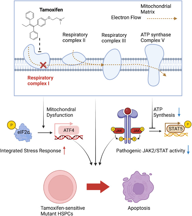
Current therapies for myeloproliferative neoplasms (MPNs) improve symptoms but have limited effect on tumor size. In preclinical studies, tamoxifen restored normal apoptosis in mutated hematopoietic stem/progenitor cells (HSPCs). TAMARIN Phase-II, multicenter, single-arm clinical trial assessed tamoxifen's safety and activity in patients with stable MPNs, no prior thrombotic events and mutated JAK2V617F, CALRins5 or CALRdel52 peripheral blood allele burden ≥20% (EudraCT 2015-005497-38). 38 patients were recruited over 112w and 32 completed 24w-treatment. The study's A'herns success criteria were met as the primary outcome ( ≥ 50% reduction in mutant allele burden at 24w) was observed in 3/38 patients. Secondary outcomes included ≥25% reduction at 24w (5/38), ≥50% reduction at 12w (0/38), thrombotic events (2/38), toxicities, hematological response, proportion of patients in each IWG-MRT response category and ELN response criteria. As exploratory outcomes, baseline analysis of HSPC transcriptome segregates responders and non-responders, suggesting a predictive signature. In responder HSPCs, longitudinal analysis shows high baseline expression of JAK-STAT signaling and oxidative phosphorylation genes, which are downregulated by tamoxifen. We further demonstrate in preclinical studies that in JAK2V617F+ cells, 4-hydroxytamoxifen inhibits mitochondrial complex-I, activates integrated stress response and decreases pathogenic JAK2-signaling. These results warrant further investigation of tamoxifen in MPN, with careful consideration of thrombotic risk.


Conflict of interest statement

C.N.H. reports funded research from Novartis; speaker fees from Novartis, Janssen, CTI, Celgene, and Medscape; and advisory board membership for Incyte, CTI, Sierra Oncology, Novartis, Celgene, Roche, AOP pharma, Geron, and AstraZeneca. A.J.M. reports funded research from Novartis, Celgene/BMS and consultancy for Abbvie, CTI, and Gilead. S.K. reports funded research and advisory board membership for Novartis. J.E. reports advisory board membership for Novartis, Incyte and Celgene/BMS. M.F.M. reports consultancies for Celgene and BMS; and advisory board membership for Novartis, Jazz, and Abbvie. S.N. reports speaker fees for Takeda, Celgene, Novartis, MSD and Alexion. D.M. reports consultancy for Incyte, Pfizer, Novartis, and Bristol-Myers Squibb. M.W.D. reports funded research from Blueprint Medicine Corporation and advisory board membership for Bristol-Myers Squibb, Novartis, Gilead, Pfizer, Jazz, Takeda, and Astellas. The remaining authors declare no competing financial interests. A.L.G. reports speaker fees from Novartis and advisory board membership AOP pharma and BMS.

Figures

Fig. 1
Fig. 1. Tamoxifen reduces mutant allele burden in a subset of MPN patients.
Swimmer plot of treatment, thrombotic events and allele burden response.
Fig. 2
Fig. 2. Distinctive transcriptomic signature of HSPCs from tamoxifen responders at baseline.
a, b Mutant allele burden change (%) in (a) responders achieving after 24w tamoxifen treatment allele burden reductions of ≥50% (n = 3, green) or ≥25%, <50% (n = 5, orange), and (b) in non-responders (n = 28, red) before treatment, and 12w or 24w after tamoxifen administration. c-d HSPCs measured as colony-forming units in culture (CFU-Cs) from study patients’ peripheral blood mononuclear cells treated with 4OH-TAM (10 mM) or vehicle for 24 h. Reduced (c) or unchanged (d) HSPC numbers upon ex vivo 4OH-TAM treatment of baseline samples are consistent with the allele burden reductions observed in the same patients after 24w tamoxifen treatment (c, green ≥ 50%, n = 2; orange≥25%, n = 1; d, red < 25%, n = 5). *p < 0.05, two-tailed unpaired t test. e CFU-C genotyping shows a reduced balance of JAK2V617F+ colonies, compared with WT colonies, by 4OH-TAM treatment in responders’, but not in non-responders’ samples (allele burden reductions after 24w are marked with green ≥ 50%, n = 1; orange ≥ 25%, n = 2; red < 25%, n = 3). **p < 0.01, two-tailed paired t test. Data are mean ± SEM. f Heatmap reveals the disparity of gene expression between responders and non-responders at baseline. g, h Integrated pathway enrichment map using gene-sets enriched in responders (g) and non-responders (h). i, l Gene set enrichment analysis (GSEA) shows a higher activation of HSPCs in responders, with increased expression of (i) STAT3-, (j) STAT5-, (k) inflammation- and (l) apoptosis-related genes. NES, normalized enrichment score. FWER, family-wise error rate.
Fig. 3
Fig. 3. JAK2V617F+ human cell lines reproduce the differential sensitivity to tamoxifen of study patients.
ad Gene set enrichment analysis (GSEA) from HSPCs from responders at baseline shows signs of integrated stress response, manifested as increased expression of genes involved in unfolded protein response (UPR) (a), response to aminoacid (AA) starvation (b), or target genes of activating transcription factors (ATF) 3 and 4 (c, d). NES, normalized enrichment score. FWER, family-wise error rate. eh 24 h treatment with the soluble tamoxifen derivative 4-hydroxytamoxifen (4OH-TAM) dose-dependently reduces the viability of HEL cells (e), SET2 cells (f) and horse-serum-starved UKE-1 cells (h), but spares UKE-1 cells (g) cultured with competent medium (n = 4 independent experiments in HEL cells, n = 3 independent experiments in SET2 and UKE-1 cells). i, j 4OH-TAM induces ATF4 translation in HEL cells (i) and horse serum-deprived UKE-1 cells (j) expressing ATF4-mScarlet. The fluorescence intensity of 4OH-TAM and thapsigargin-treated cells was normalized to the vehicle control at each time point (n = 5 independent experiments). k, l Quantification (top) and representative Western blots (bottom) of (k) eIF2α and phosphorylated (p) eIF2α after 8 h vehicle/4OH-TAM treatment or (l) ATF4 and β-tubulin (loading control) before/after 4OH-TAM treatment in UKE-1 cells cultured with/without horse serum (n = 3 independent experiments). m, n Quantification (top) and representative western blots (bottom) of (m) eIF2α and p-eIF2α after 8 h vehicle/4OH-TAM treatment, or (n) ATF4 and β-tubulin (loading control) before/after 4OH-TAM treatment in HEL cells (n = 3 independent experiments). o Integrated stress response (ISR) inhibitor (ISRIB) partially rescues 4OH-TAM-induced HEL cell death. Frequency of viable cells after 24 h treatment with 4OH-TAM and ISRIB alone or in combination (n = 3 independent experiments). eo Data are mean ± SEM. *p < 0.05, **p < 0.01, ***p < 0.001, ****p < 0.0001; One-way ANOVA and Dunnett’s test.
Fig. 4
Fig. 4. Tamoxifen downregulates OXPHOS-related genes and reduces OXPHOS-derived ATP generation in JAK2V617F-mutated human cells.
ad High enrichment of OXPHOS-related genes in sensitive cells and its marked downregulation by tamoxifen treatment. Gene set enrichment analysis (GSEA) of differentially expressed genes after 4OH-TAM treatment of sensitive cell lines (a, b, n = 3 independent experiments), HSPCs from prospective TAMARIN responders achieving after 24w tamoxifen treatment allele burden reductions of ≥50% (c, n = 3 individuals) or ≥25%, <50% (n = 3 individuals), and non-responders (n = 10 individuals) or genes selectively downregulated by tamoxifen treatment in responder HSPCs (d, n = 6 individuals). NES, normalized enrichment score. FWER, family-wise error rate. el Dose-dependent inhibition of mitochondrial respiration and its derived ATP by 4OH-TAM in HEL cells (ef, n = 8 biologically independent samples), SET-2 cells (g, h, n = 6 independent experiments) and UKE-1 cells cultured under serum starvation (i, j, n = 8 independent experiments) or (k, l, n = 9 independent experiments) with competent medium. Oxygen Consumption Rate (OCR) was measured after 2 h treatment with 4OH-TAM or vehicle, using the Cell Mito Stress kit (Agilent). Note the more severe reduction of respiratory capacity and mitochondrial ATP production in sensitive JAK2V617F-mutant cells (ej), compared with resistant cells (k, l). f, h, j, l Timeline of OCR measurement after oligomycin to inhibit ATP synthase, FCCP to disrupt the mitochondrial membrane potential and rotenone/Antimycin A to inhibit mitochondrial complex I. m-p Selective inhibition of OXPHOS-derived (not glycolysis-derived) ATP generation by 4OH-TAM in sensitive JAK2V617F-mutant cells (n = 9 independent experiments). ATP generated from OXPHOS and glycolysis was simultaneously measured in sensitive (mo) or resistant (p) cells using the ATP Real-Time rate assay kit (Agilent). ep Data are mean + SEM. *p < 0.05, **p < 0.01, ***p < 0.001, Two-way ANOVA and Dunnett’s test.
Fig. 5
Fig. 5. Tamoxifen reduces mitochondrial respiration in blood cells from responders and HSPCs from mice treated with a dose comparable to study subjects.
a, b A high capacity of mitochondrial respiration in peripheral blood mononuclear cells (PBMCs) from responders is suppressed by 4OH-TAM (10 mM) treatment (n = 5 individuals). c, d Comparatively lower baseline OXPHOS and inhibition by 4OH-TAM (10 μM) in PBMCs from non-responders (n = 5 individuals). ad Oxygen Consumption Rate (OCR) was measured in PBMCs after 24 h treatment with 4OH-TAM or vehicle and normalized to baseline. e, f Mitochondrial respiration in HSPC-enriched cells isolated from MPN mice treated with low dose tamoxifen (14 mg/kg, n = 6 animals), high dose tamoxifen (140 mg/kg, n = 5 animals) or vehicle (n = 4 animals) over two weeks (3 times/week). Note OXPHOS inhibition using a dose comparable to study patients. Data are means+SEM. *p < 0.05, **p < 0.01, ***p < 0.001, Two-way ANOVA and Dunnett’s test.
Fig. 6
Fig. 6. Tamoxifen reduces mitochondrial ATP generation by inhibiting respiratory complex I.
a Supra-resolution AiryScan2 confocal imaging of fluorescent tamoxifen derivative (FLTX1, green) colocalizing with the mitochondrial marker TOM20 (red) in HEL cells. Nucleus was stained with DAPI (blue). ImageJ plugin Colocalization Finder was used to generate a scatter plot of two selected channel intensity and calculate overlap coefficient for selected channels. R > 0.8 indicates significant colocalization. The analysis has been independently repeated three times, obtaining similar results. b 4OH-TAM dose-dependently inhibits mitochondrial respiration in permeabilized JAK2V617F-mutant cells (n = 10 independent experiments). Average oxygen consumption rate (OCR) after treatment with plasma membrane permeabilizer, 4OH-TAM or vehicle, and 5 mM Pyruvate (Pyr), 2.5 mM malate and 1 mM adenosine diphosphate (ADP). c Dose-dependent inhibition of complex I NADH:O2 oxidoreduction rate by 4OH-TAM in absence or presence of antimycin (anti) to inhibit complex III (n = 4 independent experiments). d, e Dose-dependent accumulation of 4OH-TAM in the mitochondria of MPN cell lines 24 h after 1 mM (d) and 10 mM 4OH-TAM treatment (e). Note 4-fold higher mitochondrial 4OH-TAM concentration in sensitive (serum-deprived), compared with resistant (grown in competent medium) UKE-1 cells (n = 3 independent experiments). f, g Complex II activation with monomethyl succinate rescues JAK2V617F-mutant HEL (f) and SET2 (g) cells from 4OH-TAM-induced cell death (n = 3 independent experiments). h, i OCR in HEL cells treated with 4OH-TAM or vehicle alone, or in combination with MMS (n = 8 independent experiments). The rescue of cell viability by complex II activation is explained by compensatory increase of mitochondrial respiration and ATP synthesis. d-i Data are mean ± SEM; *p < 0.05, **p < 0.01, ***p < 0.001, Two-way ANOVA and Dunnett’s test.
Fig. 7
Fig. 7. Tamoxifen preferentially inhibits oncogenic JAK-STAT signaling.
a Gene set enrichment analysis (GSEA) of HSPCs obtained at baseline from study patients showing allele burden reductions ≥50% (n = 3 individuals) or between ≥25% and 50% (n = 3 individuals) 24w after tamoxifen treatment. Interleukin-2 (IL-2)-STAT5 signatures were enriched in HSPCs from patients achieving ≥50% allele burden reductions. b Selective downregulation of JAK2-related genes in HSPCs from study patients showing allele burden reductions ≥50% (n = 3 individuals) or between ≥25% and 50% (n = 3 individuals) 24w after tamoxifen treatment. NES, normalized enrichment score. FWER, family-wise error rate. ce Mouse Ba/F3 cells expressing the erythropoietin (Epo) receptor and transduced with mutant JAK2V617F or WT JAK2 were treated with 4OH-TAM or vehicle. c Ba/F3 cells carrying JAK2V617F are twice as sensitive to 4OH-TAM-induced cell death compared with cells expressing WT JAK2 (n = 3 independent experiments). Data are mean + SEM. d, e Selective inhibition of oncogenic JAK-STAT signaling by 4OH-TAM in Ba/F3 cells. d Frequency of WT/mutant JAK2-expressing Ba/F3 cells with phosphorylated (p) STAT5 at residue Y694 at baseline or after EPO (5 U/ml) stimulation, in combination with 4OH-TAM (red) or vehicle (blue). Data are mean + SEM. e Representative flow cytometry histograms of pSTAT5 level at baseline (light blue) or after EPO (5 U/ml) stimulation (dark blue), in combination with 4OH-TAM (red) or vehicle (n = 3 independent experiments). f, g 4OH-TAM dose-dependently inhibits oncogenic JAK-STAT signaling in JAK2V617F-mutated human cells. Frequency of pSTAT5+ HEL cells or SET-2 cells (f) and representative flow cytometry histograms of pSTAT5 level (g) at baseline or after thrombopoietin (THPO, 100 ng/ml) stimulation (blue), in combination with 4OH-TAM (red) or vehicle (n = 3 independent experiments). C, control. c-d, f Data are mean ± SEM; p < 0.05, **p < 0.01, ***p < 0.001, One-way ANOVA and Tukey’s test.
Fig. 8
Fig. 8. Inhibition of pathogenic JAK-STAT signaling by tamoxifen is rescued by complex II activation or ATP supplementation.
a-b Frequency of pSTAT5+ HEL cells or SET-2 cells (a) and representative flow cytometry histograms of pSTAT5 expression (b) at baseline or after thrombopoietin (THPO) stimulation (blue), in combination with 4OH-TAM (red) or vehicle (n = 3 independent experiments). Combined treatment with the mitochondrial complex II substrate monomethyl succinate (MMS, green) rescues the reduction of pSTAT5 by 4OH-TAM. C, control. c-d Frequency of pSTAT5+ THPO-stimulated HEL cells (c) and representative flow cytometry histograms of pSTAT5 expression (d) in HEL cells treated with 4OH-TAM alone (red) or vehicle (blue), or in combination with cell-permeable ATP (5 mM 8-Br-ATP, yellow) (n = 3 independent experiments). ATP supplementation rescues the reduction of pSTAT5 by 4OH-TAM. a,c Data are mean + SEM; *p < 0.05, **p < 0.01, ***p < 0.001, One-way ANOVA and Tukey’s test. e Quantification (top) and representative Western blots (bottom) of STAT5 and phosphorylated (p) STAT5 level at baseline or after thrombopoietin (THPO) stimulation, in combination with 4 h vehicle/4OH-TAM treatment and mitochondrial complex II substrate monomethyl succinate (10 mM MMS) in HEL cell lines. Combined treatment with the mitochondrial complex II substrate monomethyl succinate (MMS) rescues the reduction of THPO-induced pSTAT5 by 4OH-TAM (n = 3 independent experiments). f-g Quantification (top) and representative Western blots (bottom) of JAK2 and phosphorylated (p) JAK2 (f, Tyr221; g, Tyr1007/8) level at baseline or after thrombopoietin (THPO) stimulation, in combination with 4 h vehicle/4OH-TAM treatment and monomethyl succinate (10 mM MMS) in HEL cell lines, showing the reduction of thrombopoietin-induced JAK2 phosphorylation by 4OH-TAM could be rescued by the addition of succinate in human JAK2V617F-mutated cell lines (n = 3 independent experiments). e-g Data are mean + SEM; *p < 0.05, **p < 0.01, ***p < 0.001, One-way ANOVA and Dunnett’s test. e-g Data are mean + SEM; *p < 0.05, **p < 0.01, ***p < 0.001, One-way ANOVA and Dunnett’s test.
Fig. 9
Fig. 9. HSPCs with high JAK-STAT signaling, integrated stress response and mitochondrial respiration are particularly sensitive to tamoxifen-induced cell death.
Tamoxifen derivatives accumulate in the mitochondria, where they bind to and inhibit complex I. Biochemical complex I inhibition adds to reduced complex I gene transcription after tamoxifen treatment. Additionally, tamoxifen further increases integrated stress response in HSPCs and tips the balance from protective (p-eIF2α-dependent) to proapoptotic (ATF4-mediated) unfolded protein response. Through transcriptional regulation and previously unrecognized direct metabolic effects, tamoxifen can eliminate cells with pathogenic JAK-STAT activation by modulating integrated sress response and inhibiting mitochondrial complex-I and ATP generation, preventing pathogenic STAT5 phosphorylation.

Similar articles

Cited by

References

    1. James C, et al. A unique clonal JAK2 mutation leading to constitutive signalling causes polycythaemia vera. Nature. 2005;434:1144–1148. doi: 10.1038/nature03546. - DOI - PubMed
    1. Kralovics R, et al. A gain-of-function mutation of JAK2 in myeloproliferative disorders. N. Engl. J. Med. 2005;352:1779–1790. doi: 10.1056/NEJMoa051113. - DOI - PubMed
    1. Levine RL, et al. Activating mutation in the tyrosine kinase JAK2 in polycythemia vera, essential thrombocythemia, and myeloid metaplasia with myelofibrosis. Cancer Cell. 2005;7:387–397. doi: 10.1016/j.ccr.2005.03.023. - DOI - PubMed
    1. Baxter EJ, et al. Acquired mutation of the tyrosine kinase JAK2 in human myeloproliferative disorders. Lancet. 2005;365:1054–1061. doi: 10.1016/S0140-6736(05)71142-9. - DOI - PubMed
    1. Klampfl T, et al. Somatic mutations of calreticulin in myeloproliferative neoplasms. N. Engl. J. Med. 2013;369:2379–2390. doi: 10.1056/NEJMoa1311347. - DOI - PubMed

Publication types

MeSH terms

Associated data