. 2023 Nov 24;14(1):7694.
doi: 10.1038/s41467-023-43465-y.

Nanoparticles exhibiting virus-mimic surface topology for enhanced oral delivery

Affiliations

Nanoparticles exhibiting virus-mimic surface topology for enhanced oral delivery

Zhentao Sang et al. Nat Commun. .

Abstract

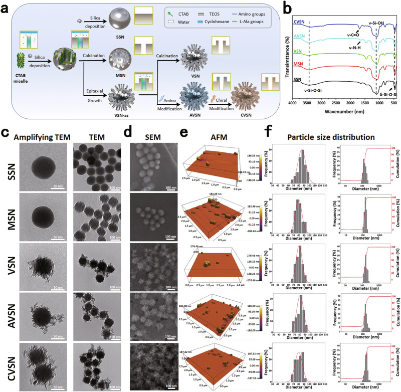
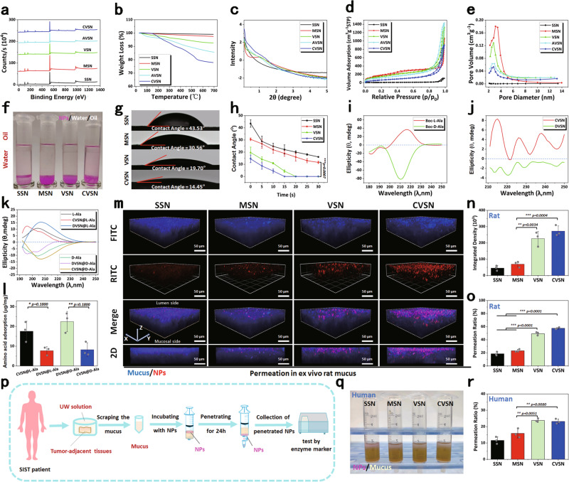
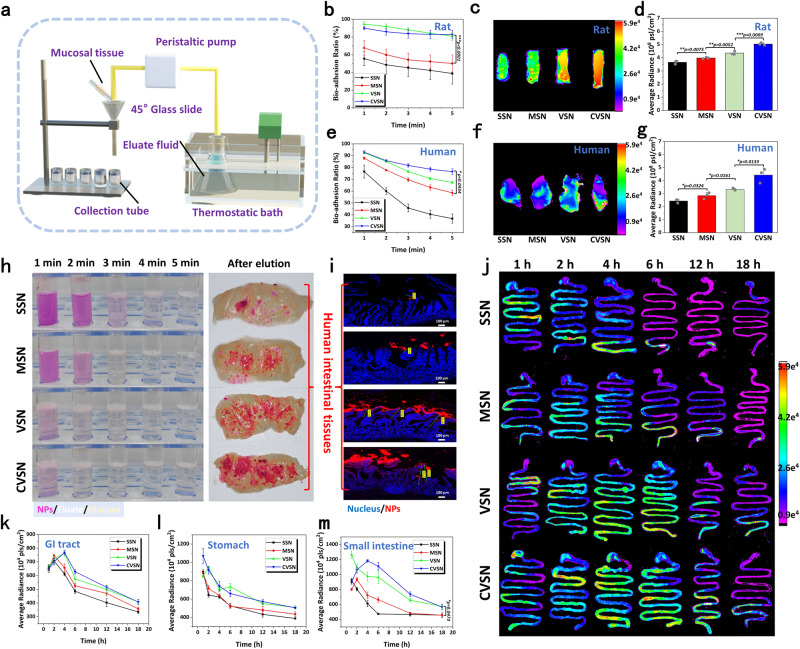
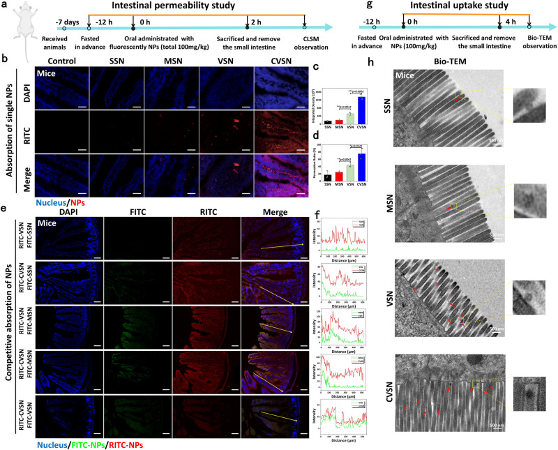
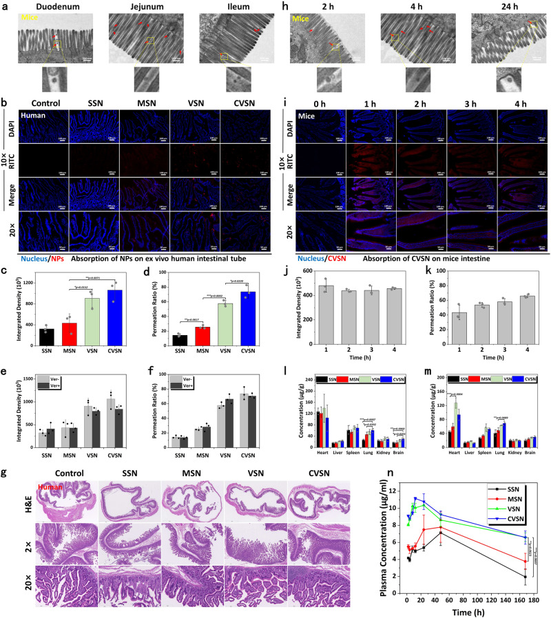
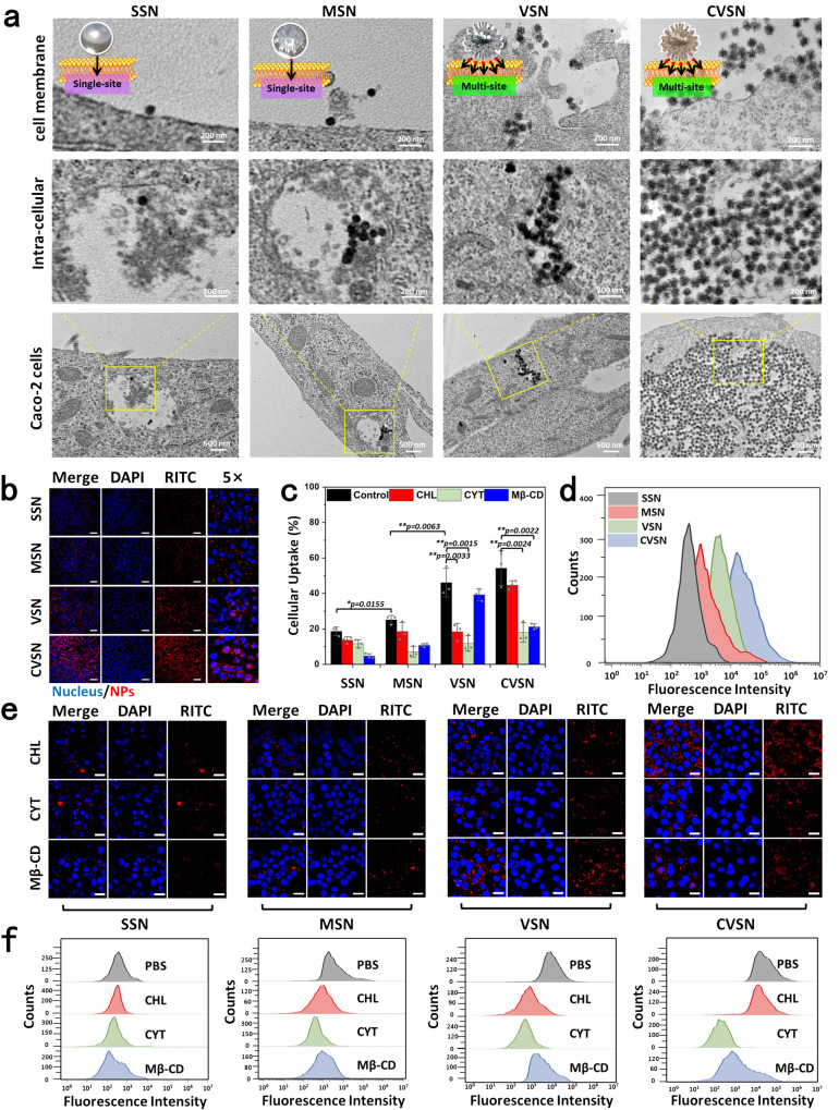
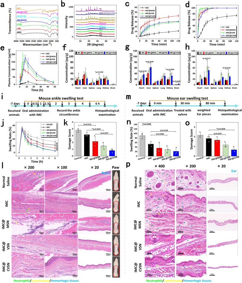
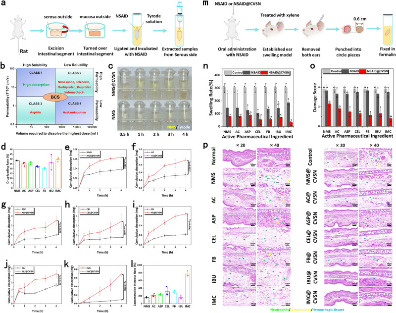
The oral delivery of nano-drug delivery systems (Nano-DDS) remains a challenge. Taking inspirations from viruses, here we construct core-shell mesoporous silica nanoparticles (NPs, ~80 nm) with virus-like nanospikes (VSN) to simulate viral morphology, and further modified VSN with L-alanine (CVSN) to enable chiral recognition for functional bionics. By comparing with the solid silica NPs, mesoporous silica NPs and VSN, we demonstrate the delivery advantages of CVSN on overcoming intestinal sequential barriers in both animals and human via multiple biological processes. Subsequently, we encapsulate indomethacin (IMC) into the nanopores of NPs to mimic gene package, wherein the payloads are isolated from bio-environments and exist in an amorphous form to increase their stability and solubility, while the chiral nanospikes multi-sited anchor and chiral recognize on the intestinal mucosa to enhance the penetrability and ultimately improve the oral adsorption of IMC. Encouragingly, we also prove the versatility of CVSN as oral Nano-DDS.


Conflict of interest statement

The authors declare no competing interests.

Figures

Fig. 1
Fig. 1. The formation and morphology of NPs.
a Synthesis steps of NPs. b Fourier transform infrared (FTIR) spectra of NPs. Source data are provided as a Source Data file. c Transmission electron microscope (TEM) images of NPs. Experiment was repeated three times independently with similar results. d Scanning electron microscope (SEM) images of NPs. Experiment was repeated three times independently with similar results. e Atomic force microscope (AFM) images of NPs. f Particle size distribution (counted from the SEM images, and measured by dynamic light scattering [DLS], respectively) of NPs. Source data are provided as a Source Data file.
Fig. 2
Fig. 2. The structure and surface properties of NPs.
a XPS spectra, b thermogravimetric analysis (TGA) curves, c Small angle X-ray scattering (SAXS) patterns d N2 adsorption/desorption isotherms and e pore size distribution curves of NPs. Source data of ae are provided as a Source Data file. f Oil-water distribution images of NPs. g Initial contact angles images of NPs. h The contact angles-time curves of NPs. Data are presented as the mean ± SD (n = 3 independent experiments). **P < 0.01 by two-tailed Student’s t-test. Source data are provided as a Source Data file. Circular dichroism (CD) spectra of i Boc-L-alanine and Boc-D-alanine (Boc-L/D-Ala), j CVSN and DVSN, which was synthesized by grafting D-Ala onto the surface of VSN. Source data of i, j are provided as a Source Data file. k CD spectra of amino acid solutions before and after incubation with CVSN and DVSN. Source data are provided as a Source Data file. l Amino acid adsorption capacity of CVSN and DVSN. Data are presented as the mean ± SD (n = 3 independent experiments). *P < 0.05 and **P < 0.01 by two-tailed Student’s t-test. Source data are provided as a Source Data file. m Three dimensions (3D) confocal fluorescence microscope (CLSM) images of NPs penetrated through the rat mucus layer; blue: mucus stained with fluorescein isothiocyanate (FITC). red: Rhodamine B isothiocyanate (RITC) labeled NPs. Scale bar: 50 μm. Depth: 70 μm. Experiment was repeated three times independently with similar results. n Fluorescence integrated density and o permeation ratio of NPs in mucus permeation. Data are presented as the mean ± SD (n = 3 independent experiments). **P < 0.01 and ***P < 0.001 by two-tailed Student’s t-test. Source data of n, o are provided as a Source Data file. p Treatment protocols on the permeation of NPs in human intestinal mucus. q Image and r permeation ratio on the permeation of NPs in human intestinal mucus. Data are presented as the mean ± SD (n = 3 independent experiments). **P < 0.01 by two-tailed Student’s t-test. Source data are provided as a Source Data file.
Fig. 3
Fig. 3. Bio-adhesion ability of NPs on mucosal tissues.
a Schematic illustration of the apparatus on bio-adhesion study. b The bio-adhesion profiles of NPs on the rat intestinal mucosa measured by weight loss. Data are presented as the mean ± SD (n = 3 independent experiments). ***P < 0.001 by two-tailed Student’s t-test. Source data are provided as a Source Data file. c Fluorescence images and d average intensity of NPs remained on the rat intestinal mucosa after elution. The color bar indicates the fluorescence intensity. Experiment was repeated three times independently with similar results. Data are presented as the mean ± SD (n = 3 independent experiments). **P < 0.01 and ***P < 0.001 by two-tailed Student’s t-test. Source data are provided as a Source Data file. e The bio-adhesion profiles of NPs on the human intestinal mucosa measured by weight loss. Data are presented as the mean ± SD (n = 3 independent experiments). *P < 0.05 by two-tailed Student’s t-test. Source data are provided as a Source Data file. f Fluorescence images and g average intensity of NPs remained on the human intestinal mucosa after elution. The color bar indicates the fluorescence intensity. Experiment was repeated three times independently with similar results. Data are presented as the mean ± SD (n = 3 independent experiments). *P < 0.05 by two-tailed Student’s t-test. Source data are provided as a Source Data file. h The bio-adhesion images of RITC labeled NPs performed on the human intestinal mucosa. Experiment was repeated three times independently with similar results. i CLSM images of RITC labeled NPs remained on the human intestinal mucosa; blue: nuclei of the intestinal mucosa stained with 4,6-diamidino-2-phenylindole (DAPI), red: RITC labeled NPs, scale bar: 100 μm. Experiment was repeated three times independently with similar results. j Fluorescence images of NPs remained on the GI tract after oral administration. Experiment was repeated three times independently with similar results. The color bar indicates the fluorescence intensity. Average intensity of NPs remained in the k GI tract, l stomach and m small intestine after oral administration. Data are presented as the mean ± SD (n = 3 independent experiments). *P < 0.05 by two-tailed Student’s t-test. Source data are provided as a Source Data file.
Fig. 4
Fig. 4. Adsorption of NPs through the intestinal villi.
a Treatment protocols on the intestinal permeability study of NPs. b CLSM images of NPs absorbed at the small intestinal villi of mice at 2 h post oral administration; blue: nuclei of the intestinal villi stained with DAPI, red: RITC labeled NPs, scale bar: 50 μm. Experiment was repeated three times independently with similar results. c Fluorescence integrated density and d permeation ratio of NPs at the small intestinal villi of mice. Data are presented as the mean ± SD (n = 3 independent experiments). *P < 0.05, *P < 0.01 and ***P < 0.001 by two-tailed Student’s t-test. Source data are provided as a Source Data file. e Competitive adsorption of NPs in the intestinal villi of mice examined by CLSM; blue: nuclei of the intestinal villi stained with DAPI, green: FITC labeled NPs, red: RITC labeled NPs, scale bar: 100 μm. Experiment was repeated three times independently with similar results. f Relative fluorescent intensity of NPs in intestinal competitive adsorption. Source data are provided as a Source Data file. g Treatment protocols on the intestinal uptake study of NPs. h Bio-TEM images of the mucosa of mice at 4 h post oral administration of NPs. Experiment was repeated three times independently with similar results.
Fig. 5
Fig. 5. Oral adsorption and distribution of NPs.
a Bio-TEM images of the mucosa from the duodenum, jejunum and ileum of mice at 4 h post oral administration of CVSN. Experiment was repeated three times independently with similar results. b CLSM images of NPs absorbed at the ex vivo small intestinal villi of human at 1.5 h post incubation without the per-treatment of verapamil (VER); blue: nuclei of the intestinal villi stained with DAPI, red: RITC labeled NPs. Experiment was repeated three times independently with similar results. c Fluorescence integrated density and d permeation ratio of NPs at the small intestinal villi of human. e Fluorescence integrated density and f permeation ratio of NPs at the small intestinal villi of human before and after pretreated with VER. All data of cf are presented as the mean ± SD (n = 3 independent experiments). *P < 0.05, **P < 0.01 and ***P < 0.001 by two-tailed Student’s t-test. Source data are provided as a Source Data file. g Hematoxylin and eosin (H&E) staining images of the small intestinal tissues of human after incubated with NPs. Experiment was repeated three times independently with similar results. h Bio-TEM images of the mucosa from the jejunum of mice at 2, 4 and 24 h post oral administration of CVSN. Experiment was repeated three times independently with similar results. i Fluorescence microscope images of CVSN absorbed at the small intestinal villi of mice at 0, 1, 2, 3 and 4 h post oral administration; blue: nuclei of the intestinal villi stained with DAPI, red: RITC labeled CVSN. Experiment was repeated three times independently with similar results. j Fluorescence integrated density and k permeation ratio of CVSN at the small intestinal villi of mice at different times Plasma concentration of Si element in main organs at l 24 h and m 7 days after oral administration of NPs. n Time-dependent plasma concentration of Si element after oral administration of NPs. All data of jn are presented as the mean ± SD (n = 3 independent experiments). *P < 0.05, **P < 0.01 and ***P < 0.001 by two-tailed Student’s t-test. Source data are provided as a Source Data file.
Fig. 6
Fig. 6. Cellular uptake of NPs on Caco-2 cells.
a bio-TEM images of the cellular uptake of NPs on Caco-2 cells. Experiment was repeated three times independently with similar results. b CLSM images of the cellular uptake of RITC labeled NPs on Caco-2 cells; blue: cell nucleus stained with DAPI, red: RITC labeled NPs, scale bar 100 μm. Experiment was repeated three times independently with similar results. c Cellular uptake amount (%) of NPs on Caco-2 cells before and after treatment with specific inhibitors semi-quantitative measured via the CLSM images. Data are presented as the mean ± SD (n = 3 independent experiments). *P < 0.05 and **P < 0.01by two-tailed Student’s t-test. Source data are provided as a Source Data file. d Flow cytometry (FCM) analysis on the cellular internalization of NPs. e CLSM images of the cellular uptake of RITC labeled NPs on Caco-2 cells after pretreatment with specific inhibitors; blue: cell nucleus stained with DAPI, red: RITC labeled NPs, scale bar 20 μm. Experiment was repeated three times independently with similar results. f FCM analysis on the cellular internalization of NPs after pretreatment with specific inhibitors. Experiment was repeated three times independently with similar results.
Fig. 7
Fig. 7. Oral delivery efficiency of IMC loaded NPs.
a FTIR spectra and b X-ray diffraction (XRD) patterns of IMC@MSN, IMC@VSN, and IMC@CVSN before and after drug loading. Source data are provided as a Source Data file. In vitro drug release of IMC loaded NPs in c SGF and d SIF. Data are presented as the mean ± SD (n = 3 independent experiments). Source data are provided as a Source Data file. e Plasma concentration-time profiles of IMC loaded NPs. Data are presented as the mean ± SD (n = 3 independent experiments). Source data are provided as a Source Data file. Distribution of IMC loaded NPs in major organs at f 1 h, g 3 h and h 6 h after oral administration. Data are presented as the mean ± SD (n = 3 independent experiments). *P < 0.05 and **P < 0.01 by two-tailed Student’s t-test. Source data are provided as a Source Data file. i Treatment protocols on the mouse ankle swelling test (MAST). j The anti-inflammatory effects of IMC loaded NPs on MAST. Data are presented as the mean ± SD (n = 3 independent experiments). *P < 0.05 by two-tailed Student’s t-test. Source data are provided as a Source Data file. k Damage score on the histopathological images of MAST. Data are presented as the mean ± SD (n = 3 independent experiments). *P < 0.05 and **P < 0.01 by two-tailed Student’s t-test. Source data are provided as a Source Data file. l Representative appearances photographs and histopathological images on the right hind paw of rats, (the green, yellow and blue arrows marked neutrophils, lymphocytes and hemorrhagic tissues, respectively). Experiment was repeated three times independently with similar results. m Treatment protocols on the mouse ear swelling test (MEST). n The anti-inflammatory effects of IMC loaded NPs on MEST. Data are presented as the mean ± SD (n = 3 independent experiments). *P < 0.05 and **P < 0.01 by two-tailed Student’s t-test. Source data are provided as a Source Data file. o Damage score on the histopathological images of MEST. Data are presented as the mean ± SD (n = 3 independent experiments). *P < 0.05 and **P < 0.01 by two-tailed Student’s t-test. Source data are provided as a Source Data file. p Histopathological images on the ear of mice, (the green, yellow and blue arrows marked neutrophils, lymphocytes and hemorrhagic tissues, respectively). Experiment was repeated three times independently with similar results.
Fig. 8
Fig. 8. Versatility of CVSN as oral Nano-DDS.
a Treatment protocols on the everted intestinal sacs model. b BCS classification of the NSAIDs. c Images of NMS and NMS@CVSN samples collected from the serosa side of everted intestine sac. Experiment was repeated three times independently with similar results. d NSAIDs loading capacity for CVSN. Data are presented as the mean ± SD (n = 3 independent experiments). Source data are provided as a Source Data file. Transport amount of e NMS, f AC, g ASP, h CEL, i FB, j IBU and k IMC measured by everted intestinal sacs model. Data are presented as the mean ± SD (n = 3 independent experiments). *P < 0.05 and **P < 0.01 by two-tailed Student’s t-test. Source data are provided as a Source Data file. l Increase rate on the cumulative intestinal transport amount of drug after loading into CVSN. Data are presented as the mean ± SD (n = 3 independent experiments). Source data are provided as a Source Data file. m Treatment protocols on the MEST. n The anti-inflammatory effects of NSAIDs loaded NPs on MEST. Data are presented as the mean ± SD (n = 3 independent experiments). Source data are provided as a Source Data file. o Damage score on the histopathological images of MEST. Data are presented as the mean ± SD (n = 3 independent experiments). Source data are provided as a Source Data file. p Histopathological images on the ear of mice, (the green, yellow and blue arrows marked granulocytes, lymphocytes and hemorrhagic tissues, respectively). Experiment was repeated three times independently with similar results.

Similar articles

Cited by

References

    1. Reinholz J, Landfester K, Mailänder V. The challenges of oral drug delivery via nanocarriers. Drug Deliv. 2018;25:1694–1705. doi: 10.1080/10717544.2018.1501119. - DOI - PMC - PubMed
    1. Li Y, Zhang W, Zhao R, Zhang X. Advances in oral peptide drug nanoparticles for diabetes mellitus treatment. Bioact. Mater. 2022;15:392–408. - PMC - PubMed
    1. Yang J, et al. Supramolecular nanomaterials based on hollow mesoporous drug carriers and macrocycle-capped CuS nanogates for synergistic chemo-photothermal therapy. Theranostics. 2020;10:615–629. doi: 10.7150/thno.40066. - DOI - PMC - PubMed
    1. Wang Y, et al. Chiral mesoporous silica nano-screws as an efficient biomimetic oral drug delivery platform through multiple topological mechanisms. Acta Pharm. Sin. B. 2022;12:1432–1446. doi: 10.1016/j.apsb.2021.08.014. - DOI - PMC - PubMed
    1. Yang Y, et al. Rapid transport of germ-mimetic nanoparticles with dual conformational polyethylene glycol chains in biological tissues. Sci. Adv. 2020;6:eaay9937. doi: 10.1126/sciadv.aay9937. - DOI - PMC - PubMed

Publication types