. 2023 Nov 24;14(1):7707.
doi: 10.1038/s41467-023-43571-x.

A self-stabilized and water-responsive deliverable coenzyme-based polymer binary elastomer adhesive patch for treating oral ulcer

Affiliations

A self-stabilized and water-responsive deliverable coenzyme-based polymer binary elastomer adhesive patch for treating oral ulcer

Chunyan Cui et al. Nat Commun. .

Abstract

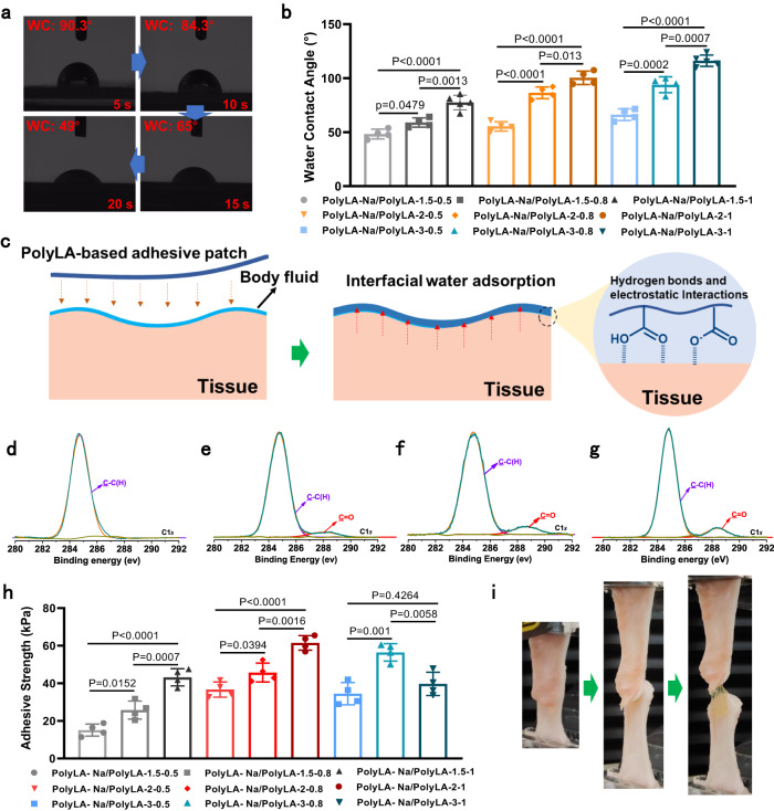
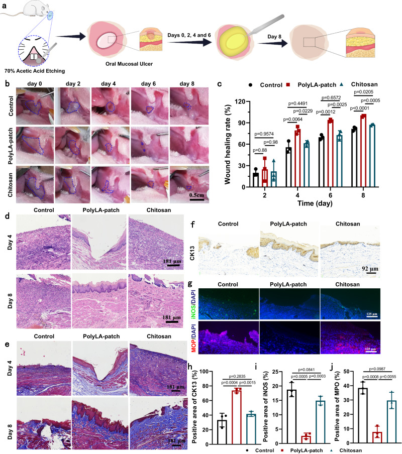
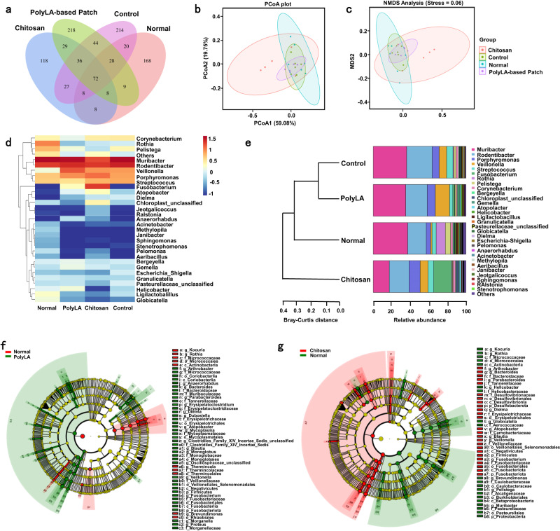
Oral ulcer can be treated with diverse biomaterials loading drugs or cytokines. However, most patients do not benefit from these materials because of poor adhesion, short-time retention in oral cavity and low drug therapeutic efficacy. Here we report a self-stabilized and water-responsive deliverable coenzyme salt polymer poly(sodium α-lipoate) (PolyLA-Na)/coenzyme polymer poly(α-lipoic acid) (PolyLA) binary synergistic elastomer adhesive patch, where hydrogen bonding cross-links between PolyLA and PolyLA-Na prevents PolyLA depolymerization and slow down the dissociation of PolyLA-Na, thus allowing water-responsive sustainable delivery of bioactive LA-based small molecules and durable adhesion to oral mucosal wound due to the adhesive action of PolyLA. In the model of mice and mini-pig oral ulcer, the adhesive patch accelerates the healing of the ulcer by regulating the damaged tissue inflammatory environment, maintaining the stability of oral microbiota, and promoting faster re-epithelialization and angiogenesis. This binary synergistic patch provided a therapeutic strategy to treat oral ulcer.


Conflict of interest statement

The authors declare no competing interests.

Figures

Fig. 1
Fig. 1. Schematic diagram of preparation and application of polyLA-based adhesive patch.
a Preparation process and structural characteristics of PolyLA; b Preparation process and structural characteristics of PolyLA-Na patch; c Preparation process and structural characteristics of PolyLA-Na/PolyLA binary synergistic adhesive patch; d Illustration of the proposed mechanism of PolyLA-Na/PolyLA patch for promoting oral ulcer healing. LA α-lipoic acid, LA-Na sodium α-lipoate, TNF-α tumor necrosis factor-α, IL-6 interleukin-6.
Fig. 2
Fig. 2. Characterizations and molecular dynamics simulation of PolyLA-Na/PolyLA binary synergistic patches.
a Pictures of dry PolyLA, dry PolyLA-Na film and PolyLA-Na/PolyLA patches with different compositions; b Raman spectra of the LA powder, dry PolyLA-Na film, and PolyLA-Na/PolyLA-1.5-1 patch; c FT-IR spectra of the LA powder, dry PolyLA, dry PolyLA-Na film and PolyLA-Na/PolyLA patches with different compositions; d 1H NMR spectra of dry PolyLA, dry PolyLA-Na, and PolyLA-Na/PolyLA-1.5-1 patch; eg Molecular dynamics simulation of potential energy of PolyLA, PolyLA-Na and PolyLA-Na/PolyLA-2-1 systems; h Total potential energy value of PolyLA, PolyLA-Na and PolyLA-Na/PolyLA-2-1 systems.
Fig. 3
Fig. 3. Crystallization behavior and mechanical properties of PolyLA-Na/PolyLA binary synergistic patches.
ae XRD patterns of the LA powder, dry PolyLA, dry PolyLA-Na film, and PolyLA-Na/PolyLA patches with different compositions; f Pictures showing the stretching process of PolyLA-Na/PolyLA-1.5-0.8 and PolyLA-Na/PolyLA-3-0.8; gi Tensile strength of PolyLA-Na/PolyLA patches with different compositions; jl Ultimate strain of PolyLA-Na/PolyLA patches with different compositions g, j: n = 4 for PolyLA/PolyLA-Na = 0.5, n = 5 for PolyLA/PolyLA-Na = 0.8 and 1; h, k n = 6 for PolyLA/PolyLA-Na = 0.5 and 0.8, n = 7 for PolyLA/PolyLA-Na = 1; i, l n = 6 for PolyLA/PolyLA-Na = 0.5, n = 5 for PolyLA/PolyLA-Na = 0.8 and 1. (All presented data are mean values ± SD. Statistics was calculated by one-way ANOVA followed by Tukey’s post-test).
Fig. 4
Fig. 4. Wet adhesion behavior of PolyLA-Na/PolyLA binary synergistic adhesive patches.
a Pictures showing the variation in the water contact angle of PolyLA-Na/PolyLA-1.5-0.5 patch at different times (WC: water contact angle); b The value of water contact angle of the PolyLA-Na/PolyLA adhesive patches with different compositions (n = 4); c Proposed adhesion mechanism of PolyLA-Na/PolyLA adhesive patch; dg X-ray photoelectron spectra of dry PolyLA-Na film and PolyLA-Na/PolyLA adhesive patches with different compositions (d PolyLA-Na; e PolyLA-Na/PolyLA-1.5-1; f PolyLA-Na/PolyLA-2-1; g PolyLA-Na/PolyLA-3-1); h Adhesion strength of PolyLA-Na/PolyLA adhesive patches with different compositions to oral mucosa tissue after soaking in artificial saliva (n = 4); i Photographs showing the lap-shear process of the PolyLA-Na/PolyLA-2-1 patch adhered to oral mucosa tissue after soaking in artificial saliva. (All presented data are mean values ± SD from the mean from n = 4 independent measurements on independent samples. Statistics were calculated by one-way ANOVA followed by Tukey’s post-test).
Fig. 5
Fig. 5. Swelling resistance, LA-Na release, antioxidant and antibacterial properties of PolyLA-Na/PolyLA binary synergistic adhesive patches.
a Swelling behavior of PolyLA-Na/PolyLA adhesive patches with different compositions in artificial saliva (n = 3); b Monomer release ratio of PolyLA-Na/PolyLA adhesive patches with different compositions in artificial saliva for different times (n = 3); c FT-IR spectra of PolyLA-Na/PolyLA adhesive patch before and after monomer release; d Scavenging effect of PolyLA-Na/PolyLA adhesive patches on OH· radicals at 0.5 h (n = 3); e ABTS radical clearance ratio of PolyLA-Na/PolyLA adhesive patches determined at different times (n = 3, ***p < 0.001); f Digital images displaying minimum inhibitory concentration of the PolyLA-Na/PolyLA adhesive patches against E.coli and S.aureus; g, h Sterilization rate of PolyLA-Na/PolyLA adhesive patches with different concentrations against E.coli and S.aureus at different times (n = 3). (All presented data are mean values ± SD from the mean from n = 3 independent measurements on independent samples. All p values were calculated using a two-sided Student’s t test).
Fig. 6
Fig. 6. In vivo adhesion and biocompatibility of PolyLA-Na/PolyLA adhesive patch.
a Adhesion of PolyLA-Na/PolyLA patch on the wet surface of the rat buccal mucosa, gingiva, palate mucosa, and tongue; b Movement-resistant adhesion of PolyLA-Na/PolyLA-2-1 patch in vivo; c Adhesion of chitosan film in oral cavity; d Durable adhesion ability of PolyLA-Na/PolyLA-2-1 patch in oral cavity; e, f Live/Dead staining and cell viability of hGFs cells co-cultured with PolyLA-Na/PolyLA patch for 1 and 3 days (n = 4); g H&E staining of buccal mucosa tissue after attachment of PolyLA-Na/PolyLA patch for 12 h (n = 3 biologically independent samples in each group); h H&E staining of main organs after PolyLA-Na/PolyLA patch treatment for 8 days (n = 3 biologically independent samples in each group). (All presented data are mean values ± SD from the mean from n = 4 independent measurements on independent samples. All p values were calculated using a two-sided Student’s t test).
Fig. 7
Fig. 7. Oral ulcer healing efficacy of PolyLA-Na/PolyLA patch in rat model.
a Scheme depicting creation of rat mucosa defect that was covered by PolyLA-Na/PolyLA patch or chitosan film to assess healing; b Photographs of buccal mucosa ulcers in rats treated by PolyLA-Na/PolyLA patch and chitosan film at the 0, 2, 4, 6, and 8 days; c Wound healing efficiency of the oral ulcers at days 2, 4, 6, and 8 days after treated with PolyLA-Na/PolyLA patch and chitosan film (n = 3); d, e H&E staining and Masson’s trichrome staining of the regenerated oral mucosa at day 4 and 8 days (n = 3 biologically independent samples in each group); f Immunohistochemistry staining of CK13 antibody in regenerated oral mucosa at 8 days; g immunofluorescence staining of iNOS (green) and MPO (red) antibody in regenerated oral mucosa at 4 days; hj Quantification of expression levels of CK13, iNOS, and MPO in regenerated oral mucosa (n = 3) CK13 cytokeratin 13, iNOS inducible nitric oxide synthase, MPO myeloperoxidase. (All presented data are mean values ± SD from the mean from n = 3 independent measurements on independent samples. All p values were calculated using a two-sided Student’s t test).
Fig. 8
Fig. 8. Effects of different treatments on oral microbiota in rats.
a Venn diagram of the oral microbiota in the normal group, control group, chitosan film group, and PolyLA-based patch group; b,c PCoA and NMDS analyses of the oral microbiota in the normal group, control group, chitosan film group, and PolyLA-based patch group; d Heat map of the oral microbiota in the normal group, control group, chitosan film group and PolyLA-Na/PolyLA patch group; e Cluster analysis of the different groups of oral microbiota at the genus level; f LEfSe analysis identifying the dominant bacteria in the normal group and PolyLA-based patch group; g LEfSe analysis identifying the dominant bacteria in the normal group and chitosan film group.
Fig. 9
Fig. 9. Oral ulcer healing efficacy of PolyLA-Na/PolyLA patch in mini-pig model.
a Scheme depicting creation of mini-pig mucosa defect that was covered by PolyLA-Na/PolyLA patch to accelerate healing; b Photographs of buccal mucosa defect in mini-pig treated by PolyLA-based patch and chitosan film at the 0, 3, 5, and 8 days; c Tracking of wound-bed closure after treating with different methods for 0, 3, 5, and 8 days; d Wound healing efficiency of the oral ulcers at days 3, 5, and 8 days after treated with PolyLA-Na/PolyLA patch and chitosan film (n = 3); e, f H&E staining and Masson’s trichrome staining of the regenerated mini-ping oral mucosa at day 8 (n = 3 biologically independent samples in each group); g Sirius red staining of type I collagen (red) and type III collagen (green) in the regenerated mini-ping oral mucosa at day 8; hj Immunofluorescence staining of CK5 (red), CD31 (red) and iNOS (red) antibody in regenerated oral mucosa at 8 days; k Analysis of the collagen I/III ratio in Sirius red staining (n = 3); ln Quantification of expression levels of CK5, CD31, and iNOS in regenerated oral mucosa (n = 3) CK5 cytokeratin 5, CD31 platelet endothelial cell adhesion molecule-1, iNOS inducible nitric oxide synthase. (All presented data are mean values ± SD from the mean from n = 3 independent measurements on independent samples. All p values were calculated using a two-sided Student’s t test).

References

    1. Dudding T, et al. Genome wide analysis for mouth ulcers identifies associations at immune regulatory loci. Nat. Commun. 2019;10:1052. doi: 10.1038/s41467-019-08923-6. - DOI - PMC - PubMed
    1. Petersen PE. World Health Organization global policy for improvement of oral health-world health assembly 2007. Int. Dent. J. 2008;58:115–121. doi: 10.1111/j.1875-595X.2008.tb00185.x. - DOI - PubMed
    1. Xing JQ, et al. Barnacle-inspired robust and aesthetic Janus patch with instinctive wet adhesive for oral ulcer treatment. Chem. Eng. J. 2022;444:136580. doi: 10.1016/j.cej.2022.136580. - DOI
    1. Zheng WP, et al. Preparation of triamcinolone acetonide-loaded chitosan/fucoidan hydrogel and its potential application as an oral mucosa patch. Corbohyd. Polym. 2021;227:118493. doi: 10.1016/j.carbpol.2021.118493. - DOI - PubMed
    1. Zhang ZJ, et al. Antibacterial, anti-inflammatory and wet-adhesive poly(ionic liquid)-based oral patch for the treatment of oral ulcers with bacterial infection. Acta Biomater. 2023;166:254–265. doi: 10.1016/j.actbio.2023.05.017. - DOI - PubMed

Publication types