. 2023 Nov 24;8(1):183.
doi: 10.1038/s41541-023-00775-y.

Conjugation of HIV-1 envelope to hepatitis B surface antigen alters vaccine responses in rhesus macaques

Affiliations

Conjugation of HIV-1 envelope to hepatitis B surface antigen alters vaccine responses in rhesus macaques

Danielle Nettere et al. NPJ Vaccines. .

Abstract

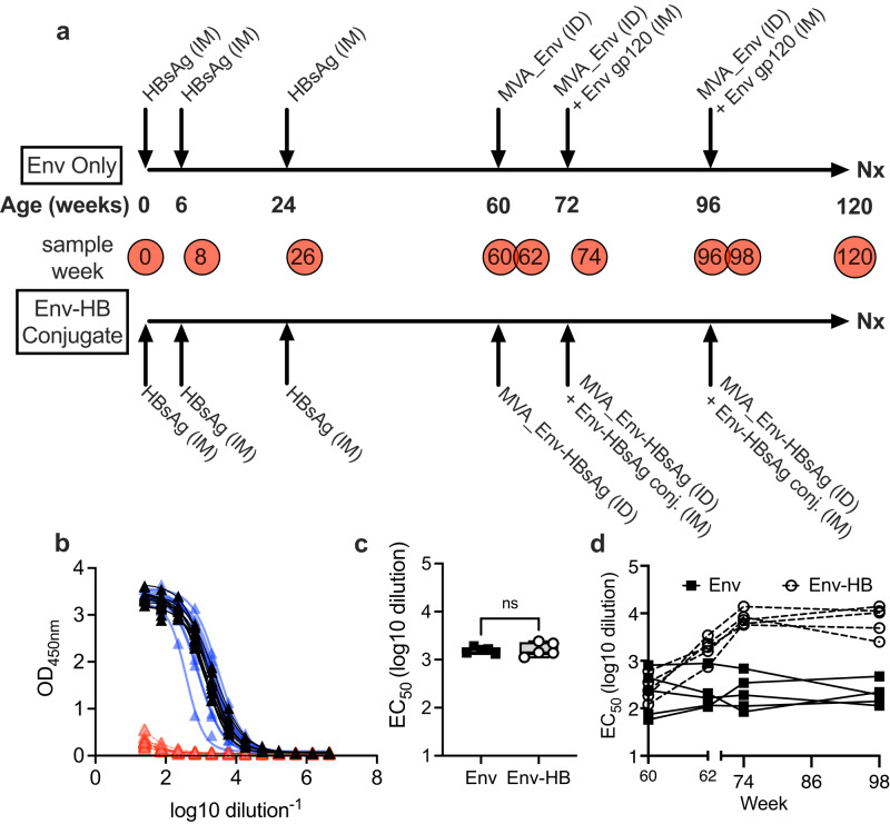
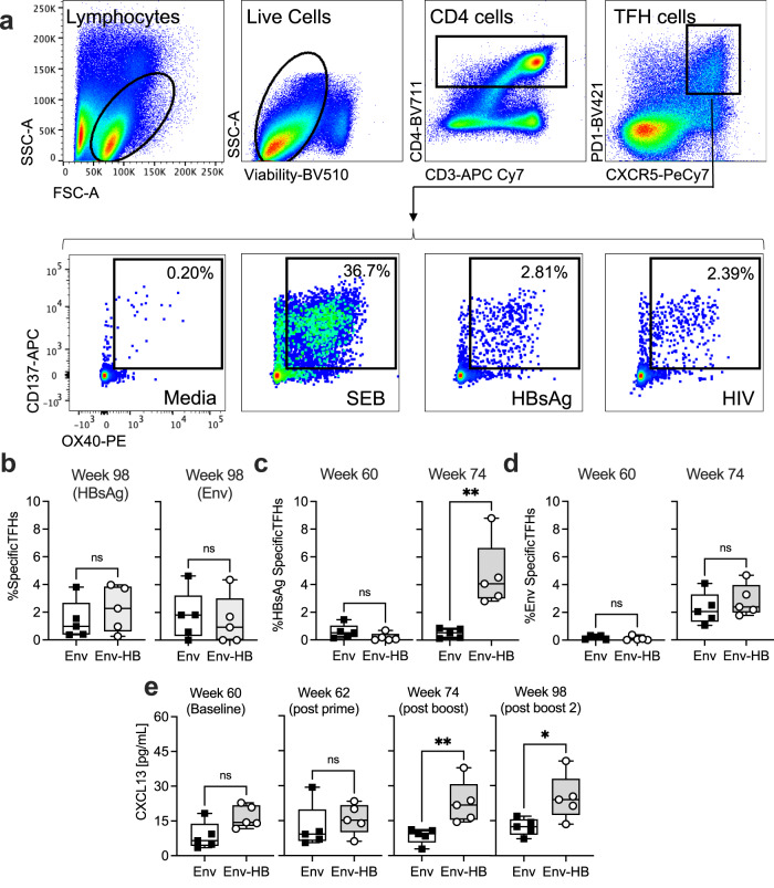
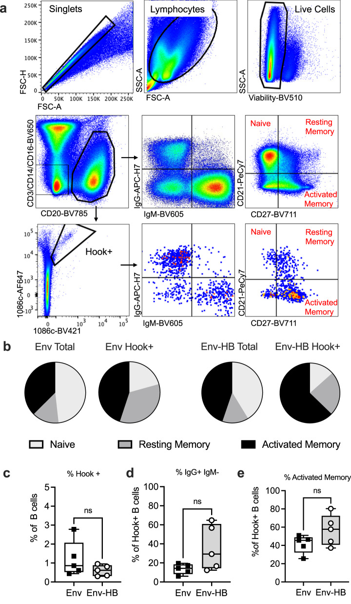
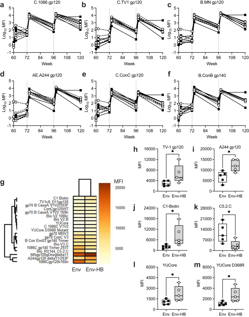
An effective HIV-1 vaccine remains a critical unmet need for ending the AIDS epidemic. Vaccine trials conducted to date have suggested the need to increase the durability and functionality of vaccine-elicited antibodies to improve efficacy. We hypothesized that a conjugate vaccine based on the learned response to immunization with hepatitis B virus could be utilized to expand T cell help and improve antibody production against HIV-1. To test this, we developed an innovative conjugate vaccine regimen that used a modified vaccinia virus Ankara (MVA) co-expressing HIV-1 envelope (Env) and the hepatitis B virus surface antigen (HBsAg) as a prime, followed by two Env-HBsAg conjugate protein boosts. We compared the immunogenicity of this conjugate regimen to matched HIV-1 Env-only vaccines in two groups of 5 juvenile rhesus macaques previously immunized with hepatitis B vaccines in infancy. We found expansion of both HIV-1 and HBsAg-specific circulating T follicular helper cells and elevated serum levels of CXCL13, a marker for germinal center activity, after boosting with HBsAg-Env conjugate antigens in comparison to Env alone. The conjugate vaccine elicited higher levels of antibodies binding to select HIV Env antigens, but we did not observe significant improvement in antibody functionality, durability, maturation, or B cell clonal expansion. These data suggests that conjugate vaccination can engage both HIV-1 Env and HBsAg specific T cell help and modify antibody responses at early time points, but more research is needed to understand how to leverage this strategy to improve the durability and efficacy of next-generation HIV vaccines.


Conflict of interest statement

S.R.P. provides individual consulting services to Moderna, Merck, Dynavax, Pfizer, GlaxoSmithKline (CMV vaccines) and HOOKIPA Biotech GmbH. Merck Vaccines and Moderna have provided grants for her Institutional sponsored programs.

Figures

Fig. 1
Fig. 1. Overview of vaccination schema and induction of HBsAg specific responses.
a HBsAg and HIV vaccination schedule and sample collection. PBMCs and serum were collected at the weeks indicated in the red circles. b All animals had no preexisting responses to HBsAg at time of birth (0 weeks, red lines and triangles) but all successfully responded to HB vaccination as evident by detection of HBsAg specific antibodies by ELISA 2 weeks after the second (8 weeks, blue lines and triangle) and third (26 weeks, black lines and triangles) HB vaccine immunization. c The titer of the HBsAg antibodies did not significantly different between vaccine groups after randomization (Mann Whitney p > 0.05). d The titer of HBsAg antibodies increased in Env-HB vaccine group but remained constant in the Env vaccine group. Box plots represent the interquartile ranges, horizontal lines indicate the medians, and error bars extend to the minimum and maximum observed values.
Fig. 2
Fig. 2. Activation induced marker assay identifies HBsAg and HIV Env specific TFH cells induced by vaccination.
a Gating strategy used to identify antigen specific TFH cells after 18 h stimulation with SEB, or HBsAg or HIV-1 peptide and protein. b Frequencies of HBsAg and Env responsive TFHs out of total lymph node TFH at week 98. c Frequencies of HBsAg responsive TFHs out of total blood TFH at week 60 and week 74. d Frequencies of HIV Env responsive TFHs out of total blood TFH at week 60 and week 74. e CXCL13 ELISA results from serum at indicated timepoints. Comparisons were made using Mann-Whitney U test, *p < 0.05, **p < 0.01. Box plots represent the interquartile ranges, horizontal lines indicate the medians, and error bars extend to the minimum and maximum observed values.
Fig. 3
Fig. 3. B cell antigen specific phenotyping in each vaccine arm.
a Gating strategy used for phenotyping and sorting Env specific B cells. b Distribution of B cell memory subsets among total B cells and antigen specific B cell from each vaccine group. c Frequency of Env hook+ (Env specific) B cells among total B cells, d frequency of class-switched Env specific B cells that were IgG + , IgM-, and e frequency of Env specific B cells that were defined as activated memory cells by surface expression of CD21 and CD27. Box plots represent the interquartile ranges, horizontal lines indicate the medians, and error bars extend to the minimum and maximum observed values.
Fig. 4
Fig. 4. Comparisons of Env-specific B cell gene expression and BCR immunogenetics across vaccine arms.
a Gene expression profiles of Env-specific B cells as determined using single cell RNA sequencing of Env hook+ cells identified 10 transcriptionally distinct B cell clusters. b Cells from Env vaccinated animals and Env-HB vaccinated animals and (c) from male and female animals showed overlapping distribution in each of these clusters. d The number of unique B cell clones per 1000 Env hook + B cells did not differ (Mann Whitney p > 0.05) when comparing the Env and Env–HB vaccine groups. e Heavy chain sequences were used to determine the BCR isotypes and subclasses, and a significant but modest increase amount of IgG1 BCR, and decrease in IgD BCR, among animals in the Env–HB group compared to those in the Env-only group was observed (Mann Whitney p < 0.01). Among the antigen specific B cells with IgG1 BCR, we saw a similar distribution of (f) CDRH3 and (g) no difference (Mann Whitney p > 0.05) in the IgG heavy chain mutation frequency when comparing the two vaccine regimens. Box plots represent the interquartile ranges, horizontal lines indicate the medians, and error bars extend to the minimum and maximum observed values.
Fig. 5
Fig. 5. Evaluation of plasma antibody binding magnitude to envelope antigens.
af Binding Antibody Multiplex Assay measured antibody binding to 6 unique gp120’s at each collection point. g Binding data for week 98 serum samples against all BAMA antigens as measured by MFI of each vaccine group and represented as a heat map. Week 98 BAMA MFI data were significantly different (Mann Whitney, p < 0.05) between the Env and Env-HB vaccine groups for vaccine antigens (h) TV-1 and (i) A244 gp120, (j) C1-Biotin, (k) C5.2C, (l) YUCore, (m) YUCore D368R. When controlling for false discovery rate (FDR) the adjusted p values for each of these observations were p = 0.100. Box plots represent the interquartile ranges, horizontal lines indicate the medians, and error bars extend to the minimum and maximum observed values.
Fig. 6
Fig. 6. Comparison of functional antibody responses between vaccine arms.
ad Max ADCC activity as measured by Granzyme B delivery to gp120 coated target cells for 4 different gp120s over the course of the study. eh ADCC antibody endpoint titer for assays performed with 4 different gp120s over the course of the study. ik Binding magnitude to HIV-infected cells over time. l Binding to TV-1 infected cells at week 98. m Max ADCC activity as measured by elimination of HIV infected cells at week 98. n Neutralization titer (ID50) of antibodies at week 98. Box plots represent the interquartile ranges, horizontal lines indicate the medians, and error bars extend to the minimum and maximum observed values.
Fig. 7
Fig. 7. Identification of immune features that differentiated the Env-only and Env-HB conjugate vaccine-elicited immune responses.
a A random forest model was used to identify immune features differentiating Env and Env-HB vaccinated macaques. The bar graph represents ranking of the measured immune variables that contributed to accuracy of the model (Variable importance) for the combined data set. The black box encompasses the 10 immune variables that contributed most to differences between vaccine groups. Bars highlighted in red were also identified as important (non-zero coefficient parameters) using Lasso-regularized logistic regression modeling. b Venn diagram representing the top 10 immunological variables that define each vaccine group by Random Forrest and Lasso-regularized logistic regression modeling.

References

    1. Supachai Rerks-Ngarm MD, et al. Vaccination with ALVAC and AIDSVAX to prevent HIV-1 infection in Thailand. N. Engl. J. Med. 2009;261:2209–2220. doi: 10.1056/NEJMoa0908492. - DOI - PubMed
    1. Haynes BF, McElrath MJ. Progress in HIV-1 vaccine development. Curr. Opin. HIV AIDS. 2013;8:326–332. - PMC - PubMed
    1. Kim J, et al. Current approaches to HIV vaccine development: a narrative review. J. Int AIDS Soc. 2021;2021:25793. doi: 10.1002/jia2.25793. - DOI - PMC - PubMed
    1. Barton F, et al. Immune correlates analysis of an HIV-1 vaccine efficacy trial. N. Engl. J. Med. 2012;366:1275–1286. doi: 10.1056/NEJMoa1113425. - DOI - PMC - PubMed
    1. Corey L, et al. Two randomized trials of neutralizing antibodies to prevent HIV-1 acquisition. N. Engl. J. Med. 2021;384:1003–1014. doi: 10.1056/NEJMoa2031738. - DOI - PMC - PubMed