. 2023 Nov 24;14(1):7717.
doi: 10.1038/s41467-023-43564-w.

Mouse models of pediatric high-grade gliomas with MYCN amplification reveal intratumoral heterogeneity and lineage signatures

Affiliations

Mouse models of pediatric high-grade gliomas with MYCN amplification reveal intratumoral heterogeneity and lineage signatures

Melanie Schoof et al. Nat Commun. .

Abstract

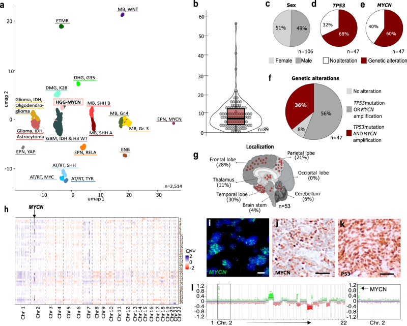
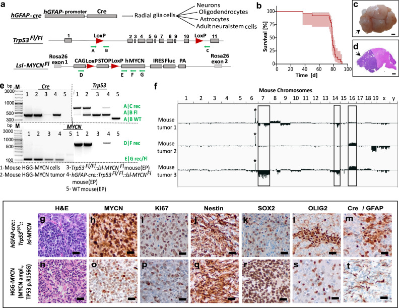
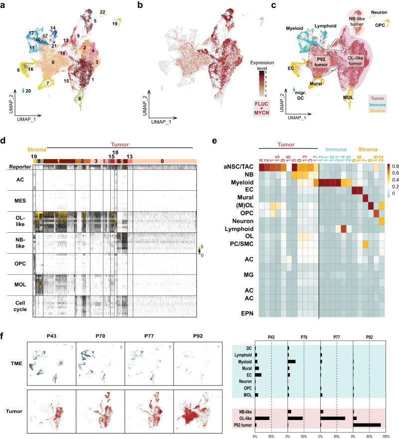
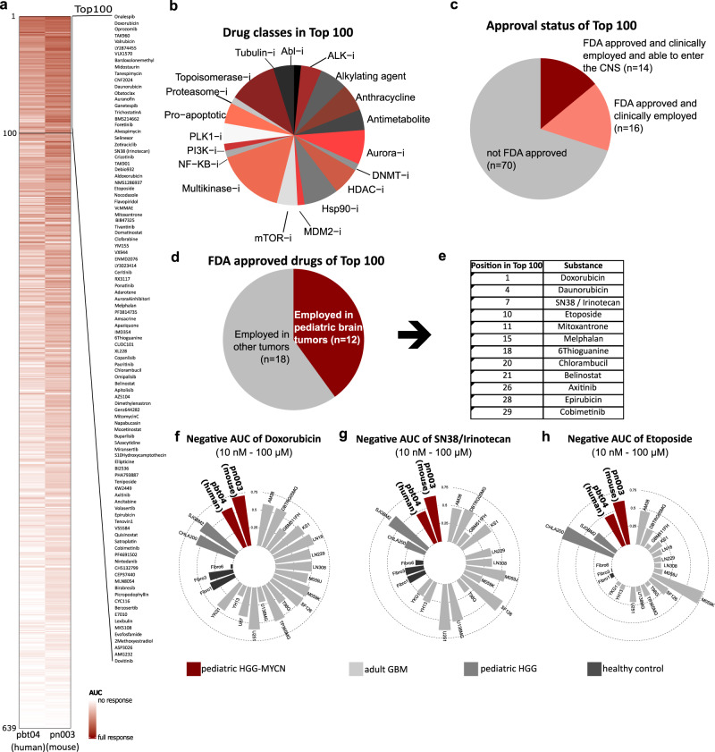
Pediatric high-grade gliomas of the subclass MYCN (HGG-MYCN) are highly aggressive tumors frequently carrying MYCN amplifications, TP53 mutations, or both alterations. Due to their rarity, such tumors have only recently been identified as a distinct entity, and biological as well as clinical characteristics have not been addressed specifically. To gain insights into tumorigenesis and molecular profiles of these tumors, and to ultimately suggest alternative treatment options, we generated a genetically engineered mouse model by breeding hGFAP-cre::Trp53Fl/Fl::lsl-MYCN mice. All mice developed aggressive forebrain tumors early in their lifetime that mimic human HGG-MYCN regarding histology, DNA methylation, and gene expression. Single-cell RNA sequencing revealed a high intratumoral heterogeneity with neuronal and oligodendroglial lineage signatures. High-throughput drug screening using both mouse and human tumor cells finally indicated high efficacy of Doxorubicin, Irinotecan, and Etoposide as possible therapy options that children with HGG-MYCN might benefit from.


Conflict of interest statement

F.M. received support for meeting attendance from Servier, AbbVie, Incyte, Gilead, Jazz Pharmaceuticals, Novartis, Teva, Pfizer and Amgen; received support for medical writing from Servier and Springer Verlag; received research grants from Apis Technologies, Daiichi Sankyo and received speaker honoraria from Servier, Jazz Pharmaceuticals, AbbVie, Astellas Pharma, Bristol-Myers Squibb, MSD Sharp & Dohme, Novartis, Pfizer, and Stemline Therapeutics. The remaining authors declare no competing interests.

Figures

Fig. 1
Fig. 1. Tumors of the DNA methylation class HGG-MYCN carry MYCN amplifications and TP53 mutations.
a UMAP of global DNA methylation profiling of 2514 cases of multiple brain tumor entities including the most common brain tumors as well as potential differential diagnoses including 47 HGG-MYCN using the 10,000 most differentially methylated CpG sites. b Age distribution of n = 89 HGG-MYCN tumors with a median age of 8 years (bounds of box = 3–13, whiskers = min:1, max:22). c Sex distribution of 106 HGG-MYCN. d 47 HGG-MYCN were screened for TP53 mutations, of which 68% carried a mutation. e The same 47 cases as in (d) were analyzed for their MYCN status. f Of the same 47 cases shown in (d) and (e), only 8% carried no TP53 or MYCN alteration, whereas 36% of those carry both alterations. g Tumors can be found throughout the entire brain with the majority of cases located in the temporal and frontal lobes. h Heatmap showing copy number variations of 47 HGG-MYCN. The copy number profile of such tumors is imbalanced with a clearly visible MYCN amplification (Chr. 2), highlighted by the arrow. MYCN amplifications can also be detected by FISH analysis (representative case shown in (i), three independent tumors showing this amplification were analyzed), while IHC may serve as a surrogate marker (representative case in (j), the same three tumors were analyzed). k Nuclear p53 accumulation indicating impaired p53 function can be detected by IHC (representative case, three independent tumors were analyzed). l Representative CNV plot of a HGG-MYCN with a magnification of chromosome 2 with the MYCN amplification (CNV plots of 47 tumors were generated). Scale bar in i = 5 μm, in j & k = 50 μm. Source data are provided as a Source Data file.
Fig. 2
Fig. 2. Mouse HGG-MYCN develop within the first 100 days of life and match their human counterparts histologically.
a Genetics of the HGG-MYCN mouse model. Cre is expressed under control of the hGFAP promoter, which targets different cell populations shown on the right. The floxed alleles of Trp53 and MYCN are depicted, loxP sites are shown as red arrowheads, and primers for genotyping and proof of recombination are shown as green arrows. b Kaplan–Meier survival curve of mice with HGG-MYCN (n = 24) as percent survival, light red area showing the asymmetrical 95% confidence interval. c Macroscopic image of a mouse brain carrying an HGG-MYCN (arrow). d Hematoxylin and Eosin (H&E) stained brain with a large forebrain tumor (arrow). e Representative PCR result of genotyping and the detection of allele recombination. Results are shown for cultured mouse tumor cells (1), fresh mouse tumor tissue (2), ear biopsy of a mouse not carrying the Cre recombinase (3), ear biopsy of a mouse with an HGG-MYCN (4), and (5) a wild-type mouse carrying none of the transgenes. Bands result from the primers indicated by green arrows in (a). The same PCR was performed for all animals generated in the study including the n = 24 animals included in the Kaplan-Meier survival analysis. f Copy number variation plots of three mouse HGG-MYCN. An amplification of the Rosa26-locus, in which the MYCN is inserted, is visible in all three samples (marked by the star). Other recurrent copy number changes are observed in at least two of the tumors (marked by the rectangle). gt Immunohistochemical comparability of mouse and a human HGG-MYCN. The pictures show representative micrographs, all stainings were performed independently on at least three samples. EP ear punch, Fl Floxed allele, M marker, Rec recombined allele, WT wild-type allele. The scale bar in c and d corresponds to 2 mm, and the scale bar in (gt) corresponds to 20 μm. Source data are provided as a Source Data file.
Fig. 3
Fig. 3. Mouse HGG-MYCN match their human counterparts molecularly.
a UMAP of global DNA methylation. Betavalues of 141 identical CpG sites between mouse and human were used for comparison of similarity. Mouse methylome data were generated with the Illumina Mouse Methylation bead chip array and compared to human data generated with the EPIC array. The three mouse tumors (in hot pink) show most similarity to the human HGG-MYCN group (pink). b UMAP of global gene expression data. Eleven mouse tumors were profiled by RNA sequencing and their gene expression profile was compared to published gene expression data of human „CNS PNET“ tumors 1. Data were normalized for interspecies differences by employing RNA Seq. data of Math1-cre::SmoM2Fl/wt mouse tumors and human SHH medulloblastoma. c Distance plot of mouse HGG-MYCN and mouse SHH-MBs and human tumors based on the 500 most significantly expressed genes. Mouse tumors showed most similarity to human HGG-MYCN and human SHH-MB, respectively. The values of the Euclidean distance are displayed, and the asterisk marks the smallest values and thereby highest similarity. d AGDEX analysis of mouse HGG-MYCN also shows the high similarity between murine and human tumors (analysis based on 14,416 orthologous genes). The values of the AGDEX analysis with their respective p-values are given. The asterisk marks the smallest p-value and thereby highest similarity. e, f Human and mouse HGG-MYCN show significantly higher MYCN expression compared to other glioma entities or control tissue (from mouse olfactory bulb (OB) or cerebellum) and comparable expression to the SHH-MB mouse model as well as highly significant enrichment for MYCN target genes. Human tumors: non-mycn, n = 40, HGG-MYCN n = 7. Mouse data: OB tissue n = 3, mouse HGG-MYCN n = 11., two-sided Welch’s-t-test, human: p = 0.0002, 95% confidence interval = 2.001 to 4.320, murine= p = 0.0002, 95% confidence interval = 2.143 to 4.085. Source data are provided as a Source Data file.
Fig. 4
Fig. 4. Mouse HGG-MYCN reveal a high intratumoral heterogeneity with oligodendroglial and neural cell populations and a time-resolved change in tumor composition.
a UMAP of single cell RNA sequencing data of seven mouse HGG-MYCN (2xP43, 2xP70, 2xP77 and 1x P92) including 24.938 cells. 23 cell clustered were identified by the Seurat algorithm. b To identify tumor cell clusters, expression of human MYCN and Luciferase (FLUC), were plotted. c, d Cell clusters were annotated by analysis of marker gene expression. This revealed immune as well as stromal cell clusters and three main superclusters of tumor cells. Tumor superclusters consist of an oligodendroglial-like, a neuronal-like, and a cluster which was only detected in the most mature mouse tumor. e Cell clusters were compared to a reference atlas of the VZ/SVZ of the mouse by logistic regression. Similarity in gene expression is displayed in red, less similarity in blue. Mouse tumor cell clusters show similarity to precursor cells of the stem cell niche, suggesting a tumor origin in this region. f UMAP and bargraph depiciting the changes in cell composition of samples of different mouse ages. The TME content is reduced in later tumor stages. Early tumors are only OL-like, during tumor development, an NB-like population appears. At P92, a unique tumor cell population is detected, showing neither OL nor NB-like features. AC astrocyte, aNSC adult neural stem cell, DC dendritic cell, EC endothelial cells, EPN ependymal cells, MES mesenchymal, MG microglia, migr. DC migratory dendritc cells, MOL mature oligodendrocyte, mural fibroblasts, pericytes, smooth muscle cells etc., NB neuroblast, OL oligodendrocytic, OPC oligodendrocytic precursor cell, PC pericytes, SMC smooth muscle cells, TAC transit amplifying cell, TME tumor microenvironment, UMAP uniform manifold approximation and projection.
Fig. 5
Fig. 5. High throughput drug screen indicates efficacy of Doxorubicin, Etoposide, and Irinotecan for the treatment of HGG-MYCN.
a Heatmap of the AUC in a drug screen of 639 substances in human (pbt-04) and murine (pn003) HGGMYCN cell lines. A darker color indicates a stronger response. b Among the top 100 substances of the drug screen, we detected 18 drug classes with at least two substances. The most prominent were anthracyclins, aurora inhibitors, Hsp90-inhibitors, multikinase inhibitors, and topoisomerase inhibitors. c Of the top 100 most effective substances in the screen, 30 were FDA approved. Of those, 14 have been described to be delivered into the CNS. d Of the 30 FDA-approved substances, 12 have been used to treat pediatric brain tumor patients as depicted in (e). fh The top three substances Doxorubicin, SN-38 (active metabolite of Irinotecan) and Etoposide are efficiently impairing growth of HGG-MYCN cells, while showing a mixed effect on other glioma cell lines and almost no effect on healthy fibroblasts. Depicted is the negative AUC (1 minus the respective AUC). AUC area under the curve. AUC values are supplied in Supplementary data 1. Source data are provided as a Source Data file.

References

    1. Sturm D, et al. New brain tumor entities emerge from molecular classification of CNS-PNETs. Cell. 2016;164:1060–1072. doi: 10.1016/j.cell.2016.01.015. - DOI - PMC - PubMed
    1. Korshunov A, et al. H3-/IDH-wild type pediatric glioblastoma is comprised of molecularly and prognostically distinct subtypes with associated oncogenic drivers. Acta Neuropathol. 2017;134:507–516. doi: 10.1007/s00401-017-1710-1. - DOI - PubMed
    1. Sturm D, et al. Multiomic neuropathology improves diagnostic accuracy in pediatric neuro-oncology. Nat. Med. 2023;29:917–926. doi: 10.1038/s41591-023-02255-1. - DOI - PMC - PubMed
    1. Schoof M, et al. Malignant gliomas with H3F3A G34R mutation or MYCN amplification in pediatric patients with Li Fraumeni syndrome. Acta Neuropathol. 2021;142:591–593. doi: 10.1007/s00401-021-02346-8. - DOI - PMC - PubMed
    1. Guerrini-Rousseau L, et al. Pediatric high-grade glioma MYCN is frequently associated with Li-Fraumeni syndrome. Acta Neuropathol. Commun. 2023;11:3. doi: 10.1186/s40478-022-01490-w. - DOI - PMC - PubMed

Publication types

Substances

Associated data