. 2024 Jan;31(1):28-39.
doi: 10.1038/s41418-023-01233-x. Epub 2023 Nov 24.

LUBAC is required for RIG-I sensing of RNA viruses

Affiliations

LUBAC is required for RIG-I sensing of RNA viruses

Helena C Teague et al. Cell Death Differ. 2024 Jan.

Abstract

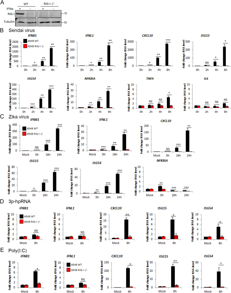
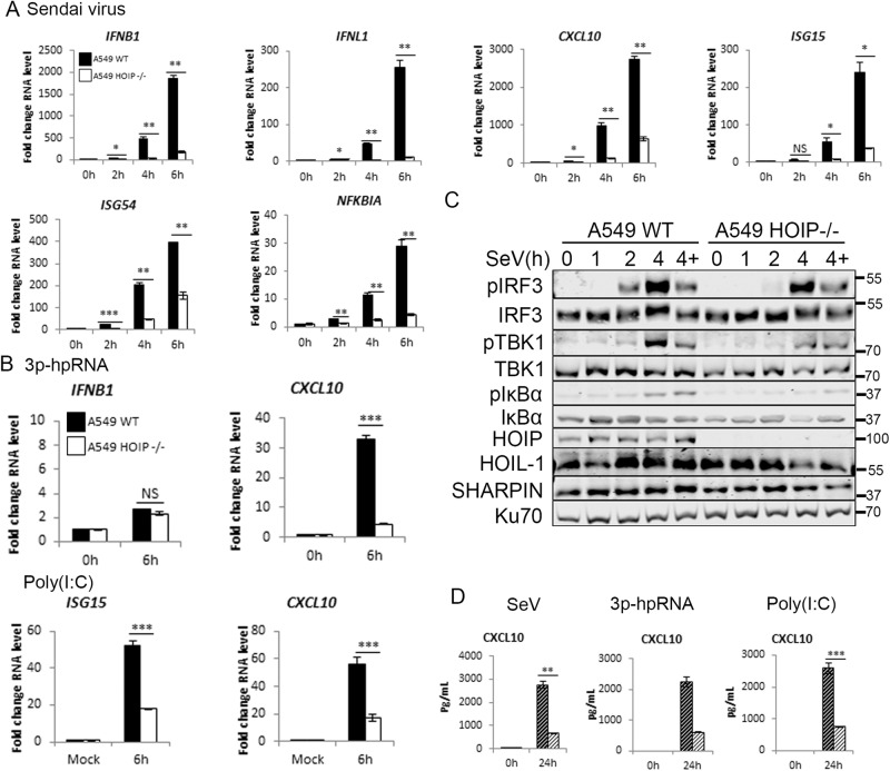
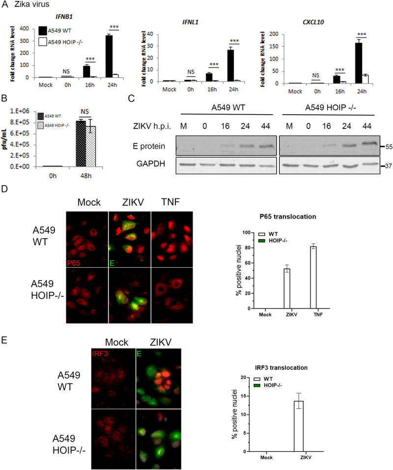
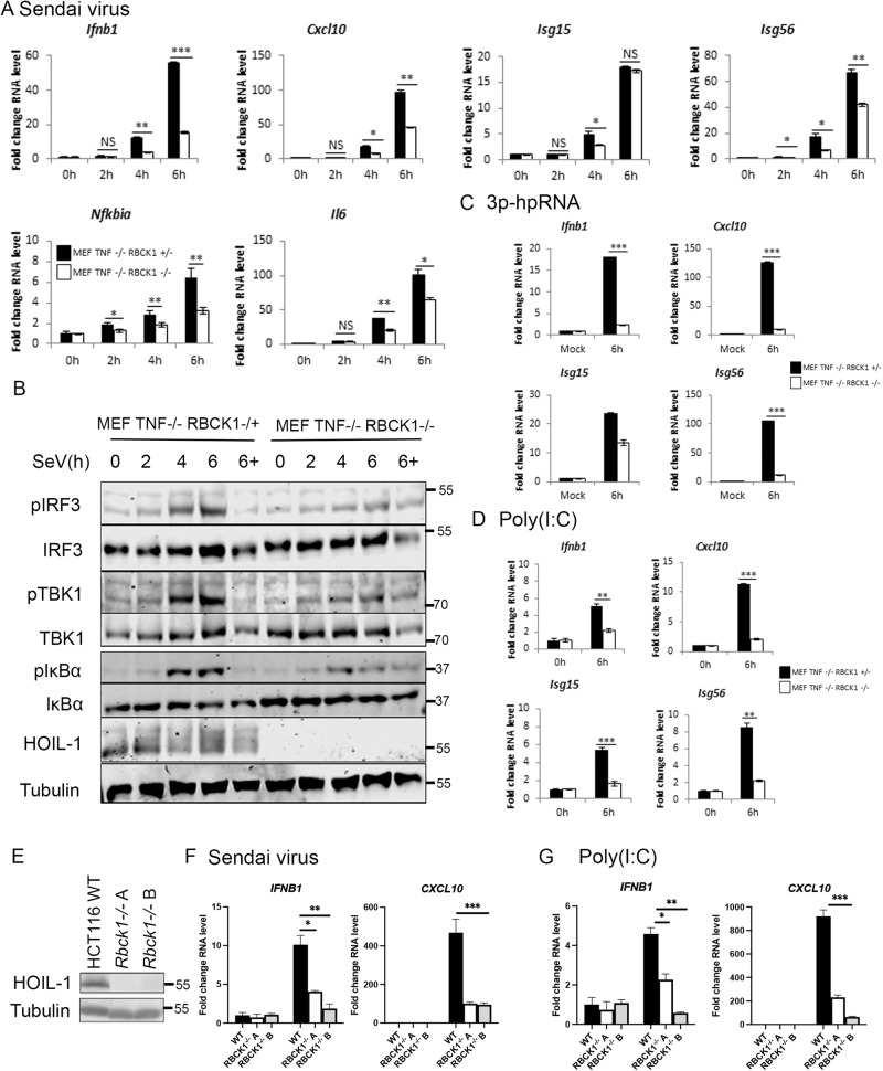
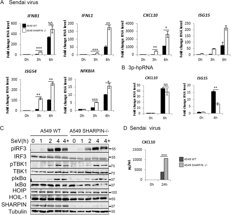
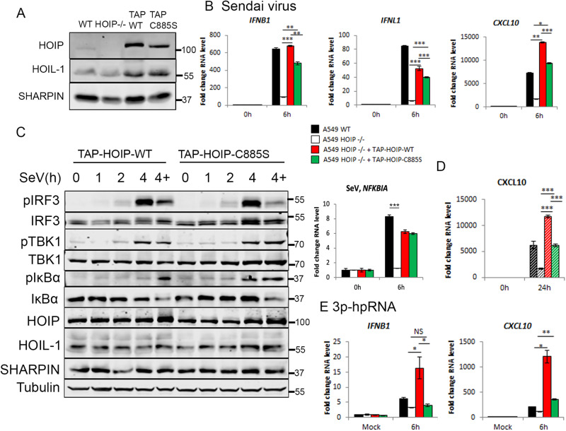
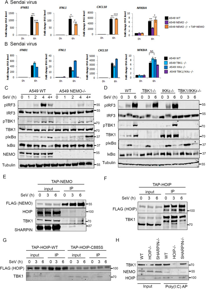
The ability of cells to mount an interferon response to virus infections depends on intracellular nucleic acid sensing pattern recognition receptors (PRRs). RIG-I is an intracellular PRR that binds short double-stranded viral RNAs to trigger MAVS-dependent signalling. The RIG-I/MAVS signalling complex requires the coordinated activity of multiple kinases and E3 ubiquitin ligases to activate the transcription factors that drive type I and type III interferon production from infected cells. The linear ubiquitin chain assembly complex (LUBAC) regulates the activity of multiple receptor signalling pathways in both ligase-dependent and -independent ways. Here, we show that the three proteins that constitute LUBAC have separate functions in regulating RIG-I signalling. Both HOIP, the E3 ligase capable of generating M1-ubiquitin chains, and LUBAC accessory protein HOIL-1 are required for viral RNA sensing by RIG-I. The third LUBAC component, SHARPIN, is not required for RIG-I signalling. These data cement the role of LUBAC as a positive regulator of RIG-I signalling and as an important component of antiviral innate immune responses.


Conflict of interest statement

The authors declare no competing interests.

Figures

Fig. 1
Fig. 1. RIG-I dependent RNA and RNA virus sensing in A549 cells.
A Western blotting analysis of A549 WT and RIG-I−/− cells with and without stimulation with IFNα for 24 h. qPCR to measure transcription of indicated genes in A549 WT and RIG-I−/− cells stimulated by B SeV infection at 1:300 dilution, C Zika virus infection at MOI 3, D transfection with 1 μg 3p-hpRNA and E transfection with 1 μg poly(I:C).
Fig. 2
Fig. 2. HOIP is required for RIG-I-driven transcription, chemokine secretion and signalling pathway activation.
A A549 WT and HOIP−/− were infected with SeV at 1:300 dilution and qPCR used to measure transcription of indicated genes. B qPCR to measure transcription of indicated genes in A549 WT and HOIP−/− cells transfected with 1 μg 3p-hpRNA or poly(I:C). C Western blotting analysis of signalling protein activation in the presence and absence of 10 µM MG-132 ('4+'). D ELISA to measure CXCL10 secretion in A549 WT and HOIP−/− cells infected with SeV at 1:300 dilution or transfected with 1 μg 3p-hpRNA or 1 μg poly(I:C).
Fig. 3
Fig. 3. HOIP is required for ZIKV-driven interferon responses.
A549 WT and HOIP−/− cells infected with ZIKV at MOI 3 and A qPCR to measure transcription of indicated genes, B ZIKV replication measured by plaque assay on Vero cells and C Western blotting analysis. Quantification of nuclear translocation of D NF-κB P65 and E IRF3 in A549 WT and HOIP−/− cells infected with ZIKV at MOI 1 for 24 h or stimulated with 100 ng/mL TNF, analysed by immunofluorescence (left panels) and quantified by scoring cells with nuclear staining (right panels).
Fig. 4
Fig. 4. HOIL-1 is required for RIG-I immune response to SeV and synthetic RNAs.
MEF Tnf−/− Rbck+/− and Tnf−/− Rbck−/− cells infected with SeV at a 1:300 dilution and A qPCR to measure transcription of indicated genes and B Western blotting analysis of signalling protein activation in the presence and absence of 10 µM MG-132 (‘6+’). qPCR to measure transcription of indicated genes in MEF TNF−/− HOIL+/− and TNF−/− HOIL−/− cells transfected with C 1 μg 3p-hpRNA and D 1 μg Poly(I:C). E Western blot of WT and HOIL-1−/− HCT116 cells indicating successful generation of two knockout clones. qPCR to measure transcription of indicated genes in HCT116 WT and HOIL−/− cells F infected with SeV or G transfected with 1 μg poly(I:C).
Fig. 5
Fig. 5. SHARPIN is not required for RIG-I immune response to SeV and synthetic RNAs in A549 cells.
A549 WT and SHARPIN−/− cells A infected with SeV at a 1:300 dilution or B stimulated with 1 μg 3p-hpRNA were analysed by qPCR to measure transcription of indicated genes, C Cells were infected with SeV and analysed by Western blotting for signalling protein activation in the presence and absence of 10 µM MG-132 (‘4+’) and D CXCL10 secretion measured by ELISA.
Fig. 6
Fig. 6. The E3 ligase activity of LUBAC is partially required for its function in RIG-I signalling.
A Western blotting analysis of A549 WT, HOIP−/−, TAP-HOIP-WT and TAP-HOIP-C885S cells. A549 WT, HOIP−/−, TAP-HOIP-WT and TAP-HOIP-C885S cells infected with SeV at a 1:300 dilution and B qPCR to measure transcription of indicated genes. C Western blotting analysis of signalling protein activation in A549 TAP-HOIP-WT and TAP-HOIP-C885S cells infected with SeV at a 1:300 dilution in the presence and absence of 10 µM MG-132 (labelled as ‘4+’). D ELISA to measure CXCL10 secretion following infection with SeV. E qPCR to measure transcription of indicated genes in A549 WT, HOIP−/−, TAP-HOIP-WT and TAP-HOIP-C885S cells transfected with 1 μg 3p-hpRNA.
Fig. 7
Fig. 7. LUBAC interacts with TBK1 and NEMO downstream of RIG-I activation.
qPCR to measure transcription of indicated genes during SeV infection at 1:300 dilution in A A549 WT, NEMO−/−, NEMO−/− + TAP-NEMO cells and B A549 WT, TBK1−/−, IKKε−/− and TBK1/IKKε−/− cells. Western blotting analysis of signalling protein phosphorylation during SeV infection at 1:300 dilution in the presence and absence of 10 µM MG-132 (‘4+’) in C A549 WT, NEMO−/−, NEMO−/− + TAP-NEMO cells and D A549 WT, TBK1−/−, IKKε−/− and TBK1/IKKε−/− cells. Western blotting analysis of Flag-M2 IP in E A549 TAP-NEMO and F A549 TAP-HOIP-WT G A549 TAP-HOIP-WT or A549 TAP-HOIP-C885S cells infected with SeV at a 1:300 dilution. H Western blotting analysis of affinity-purified (AP) biotin-poly(I:C) from A549 WT, HOIP−/− or SHARPIN−/− cells.

Similar articles

Cited by

References

    1. McNab F, Mayer-Barber K, Sher A, Wack A, O’Garra A. Type I interferons in infectious disease. Nat Rev Immunol. 2015;15:87–103. doi: 10.1038/nri3787. - DOI - PMC - PubMed
    1. Rehwinkel J, Tan CP, Goubau D, Schulz O, Pichlmair A, Bier K, et al. RIG-I detects viral genomic RNA during negative-strand RNA virus infection. Cell. 2010;140:397–408. doi: 10.1016/j.cell.2010.01.020. - DOI - PubMed
    1. Yoneyama M, Kikuchi M, Natsukawa T, Shinobu N, Imaizumi T, Miyagishi M, et al. The RNA helicase RIG-I has an essential function in double-stranded RNA-induced innate antiviral responses. Nat Immunol. 2004;5:730–7. doi: 10.1038/ni1087. - DOI - PubMed
    1. Hou F, Sun L, Zheng H, Skaug B, Jiang Q-X, Chen ZJ. MAVS forms functional prion-like aggregates to activate and propagate antiviral innate immune response. Cell. 2011;146:448–61. doi: 10.1016/j.cell.2011.06.041. - DOI - PMC - PubMed
    1. Reikine S, Nguyen JB, Modis Y. Pattern recognition and signaling mechanisms of RIG-I and MDA5. Front Immunol. 2014;0:342. - PMC - PubMed

Publication types

MeSH terms