

Near infrared II excitation nanoplatform for photothermal/chemodynamic/antibiotic synergistic therapy combating bacterial biofilm infections
- PMID: 38001486
- PMCID: PMC10668414
- DOI: 10.1186/s12951-023-02212-7
Near infrared II excitation nanoplatform for photothermal/chemodynamic/antibiotic synergistic therapy combating bacterial biofilm infections
Abstract
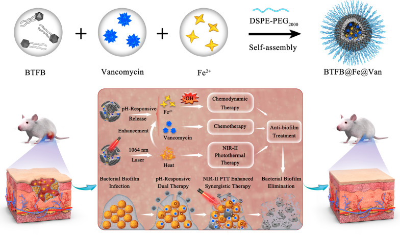
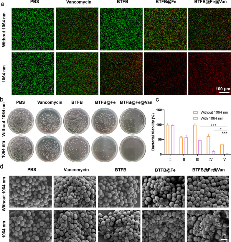
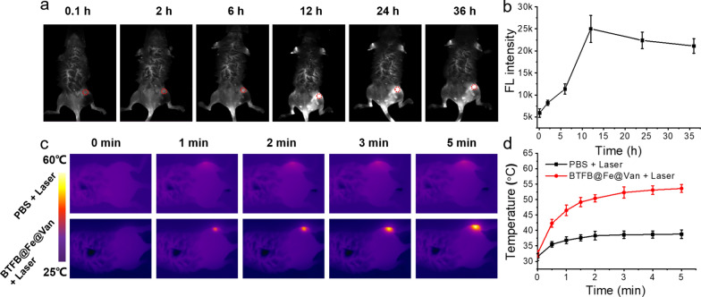
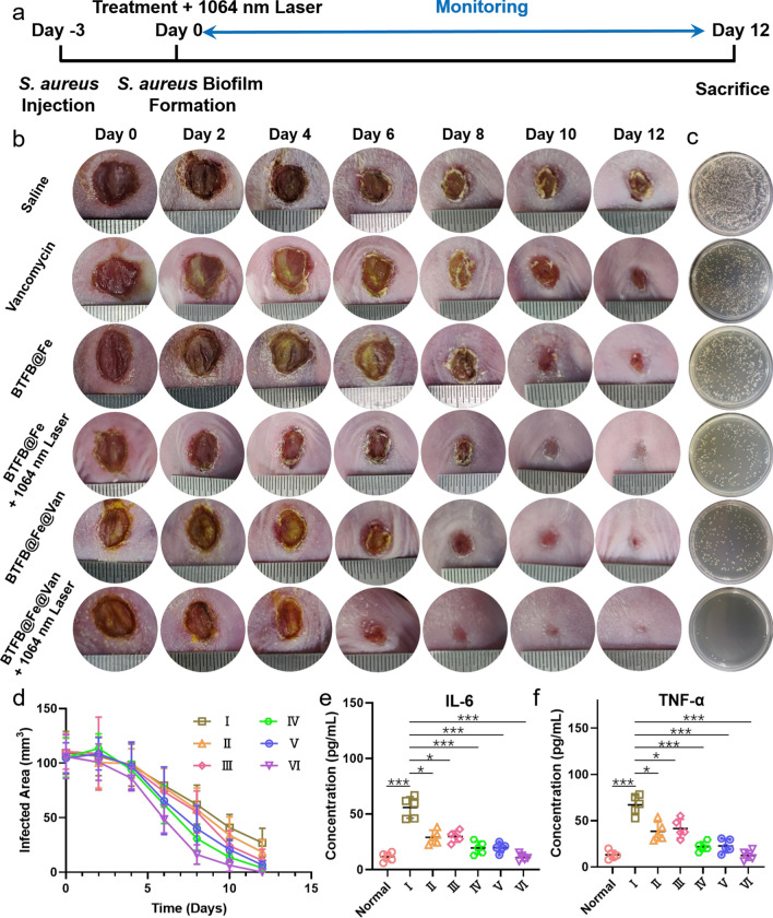
Drug-resistant bacterial biofilm infections (BBIs) are refractory to elimination. Near-infrared-II photothermal therapy (NIR-II PTT) and chemodynamic therapy (CDT) are emerging antibiofilm approaches because of the heavy damage they inflict upon bacterial membrane structures and minimal drug-resistance. Hence, synergistic NIR-II PTT and CDT hold great promise for enhancing the therapeutic efficacy of BBIs. Herein, we propose a biofilm microenvironment (BME)-responsive nanoplatform, BTFB@Fe@Van, for use in the synergistic NIR-II PTT/CDT/antibiotic treatment of BBIs. BTFB@Fe@Van was prepared through the self-assembly of phenylboronic acid (PBA)-modified small-molecule BTFB, vancomycin, and the CDT catalyst Fe2+ ions in DSPE-PEG2000. Vancomycin was conjugated with BTFB through a pH-sensitive PBA-diol interaction, while the Fe2+ ions were bonded to the sulfur and nitrogen atoms of BTFB. The PBA-diol bonds decomposed in the acidic BME, simultaneously freeing the vancomycin and Fe2+ irons. Subsequently, the catalytic product hydroxyl radical was generated by the Fe2+ ions in the oxidative BME overexpressed with H2O2. Moreover, under 1064 nm laser, BTFB@Fe@Van exhibited outstanding hyperthermia and accelerated the release rate of vancomycin and the efficacy of CDT. Furthermore, the BTFB@Fe@Van nanoplatform enabled the precise NIR-II imaging of the infected sites. Both in-vitro and in-vivo experiments demonstrated that BTFB@Fe@Van possesses a synergistic NIR-II PTT/CDT/antibiotic mechanism against BBIs.
Keywords: Bacterial biofilm infection; Chemodynamic therapy; NIR-II light excitation; NIR-II photothermal therapy; pH-responsive nanoplatform.
© 2023. The Author(s).
Conflict of interest statement
The authors declare no competing interests.
Figures


Similar articles
-
Fe-Doped Polyoxometalate as Acid-Aggregated Nanoplatform for NIR-II Photothermal-Enhanced Chemodynamic Therapy.Adv Healthc Mater. 2020 May;9(9):e2000005. doi: 10.1002/adhm.202000005. Epub 2020 Mar 17. Adv Healthc Mater. 2020. PMID: 32181991
-
A biofilm microenvironment-responsive one-for-all bactericidal nanoplatform for photothermal-augmented multimodal synergistic therapy of pathogenic bacterial biofilm infection.J Mater Chem B. 2022 Oct 5;10(38):7744-7759. doi: 10.1039/d2tb01200f. J Mater Chem B. 2022. PMID: 36056708
-
Glucose-responsive enzymatic biomimetic nanodots for H2O2 self-supplied catalytic photothermal/chemodynamic anticancer therapy.Acta Biomater. 2023 Dec;172:441-453. doi: 10.1016/j.actbio.2023.10.001. Epub 2023 Oct 4. Acta Biomater. 2023. PMID: 37802309
-
Recent Advances on NIR-II Light-Enhanced Chemodynamic Therapy.Adv Healthc Mater. 2024 Apr;13(10):e2303451. doi: 10.1002/adhm.202303451. Epub 2023 Nov 27. Adv Healthc Mater. 2024. PMID: 37983596 Review.
-
Recent Advances in Single Fe-Based Nanoagents for Photothermal-Chemodynamic Cancer Therapy.Biosensors (Basel). 2022 Jan 31;12(2):86. doi: 10.3390/bios12020086. Biosensors (Basel). 2022. PMID: 35200346 Free PMC article. Review.
Cited by
-
Advanced biomaterials for targeting mature biofilms in periodontitis therapy.Bioact Mater. 2025 Feb 27;48:474-492. doi: 10.1016/j.bioactmat.2025.02.026. eCollection 2025 Jun. Bioact Mater. 2025. PMID: 40093304 Free PMC article. Review.
-
Recent advances in NIR-II photothermal and photodynamic therapies for drug-resistant wound infections.Mater Today Bio. 2025 May 14;32:101871. doi: 10.1016/j.mtbio.2025.101871. eCollection 2025 Jun. Mater Today Bio. 2025. PMID: 40496729 Free PMC article.
-
Review of light activated antibacterial nanomaterials in the second biological window.J Nanobiotechnology. 2025 Apr 15;23(1):293. doi: 10.1186/s12951-025-03333-x. J Nanobiotechnology. 2025. PMID: 40229882 Free PMC article. Review.
References
-
- Tian S, Su L, Liu Y, Cao J, Yang G, Ren Y, Huang F, Liu J, An Y, van der Mei HC, Busscher HJ, Shi L. Self-targeting, zwitterionic micellar dispersants enhance antibiotic killing of infectious biofilms—an intravital imaging study in mice. Sci Adv. 2020;6:1112. doi: 10.1126/sciadv.abb1112. - DOI - PMC - PubMed
MeSH terms
Substances
Grants and funding
LinkOut - more resources
Full Text Sources
Other Literature Sources
Medical
Miscellaneous
