

Multiparametric Ultrasound for Diagnosing Testicular Lesions: Everything You Need to Know in Daily Clinical Practice
- PMID: 38001591
- PMCID: PMC10670367
- DOI: 10.3390/cancers15225332
Multiparametric Ultrasound for Diagnosing Testicular Lesions: Everything You Need to Know in Daily Clinical Practice
Abstract
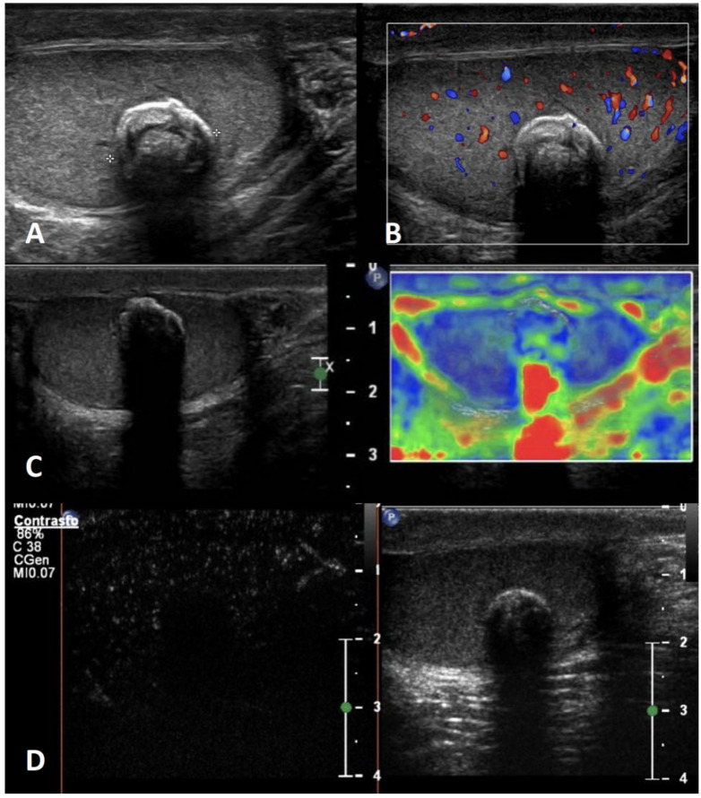
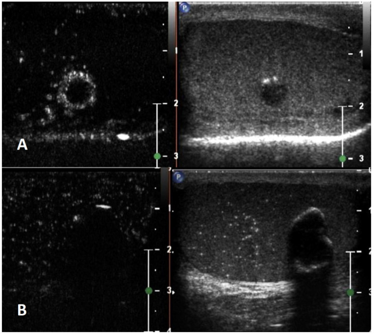
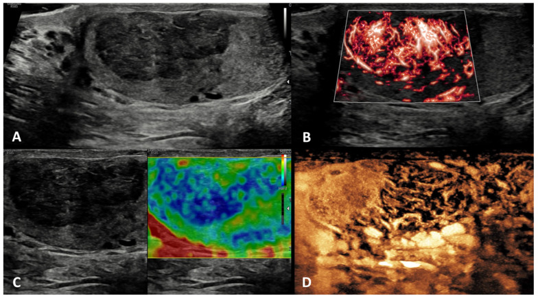
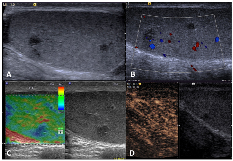
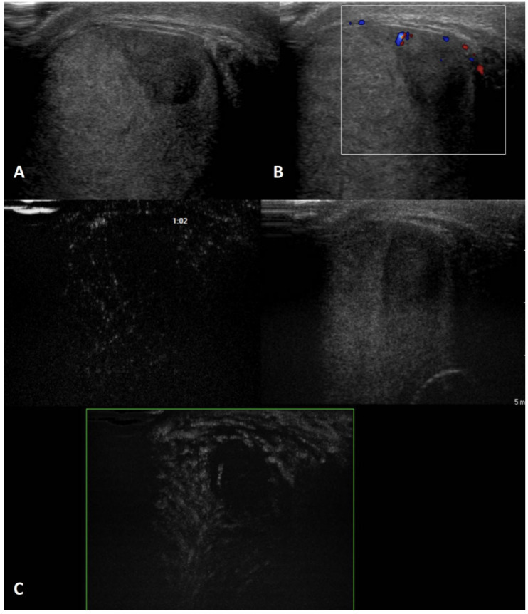
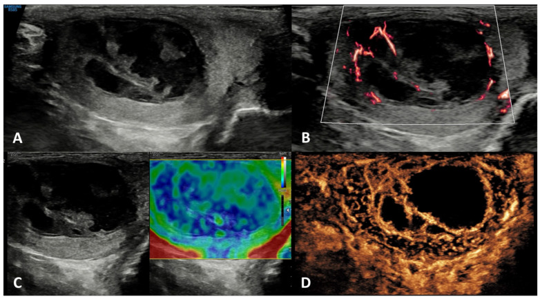
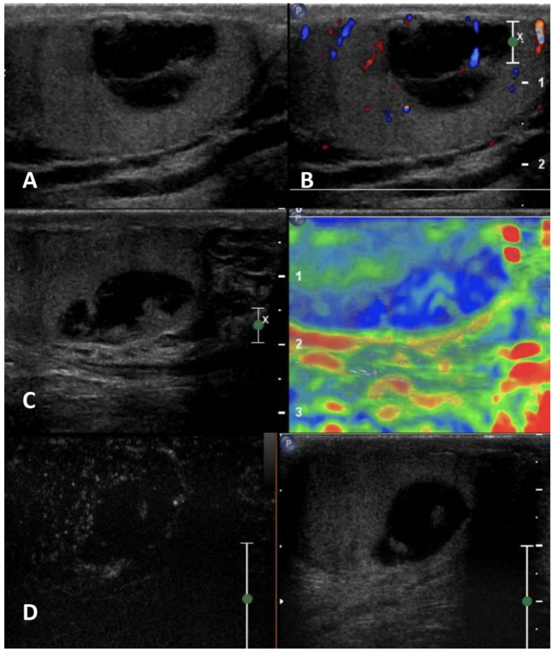
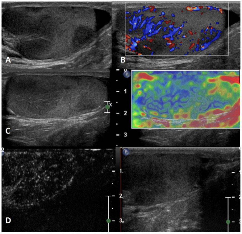
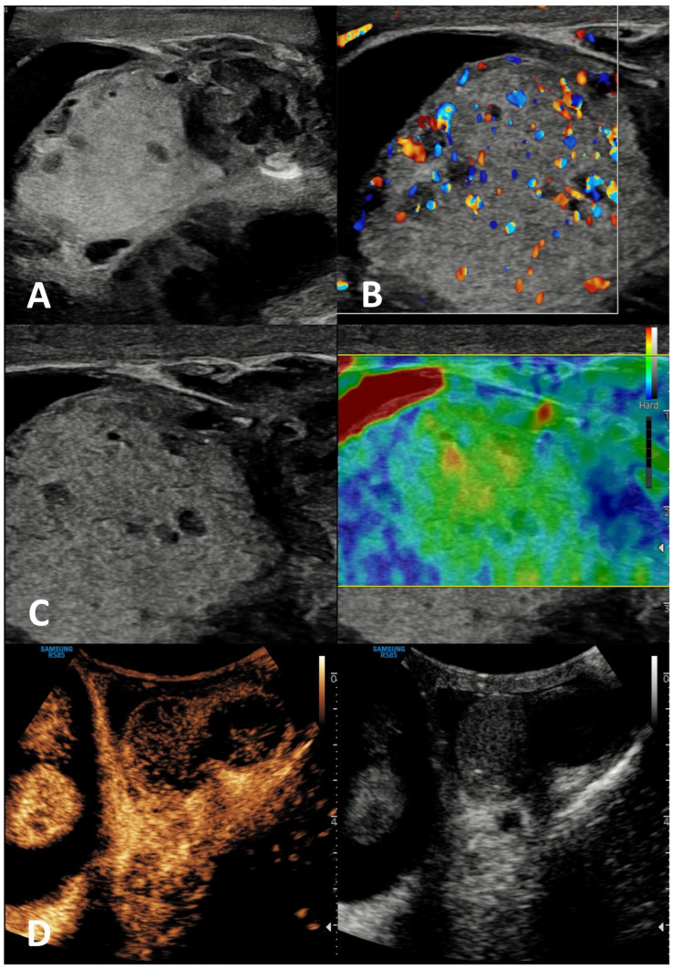
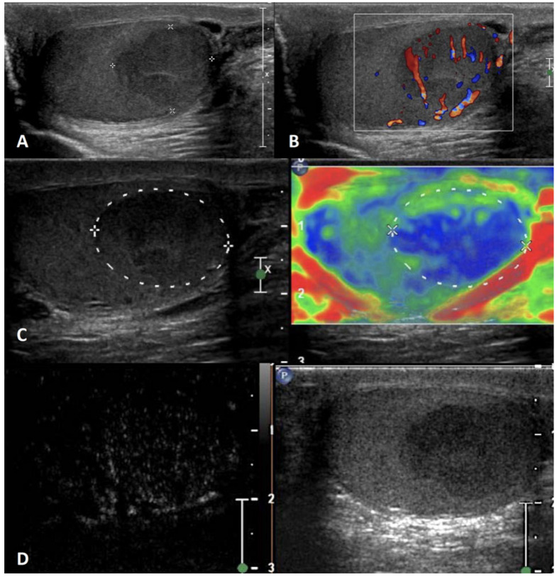
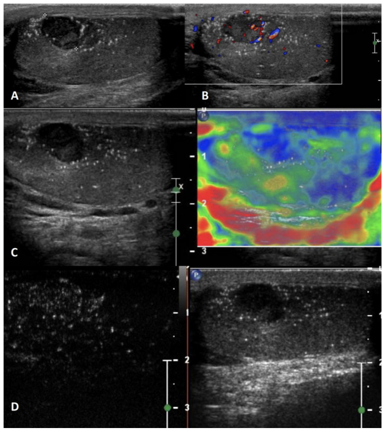
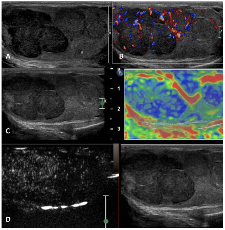
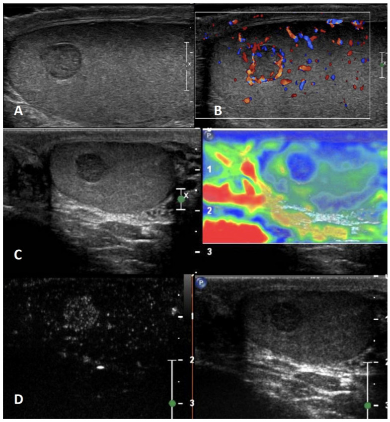
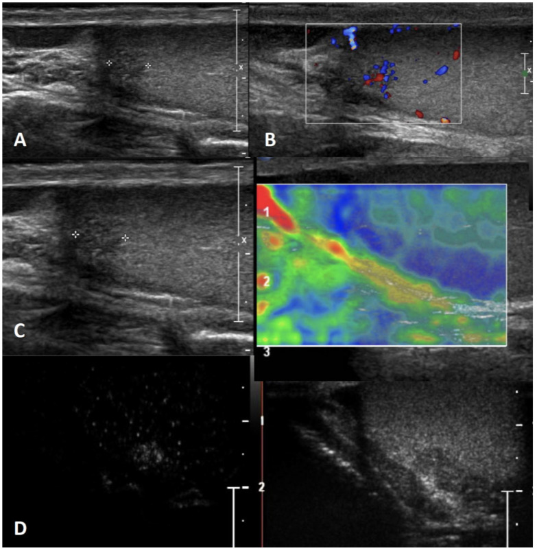
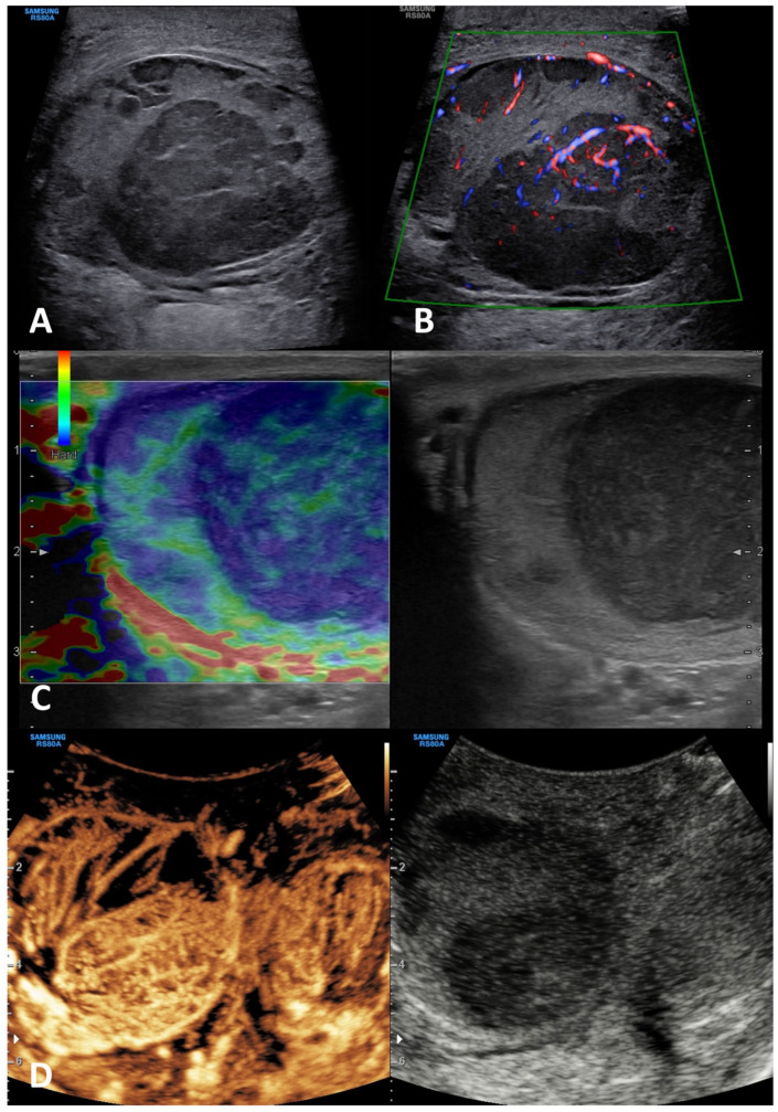
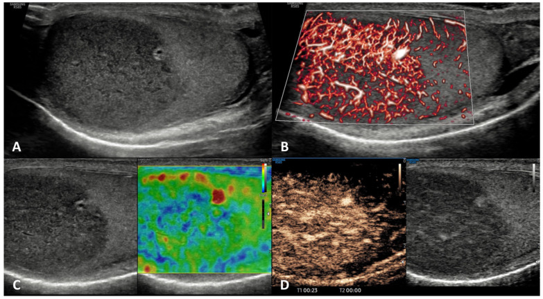
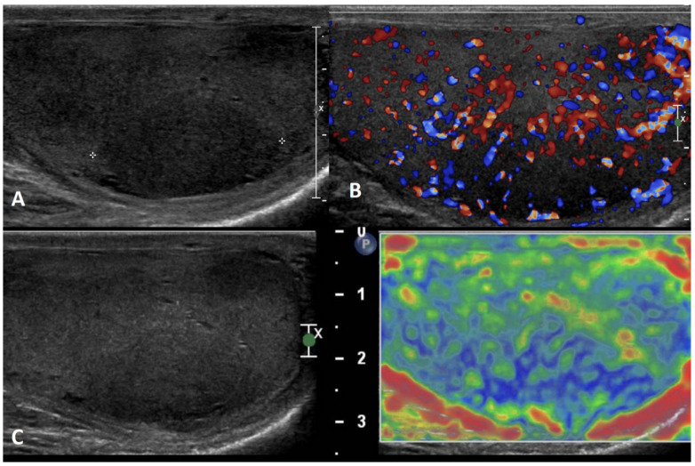
Background: Ultrasonography (US) represents the gold standard imaging method for the assessment of testicular lesions (TL). The gray-scale (GSUS) and color-Doppler (CDUS) ultrasound examination allow sonographers to investigate the size, margins, echotexture, and vascular features of TLs with the aim to differentiate benign from malignant lesions. Recently, the use of contrast-enhanced US (CEUS) and sonoelastography (SE) has led to further improvements in the differential diagnosis of TL. Although GSUS and CDUS are often sufficient to suggest the benign or malignant nature of the TL, CEUS can be decisive in the differential diagnosis of unclear findings, while SE can help to strengthen the diagnosis. The contemporary combination of GSUS, CDUS, CEUS, and SE has led to a new diagnostic paradigm named multiparametric US (mp-US), which is able to provide a more detailed characterization of TLs than single techniques alone. This narrative and pictorial review aimed to describe the mp-US appearance of several TLs.
Methods: An extensive Medline search was performed to identify studies in the English language focusing on the mp-US evaluation of TLs.
Results: A practical mp-US "identity card" and iconographic characterization of several benign and malignant TLs is provided herein.
Conclusions: The mp-US characterization of TL reported herein can be useful in daily clinical practice.
Keywords: color-Doppler ultrasound (CDUS); contrast-enhanced ultrasound (CEUS); differential diagnosis; gray-scale ultrasound (GSUS); multi-parametric ultrasound (mp-US); sonoelastography (SE); testicular lesions; testicular tumors; ultrasound (US).
Conflict of interest statement
The authors declare no conflict of interest.
Figures
Similar articles
-
Diagnostic performances of various gray-scale, color Doppler, and contrast-enhanced ultrasonography findings in predicting malignant thyroid nodules.Thyroid. 2014 Feb;24(2):355-63. doi: 10.1089/thy.2013.0150. Epub 2013 Nov 14. Thyroid. 2014. PMID: 23978252
-
Multiparametric Ultrasound for Focal Testicular Pathology: A Ten-Year Retrospective Review.Cancers (Basel). 2024 Jun 24;16(13):2309. doi: 10.3390/cancers16132309. Cancers (Basel). 2024. PMID: 39001372 Free PMC article.
-
Diagnosis of benign and malignant portal vein thrombosis in cirrhotic patients with hepatocellular carcinoma: color Doppler US, contrast-enhanced US, and fine-needle biopsy.Abdom Imaging. 2006 Sep-Oct;31(5):537-44. doi: 10.1007/s00261-005-0150-x. Abdom Imaging. 2006. PMID: 16865315
-
Use of contrast enhanced ultrasound in testicular diseases: A comprehensive review.Andrology. 2021 Sep;9(5):1369-1382. doi: 10.1111/andr.13057. Epub 2021 Jun 11. Andrology. 2021. PMID: 34043256 Free PMC article. Review.
-
Ultrasound 2020 - Diagnostics & Therapy: On the Way to Multimodal Ultrasound: Contrast-Enhanced Ultrasound (CEUS), Microvascular Doppler Techniques, Fusion Imaging, Sonoelastography, Interventional Sonography.Rofo. 2021 Jan;193(1):23-32. doi: 10.1055/a-1217-7400. Epub 2020 Jul 30. Rofo. 2021. PMID: 32731265 Review. English, German.
Cited by
-
Expanding Role of Contrast-Enhanced Ultrasound and Elastography in the Evaluation of Abdominal Pathologies in Children.Diagnostics (Basel). 2025 Jul 1;15(13):1680. doi: 10.3390/diagnostics15131680. Diagnostics (Basel). 2025. PMID: 40647679 Free PMC article. Review.
-
Testicular ultrasound: an emergency medicine perspective.Intern Emerg Med. 2025 Jun;20(4):1153-1163. doi: 10.1007/s11739-025-03864-z. Epub 2025 Mar 18. Intern Emerg Med. 2025. PMID: 40102331 Free PMC article. Review.
-
Testicular ultrasonographic features predict future risk for bilateral testicular germ cell tumour: A long-term single centre follow-up study.Andrology. 2025 Mar;13(3):587-599. doi: 10.1111/andr.13704. Epub 2024 Jul 30. Andrology. 2025. PMID: 39078248 Free PMC article.
-
"Management of andrological disorders from childhood and adolescence to transition age: guidelines from the Italian Society of Andrology and Sexual Medicine (SIAMS) in collaboration with the Italian Society for Pediatric Endocrinology and Diabetology (SIEDP)-Part-1".J Endocrinol Invest. 2025 Jan;48(1):1-22. doi: 10.1007/s40618-024-02435-x. Epub 2024 Aug 10. J Endocrinol Invest. 2025. PMID: 39126560 Free PMC article.
-
Development and Validation of an Ultrasound Imaging Algorithm for Structured Reporting in Testicular Pathology.Diagnostics (Basel). 2025 Apr 9;15(8):951. doi: 10.3390/diagnostics15080951. Diagnostics (Basel). 2025. PMID: 40310366 Free PMC article.
References
-
- Isidori A.M., Lenzi A. Ultrasound of the Testis for the Andrologist: Morphological and Functional Atlas. Springer; Berlin/Heidelberg, Germany: 2018.
Publication types
LinkOut - more resources
Full Text Sources
