Review
. 2023 Nov 13;15(22):5393.
doi: 10.3390/cancers15225393.

Leukemia Cutis-The Current View on Pathogenesis, Diagnosis, and Treatment

Affiliations
Review

Leukemia Cutis-The Current View on Pathogenesis, Diagnosis, and Treatment

Ewa Robak et al. Cancers (Basel). .

Abstract

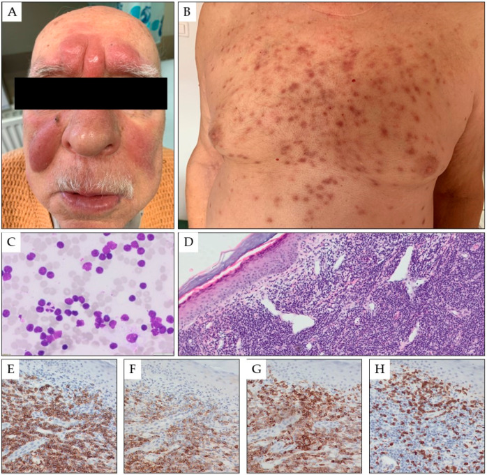
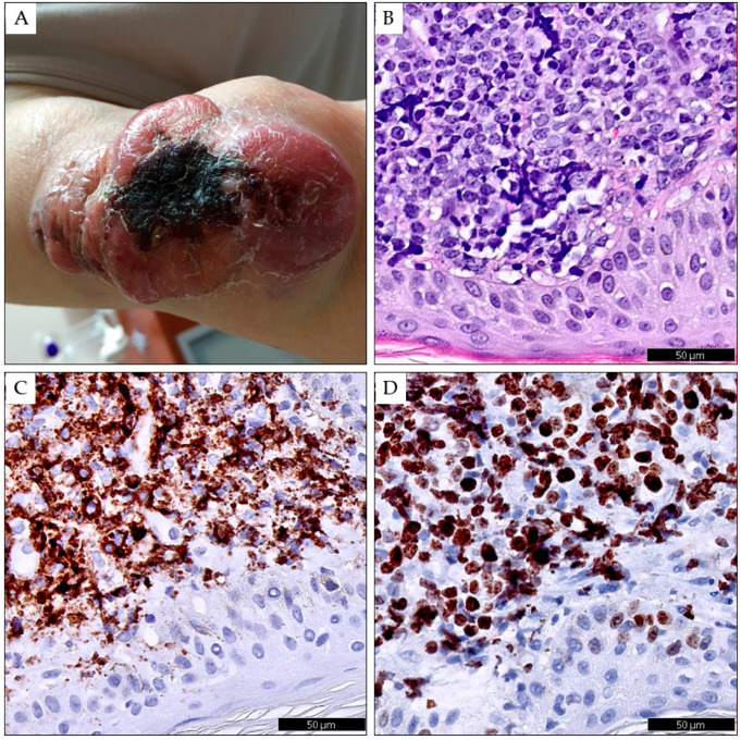
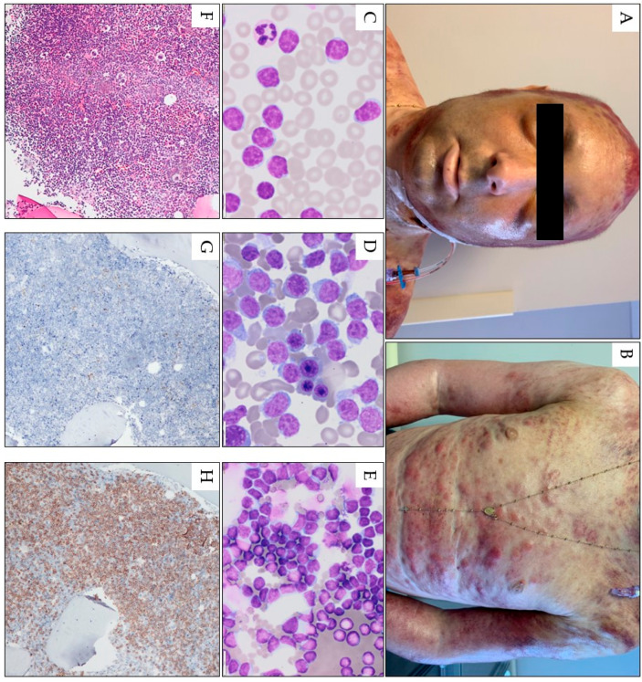
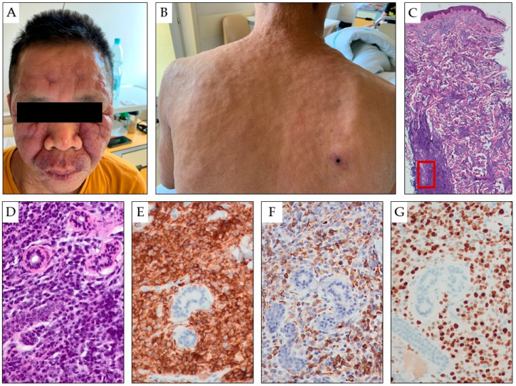
Leukemia cutis (LC) is defined as the leukemic infiltration of the epidermis, the dermis, and the subcutaneous tissue. Leukemia cutis may follow or occur simultaneously with the diagnosis of systemic leukemia. However, cutaneous lesions are occasionally diagnosed as the primary manifestation of leukemia. Leukemic skin infiltrations demonstrate considerable variation regarding a number of changes, distribution, and morphology. The highest incidence of LC is observed in chronic lymphocytic leukemia, monocytic and myelomonocytic acute myeloid leukemia, and T-cell lineage leukemia. Although the pathogenic mechanism of the invasion of leukemic cells into the skin is not well understood, chemokine receptors and adhesion molecules as well as the genetic characteristics of leukemia are thought to play a role. Leukemic skin lesions may be localized or disseminated and may occur alone or in combination on any site of the skin, most frequently in the trunk and extremities. The most common clinical presentations of leukemia cutis are papules, nodules, macules, plaques, and ulcers. In most patients, the complete or partial resolution of cutaneous infiltrations occurs simultaneously with hematologic remission. However, in patients with resistant disease or recurrent skin infiltration, local radiotherapy can be used. This review presents recent data on the pathogenesis, diagnosis, and treatment of leukemic skin involvement in different types of leukemia.

Keywords: Richter transformation; acute leukemia; chronic leukemia; cutis; diagnosis; pathogenesis; prolympchocytic leukemia; skin lesions; treatment.


Conflict of interest statement

The authors declare no conflict of interest. The funders had no role in the design of this study; interpretation of data, writing of the manuscript, or in the decision to publish the review.

Figures

Figure 1
Figure 1
Multiple red-brown tumors and nodules on the skin of the central surface of the forehead and the cheeks of a 78-year-old male patient with CLL in the form of symmetrical nodular infiltrates (A). Moreover, numerous, scattered red-blue papules and nodules with a hemorrhagic reaction are present on the torso (B). Fine needle biopsy of the forehead tumor (1000× magnification) (C) and biopsy of the skin torso infiltration in hematoxylin and eosin staining (25× magnification, panel (D)), and in immunohistochemistry for CD20 (200× magnification, panel (E)), CD23 (200× magnification, panel (F)), CD5 (200× magnification, panel (G)), and CD3 (200× magnification, panel (H)).
Figure 2
Figure 2
Richter transformation in the skin of the left upper limb in a 78-year-old female patient with CLL lasting four years. The skin shows a conglomerate of merging red-blue tumors of various sizes with a necrotic scab in the center (A). Skin biopsy showing infiltration by diffuse large B-cell lymphoma in hematoxylin and eosin staining (B), in immunohistochemistry for CD20 (C), and Ki67 (D). Digital scans were obtained using Phillips IntelliSite UltraFast Scanner. (Philips, Amsterdam, The Netherlands.)
Figure 3
Figure 3
Pink-red papules, nodules, and tumors scattered on the face torso, and upper limbs in a 32-year-old male with T-PLL. A confluent erythema with a hemorrhagic reaction is also visible in the area of the scalp. Generalized skin edema, scattered petechiae, and conjunctivitis are also visible (A,B). Peripheral blood smear (1000× magnification) (C) and bone marrow smear (1000× magnification) (D) and fine-needle skin aspiration smear (1000× magnification) (E) show numerous atypical prolymphocytes. In bone marrow trephine, a homogenous infiltrate by CD3-positive, TdT-negative prolymphocytes is seen; hematoxylin and eosin staining image under 100× magnification (F) and in immunohistochemistry for TDT (100× magnification, (G)), and CD3 (100× magnification, (H)).
Figure 4
Figure 4
Asymptomatic, multiple, variable-sized, erythematous tumors and papules are present on the face (A) and torso (B) of a 49-year-old patient with AML. The entire skin was thickened and infiltrated, with numerous red-blue tumors and nodules scattered over the entire surface. A particularly large, hard infiltrate is visible on the facial skin, where erythematous and exfoliative foci are also present on the basis of the nodular infiltrate (A,B). Biopsy of the skin showing infiltration by myeloblasts (hematoxylin and eosin staining in panels (C,D) under 25× and 200× magnification, respectively), which are positive for CD45 (LCA, 200× magnification, panel (E)), myeloperoxidase (200× magnification, panel (F)), and Ki-67 proliferation marker (200× magnification, panel (G)). The red box indicates area shown under larger magnification in panels (DG).

References

    1. Su W.D., Buechner S., Li C.-Y. Clinicopathologic correlations in leukemia cutis. J. Am. Acad. Dermatol. 1984;11:121–128. - PubMed
    1. Wagner G., Fenchel K., Back W., Schulz A., Sachse M.M. Leukemia cutis—Epidemiology, clinical presentation, and differential diagnoses. J. Dtsch. Dermatol. Ges. 2012;10:27–36. doi: 10.1111/j.1610-0387.2011.07842.x. - DOI - PubMed
    1. Yook H.J., Son J.H., Kim Y.H., Han J.H., Lee J.H., Park Y.M., Chung N.G., Kim H.J., Bang C.H. Leukaemia Cutis: Clinical Features and Outcomes of 56 Patients. Acta Derm. Venereol. 2022;102:adv00647. doi: 10.2340/actadv.v102.1123. - DOI - PMC - PubMed
    1. Jang K.A., Chi D.H., Choi J.H., Sung K.J., Moon K.C., Koh J.K. Leukemia cutis:a clinico-pathologic study of 23 patients. Korean J. Dermatol. 2000;38:15–22.
    1. Chang Y.W., Lee C.H., Tseng H.C. Leukemia cutis in a medical center in southern Taiwan: A retrospective study of 42 patients. Pt 1J. Formos. Med. Assoc. 2021;120:226–233. doi: 10.1016/j.jfma.2020.04.025. - DOI - PubMed

LinkOut - more resources