

Detection of Liver Lesions in Colorectal Cancer Patients Using 18F-FDG PET/CT Dual-Time-Point Scan Imaging
- PMID: 38001662
- PMCID: PMC10670707
- DOI: 10.3390/cancers15225403
Detection of Liver Lesions in Colorectal Cancer Patients Using 18F-FDG PET/CT Dual-Time-Point Scan Imaging
Abstract
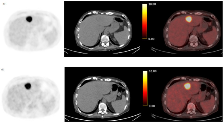
Objective: The aim of this study was to evaluate the diagnostic performance of dual-time-point fluorine-18-fluorodeoxyglucose positron emission computed tomography/computed tomography (18F-FDG PET/CT) compared to conventional early imaging for detecting colorectal liver metastases (CRLM) in colorectal cancer (CRC) patients.
Methods: One hundred twenty-four consecutive CRC patients underwent dual-time-point imaging scans on a retrospective basis. Histopathological confirmation and/or clinical follow-up were accepted as the gold standard. Standard uptake values (SUV), signal-to-noise ratio (SNR), retention index (RI), tumor-to-normal liver ratio (TNR), and lesion sizes were measured for early and delayed PET scans. The diagnostic performance of early and delayed images was calculated on a per-patient basis and compared using McNemar's test.
Results: Among the 124 patients, 57 (46%) had CRLM, 6 (4.8%) had benign lesions, and 61 (49.2%) had no concerning lesions detected. Smaller CRLM lesions (<5 cm3) showed significantly higher uptake in the delayed scans relative to early imaging (p < 0.001). The SUV and TNR increased significantly in delayed imaging of all metastatic lesions (p < 0.001). The retention index of all CRLM was high (40.8%), especially for small lesions (54.8%). A total of 177 lesions in delayed images and 124 in standard early images were identified. In a per-patient analysis, delayed imaging had significantly higher sensitivity (100% vs. 87.7%) and specificity (91.0% vs. 94.0%) compared to early imaging (p-value = 0.04).
Conclusions: The detection of liver lesions using dual-time-point PET/CT scan improves the sensitivity and specificity for the detection of colorectal liver metastasis.
Keywords: 18F-FDG PET/CT; colorectal cancer; colorectal liver metastases; delayed scan; early scan.
Conflict of interest statement
The authors declare no conflict of interest.
Figures


Similar articles
-
The added value of dual-time-point 18F-FDG PET/CT imaging in the diagnosis of colorectal cancer liver metastases.Abdom Radiol (NY). 2020 Apr;45(4):1075-1081. doi: 10.1007/s00261-019-02396-3. Abdom Radiol (NY). 2020. PMID: 31927618
-
More advantages in detecting bone and soft tissue metastases from prostate cancer using 18F-PSMA PET/CT.Hell J Nucl Med. 2019 Jan-Apr;22(1):6-9. doi: 10.1967/s002449910952. Epub 2019 Mar 7. Hell J Nucl Med. 2019. PMID: 30843003
-
Analysis of the best time-point for 18F-FDG PET/CT delayed imaging in patients of small colorectal cancer liver metastasis with hypothyroidism based on diagnostic efficacy and image standardized uptake values.Ann Nucl Med. 2025 Jul;39(7):707-715. doi: 10.1007/s12149-025-02045-4. Epub 2025 Apr 2. Ann Nucl Med. 2025. PMID: 40172768
-
Relationship between KRAS mutations and dual time point 18F-FDG PET/CT imaging in colorectal liver metastases.Abdom Radiol (NY). 2019 Jun;44(6):2059-2066. doi: 10.1007/s00261-018-1740-8. Abdom Radiol (NY). 2019. PMID: 30143816
-
Dual-time point 18F-FDG-PET and PET/CT for Differentiating Benign From Malignant Musculoskeletal Lesions: Opportunities and Limitations.Semin Nucl Med. 2017 Jul;47(4):373-391. doi: 10.1053/j.semnuclmed.2017.02.009. Epub 2017 Apr 19. Semin Nucl Med. 2017. PMID: 28583277 Review.
Cited by
-
Comparative study of total-body PET and PET/MR in the diagnosis of liver metastases.Front Oncol. 2025 May 14;15:1519107. doi: 10.3389/fonc.2025.1519107. eCollection 2025. Front Oncol. 2025. PMID: 40438693 Free PMC article.
-
Detection of Hepatic Metastasis from Early Delayed Images of Modified Dual-Time-Point F-18 FDG PET/CT Images in a Patient with Breast Cancer.Diagnostics (Basel). 2024 Jul 31;14(15):1653. doi: 10.3390/diagnostics14151653. Diagnostics (Basel). 2024. PMID: 39125529 Free PMC article.
-
Distinguishing benign lesions from malignant ones using FAPI-based tracers: will we need to bid farewell to dual-time points imaging?Eur J Nucl Med Mol Imaging. 2025 Sep;52(11):3935-3937. doi: 10.1007/s00259-025-07254-7. Epub 2025 Apr 9. Eur J Nucl Med Mol Imaging. 2025. PMID: 40202685 No abstract available.
References
-
- Zarour L.R., Anand S., Bilingsley K.G., Bisson W.H., Cercek A., Clarke M.F., Coussens L.M., Gast C.E., Geltzeiller C.B., Hansen L., et al. Colorectal Cancer Liver Metastasis: Evolving Paradigms and Future Directions. Cell. Mol. Gastroenterol. Hepatol. 2017;3:163–173. doi: 10.1016/j.jcmgh.2017.01.006. - DOI - PMC - PubMed
Grants and funding
LinkOut - more resources
Full Text Sources
