

Ring Chromosomes in Hematological Malignancies Are Associated with TP53 Gene Mutations and Characteristic Copy Number Variants
- PMID: 38001699
- PMCID: PMC10670249
- DOI: 10.3390/cancers15225439
Ring Chromosomes in Hematological Malignancies Are Associated with TP53 Gene Mutations and Characteristic Copy Number Variants
Abstract
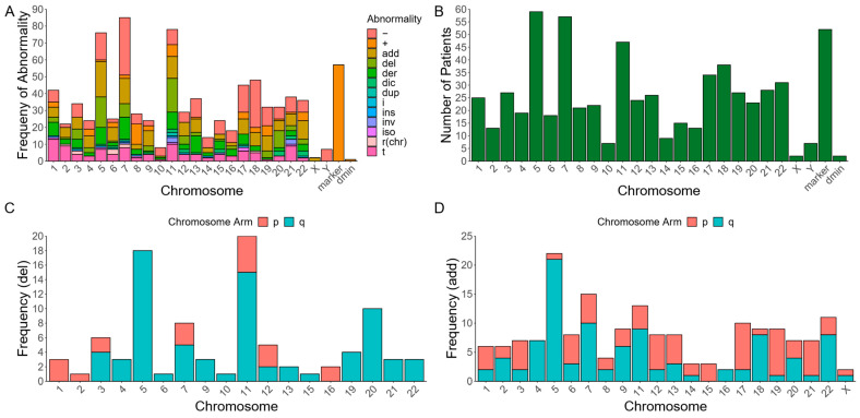
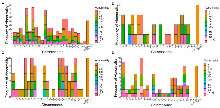
Ring chromosomes (RC) are present in <10% of patients with hematological malignancies and are associated with poor prognosis. Until now, only small cohorts of patients with hematological neoplasms and concomitant RCs have been cytogenetically characterized. Here, we performed a conventional chromosome analysis on metaphase spreads from >13,000 patients diagnosed with hematological malignancies at the Johns Hopkins University Hospital and identified 98 patients with RCs-90 with myeloid malignancies and 8 with lymphoid malignancies. We also performed a targeted Next-Generation Sequencing (NGS) assay, using a panel of 642 cancer genes, to identify whether these patients harbor relevant pathogenic variants. Cytogenetic analyses revealed that RCs and marker chromosomes of unknown origin are concurrently present in most patients by karyotyping, and 93% of patients with NGS data have complex karyotypes. A total of 72% of these individuals have pathogenic mutations in TP53, most of whom also possess cytogenetic abnormalities resulting in the loss of 17p, including the loss of TP53. All patients with a detected RC and without complex karyotypes also lack TP53 mutations but have pathogenic mutations in TET2. Further, 70% of RCs that map to a known chromosome are detected in individuals without TP53 mutations. Our data suggest that RCs in hematological malignancies may arise through different mechanisms, but ultimately promote widespread chromosomal instability.
Keywords: complex karyotype; copy number variants; gene mutation; myeloid malignancies; ring chromosomes.
Conflict of interest statement
The authors declare no conflict of interest.
Figures


Similar articles
-
Copy number neutral loss of heterozygosity at 17p and homozygous mutations of TP53 are associated with complex chromosomal aberrations in patients newly diagnosed with myelodysplastic syndromes.Leuk Res. 2016 Mar;42:7-12. doi: 10.1016/j.leukres.2016.01.009. Epub 2016 Jan 24. Leuk Res. 2016. PMID: 26851439
-
Marker chromosomes can arise from chromothripsis and predict adverse prognosis in acute myeloid leukemia.Blood. 2017 Mar 9;129(10):1333-1342. doi: 10.1182/blood-2016-09-738161. Epub 2017 Jan 24. Blood. 2017. PMID: 28119329 Clinical Trial.
-
TP53 Expression and Mutational Analysis in Hematological Malignancy in Jeddah, Saudi Arabia.Diagnostics (Basel). 2022 Mar 16;12(3):724. doi: 10.3390/diagnostics12030724. Diagnostics (Basel). 2022. PMID: 35328276 Free PMC article.
-
TP53 mutations in myelodysplastic syndrome.Leuk Lymphoma. 1996 Nov;23(5-6):417-22. doi: 10.3109/10428199609054848. Leuk Lymphoma. 1996. PMID: 9031070 Review.
-
Germline CHEK2 and ATM Variants in Myeloid and Other Hematopoietic Malignancies.Curr Hematol Malig Rep. 2022 Aug;17(4):94-104. doi: 10.1007/s11899-022-00663-7. Epub 2022 Jun 8. Curr Hematol Malig Rep. 2022. PMID: 35674998 Review.
Cited by
-
Myelodysplastic syndrome with ring chromosomes in a case of dehydrated hereditary stomatocytosis 1 (DHS1).Int J Hematol. 2025 Jun;121(6):857-861. doi: 10.1007/s12185-025-03967-5. Epub 2025 Mar 14. Int J Hematol. 2025. PMID: 40085347
-
An Integrated Approach Including CRISPR/Cas9-Mediated Nanopore Sequencing, Mate Pair Sequencing, and Cytogenomic Methods to Characterize Complex Structural Rearrangements in Acute Myeloid Leukemia.Biomedicines. 2024 Mar 7;12(3):598. doi: 10.3390/biomedicines12030598. Biomedicines. 2024. PMID: 38540211 Free PMC article.
-
Ring Chromosomes in Patients with Myeloid Neoplasms Are Associated with a Poor Response to Therapy.Acta Haematol. 2025 Jun 12:1-10. doi: 10.1159/000546757. Online ahead of print. Acta Haematol. 2025. PMID: 40505637 Free PMC article.
-
Optical Genome Mapping Reveals Complex and Cryptic Rearrangement Involving PML::RARA Fusion in Acute Promyelocytic Leukemia.Genes (Basel). 2024 Oct 30;15(11):1402. doi: 10.3390/genes15111402. Genes (Basel). 2024. PMID: 39596602 Free PMC article.
-
Can O-GIcNAc Transferase (OGT) Complex Be Used as a Target for the Treatment of Hematological Malignancies?Pharmaceuticals (Basel). 2024 May 22;17(6):664. doi: 10.3390/ph17060664. Pharmaceuticals (Basel). 2024. PMID: 38931332 Free PMC article. Review.
References
-
- Guilherme R.S., Meloni V.F.A., Kim C.A., Pellegrino R., Takeno S.S., Spinner N.B., Conlin L.K., Christofolini D.M., Kulikowski L.D., Melaragno M.I. Mechanisms of Ring Chromosome Formation, Ring Instability and Clinical Consequences. BMC Med. Genet. 2011;12:171. doi: 10.1186/1471-2350-12-171. - DOI - PMC - PubMed
Grants and funding
LinkOut - more resources
Full Text Sources
Research Materials
Miscellaneous
