. 2023 Nov 17;15(22):5454.
doi: 10.3390/cancers15225454.

The m7G Reader NCBP2 Promotes Pancreatic Cancer Progression by Upregulating MAPK/ERK Signaling

Affiliations

The m7G Reader NCBP2 Promotes Pancreatic Cancer Progression by Upregulating MAPK/ERK Signaling

Jiancong Xie et al. Cancers (Basel). .

Abstract

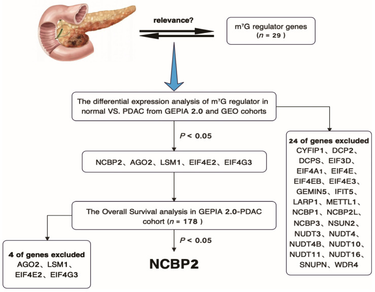
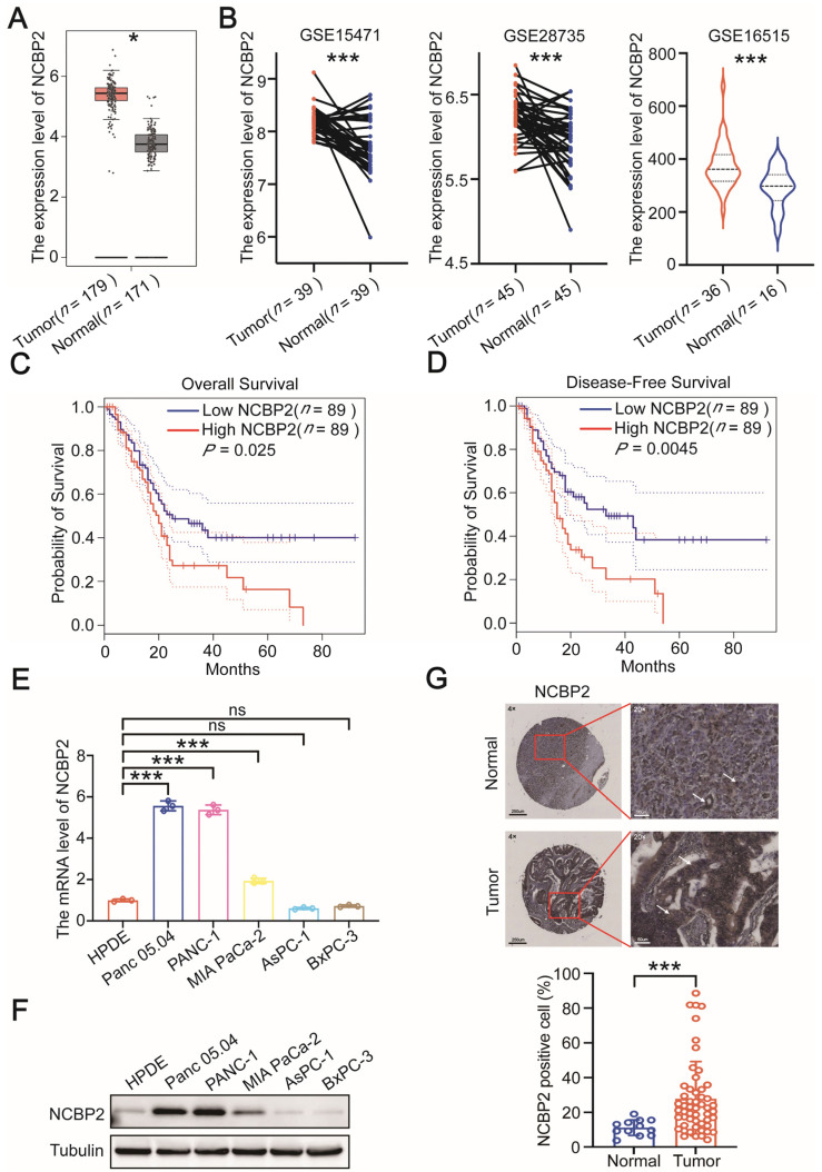
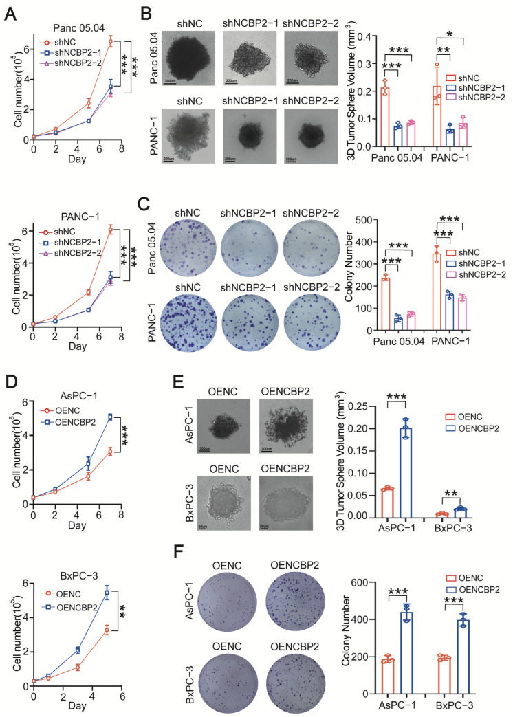
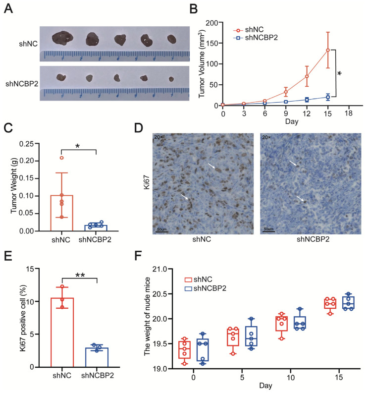
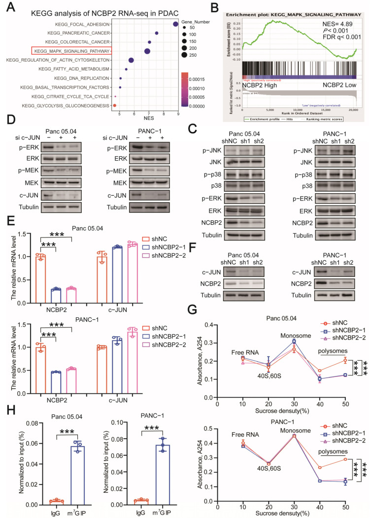
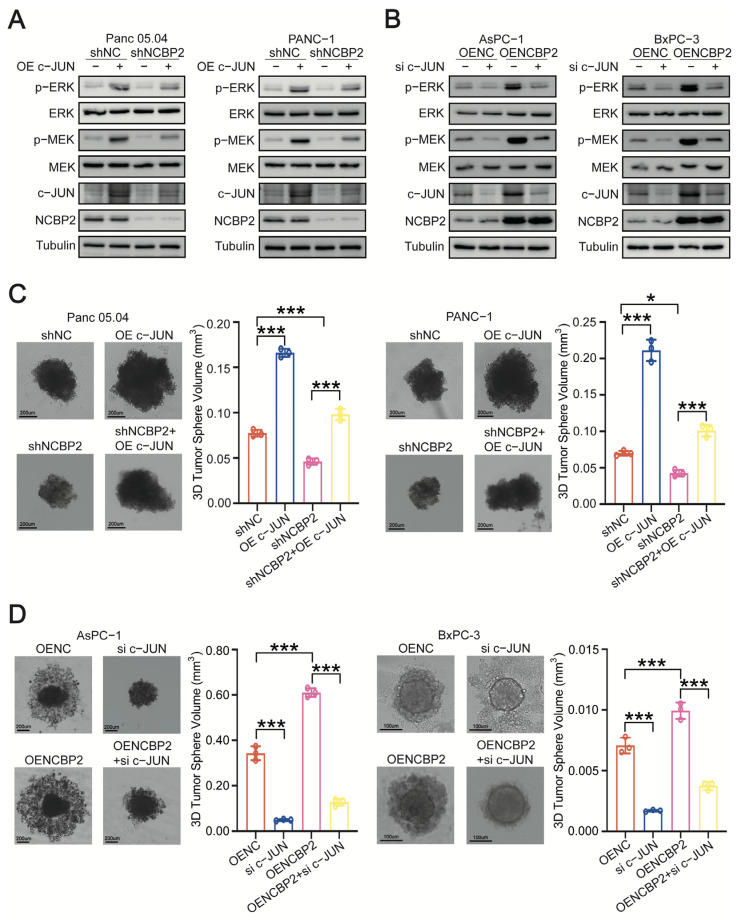
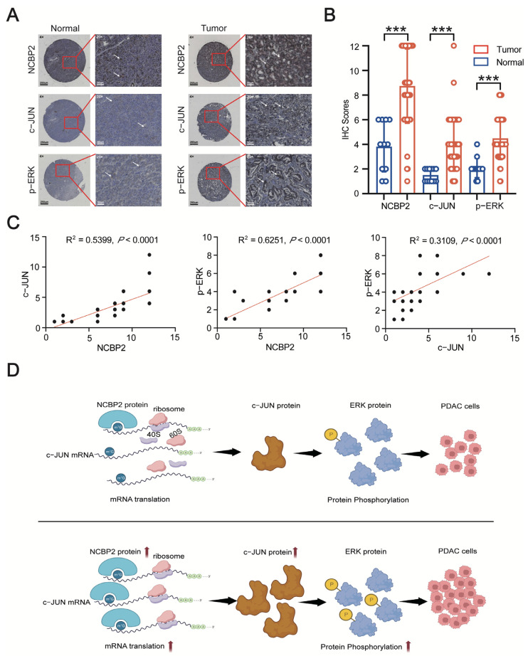
PDAC is one of the most common malignant tumors worldwide. The difficulty of early diagnosis and lack of effective treatment are the main reasons for its poor prognosis. Therefore, it is urgent to identify novel diagnostic and therapeutic targets for PDAC patients. The m7G methylation is a common type of RNA modification that plays a pivotal role in regulating tumor development. However, the correlation between m7G regulatory genes and PDAC progression remains unclear. By integrating gene expression and related clinical information of PDAC patients from TCGA and GEO cohorts, m7G binding protein NCBP2 was found to be highly expressed in PDAC patients. More importantly, PDAC patients with high NCBP2 expression had a worse prognosis. Stable NCBP2-knockdown and overexpression PDAC cell lines were constructed to further perform in-vitro and in-vivo experiments. NCBP2-knockdown significantly inhibited PDAC cell proliferation, while overexpression of NCBP2 dramatically promoted PDAC cell growth. Mechanistically, NCBP2 enhanced the translation of c-JUN, which in turn activated MEK/ERK signaling to promote PDAC progression. In conclusion, our study reveals that m7G reader NCBP2 promotes PDAC progression by activating MEK/ERK pathway, which could serve as a novel therapeutic target for PDAC patients.

Keywords: MEK/ERK; NCBP2; c-JUN; m7G methylation; pancreatic adenocarcinoma.


Conflict of interest statement

The authors declare no conflict of interest.

Figures

Figure 1
Figure 1
Flow chart of target m7G regulator gene screening process in PDAC.
Figure 2
Figure 2
NCBP2 is significantly expressed in PDAC patients and correlated with poor prognosis. (A) Analysis of NCBP2 transcript levels in PDAC and normal tissues based on the RNA-seq data from GEPIA 2.0 cohort. (B) Analysis of NCBP2 transcript levels in PDAC and normal tissues based on DNA microarray data from GEO cohorts (GSE15471, GSE28735, GSE16515). (C,D) Overall survival and disease-free survival curves for PDAC patients with high or low NCBP2 expression in GEPIA 2.0 cohort. (E,F) The RNA and protein expression level of NCBP2 in HPDE and five PDAC cell lines. (G) The expression level of NCBP2 in PDAC and para-carcinoma tissues from tissue microarray, * p < 0.05 and *** p < 0.001.
Figure 3
Figure 3
NCBP2 promotes PDAC cell growth in vitro. (AC). The cell counting, 3D sphere, and colony formation assay results in control and NCBP2-knockdown Panc 05.04 and PANC-1 cells. (DF). The cell counting, 3D sphere and colony formation assay results in control and NCBP2-overexpression AsPC-1 and BxPC-3 cells, * p < 0.05, ** p < 0.01, and *** p < 0.001.
Figure 4
Figure 4
NCBP2 promotes PDAC cell growth in-vivo. (A,B) Tumor growth curves of xenograft models established from control or stable NCBP2-knockdown Panc 05.04 cells. (C) Assessment of tumor weight from control and NCBP2-knockdown groups. (D,E) The expression level of Ki-67 in tumor tissues from control and NCBP2-knockdown groups. (F) Body weight of nude mice in control and NCBP2-knockdown group, * p < 0.05, and ** p < 0.01.
Figure 5
Figure 5
NCBP2 upregulates c-JUN to activate MEK/ERK signaling in a m7G-dependent manner. (A) KEGG analysis of RNA-seq data in PDAC patients with high or low NCBP2 expression from TCGA cohort. (B) The GSEA enrichment plot of “MAPK signaling pathway” in PDAC patients with high or low NCBP2 expression from TCGA cohort. (C) Immunoblotting for protein levels of total JNK/phosphorylated JNK (Thr183/Tyr185), total p38/phosphorylated p38 (Thr180/Tyr182), and total ERK/phosphorylated ERK (Thr202/Tyr204) in control and NCBP2-knockdown PDAC cells. Tubulin was used as the internal control. (D) Immunoblotting for protein levels of total/phosphorylated MEK and total/phosphorylated ERK (Thr202/Tyr204) in control and c-JUN-knockdown Panc 05.04 and PANC-1 cells. Tubulin was used as the internal control. (E) The mRNA expression levels of NCBP2 and c-JUN in control and NCBP2-knockdown Panc 05.04 and PANC-1 cells. (F) Immunoblotting for protein levels of NCBP2 and c-JUN in control and NCBP2-knockdown Panc 05.04 and PANC-1 cells. Tubulin was used as the internal control. (G) Polysome profiling results of the control and NCBP2-knockdown Panc 05.04 and PANC-1 cells. (H) Gene-specific m7G qPCR results for the m7G methylation levels of c-JUN in PANC 05.04 and PANC-1 cells, *** p < 0.001.
Figure 6
Figure 6
NCBP2 promotes PDAC progression by activating c-JUN/MEK/ERK signaling pathway. (A) Immunoblotting for protein levels of NCBP2, c-JUN, total/phosphorylated MEK, and total/phosphorylated ERK (Thr202/Tyr204) in control and NCBP2-knockdown PDAC cells overexpressing c-JUN. Tubulin was used as the internal control. (B) Immunoblotting for protein levels of NCBP2, c-JUN, total/phosphorylated MEK, and total/phosphorylated ERK (Thr202/Tyr204) in control and NCBP2-overexpression PDAC cells with c-JUN silenced. Tubulin was used as the internal control. (C) 3D sphere assays were performed after overexpressing c-JUN in control and NCBP2-knockdown PDAC cells. (D) 3D sphere assays were performed after c-JUN-knockdown in control and NCBP2-overexpressed PDAC cells, * p < 0.05 and *** p < 0.001.
Figure 7
Figure 7
NCBP2 and c-JUN/MEK/ERK signaling pathways are significantly upregulated in PDAC patients. (A) The expression level of NCBP2, c-JUN, and p-ERK was determined by IHC analysis in tissue microarray. (B) The IHC scores of NCBP2, c-JUN, and p-ERK between PDAC and para-carcinoma tissues. (C) The correlation analysis results between NCBP2, c-JUN, and p-ERK based on their IHC scores in PDAC tissues. (D) A schematic model of NCBP2 function in PDAC. NCBP2 upregulates c-JUN-mediated MEK/ERK signaling activation in an m7G methylation-dependent manner, *** p < 0.001.

Similar articles

Cited by

References

    1. GBD 2017 Pancreatic Cancer Collaborators The global, regional, and national burden of pancreatic cancer and its attributable risk factors in 195 countries and territories, 1990–2017: A systematic analysis for the Global Burden of Disease Study 2017. Lancet Gastroenterol. Hepatol. 2019;4:934–947. doi: 10.1016/s2468-1253(19)30347-4. Erratum in Lancet Gastroenterol. Hepatol. 2020, 5, e2. - DOI - PMC - PubMed
    1. Chauhan V.P., Martin J.D., Liu H., Lacorre D.A., Jain S.R., Kozin S.V., Stylianopoulos T., Mousa A.S., Han X., Adstamongkonkul P., et al. Angiotensin inhibition enhances drug delivery and potentiates chemotherapy by decompressing tumour blood vessels. Nat. Commun. 2013;4:2516. doi: 10.1038/ncomms3516. - DOI - PMC - PubMed
    1. Okada S. Review of tuberculosis control measures. 4. Studies on high risk groups at the present in Japan and on the effects of chemoprophylaxis. Kekkaku. 1968;43:239–242. (In Japanese) - PubMed
    1. Johnson B.A., 3rd, Yarchoan M., Lee V., Laheru D.A., Jaffee E.M. Strategies for Increasing Pancreatic Tumor Immunogenicity. Clin. Cancer Res. 2017;23:1656–1669. doi: 10.1158/1078-0432.CCR-16-2318. - DOI - PMC - PubMed
    1. Roundtree I.A., Evans M.E., Pan T., He C. Dynamic RNA Modifications in Gene Expression Regulation. Cell. 2017;169:1187–1200. doi: 10.1016/j.cell.2017.05.045. - DOI - PMC - PubMed

LinkOut - more resources