. 2023 Nov 19;15(22):5472.
doi: 10.3390/cancers15225472.

CX3CR1-Expressing Immune Cells Infiltrate the Tumor Microenvironment and Promote Radiation Resistance in a Mouse Model of Lung Cancer

Affiliations

CX3CR1-Expressing Immune Cells Infiltrate the Tumor Microenvironment and Promote Radiation Resistance in a Mouse Model of Lung Cancer

Tamar Ben-Mordechai et al. Cancers (Basel). .

Abstract

Introduction: Chemokine (C-X3-C Motif) Receptor 1 (CX3CR1) is present in a subset of the immune cells in the tumor microenvironment (TME) and plays an essential and diverse role in cancer progression. However, its potential function in the irradiated TME remains unknown.

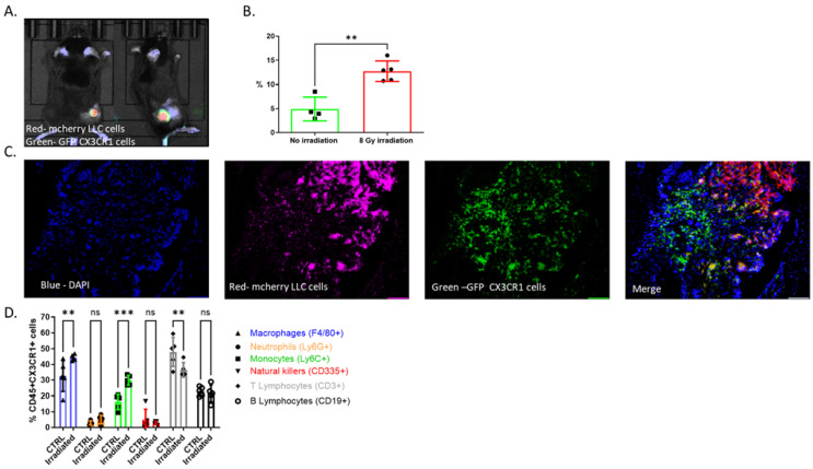
Materials and methods: A mouse lung cancer model was performed by subcutaneously inoculating Lewis Lung Carcinoma (LLC) cells expressing luciferase (Luc-2) and mCherry cells in CX3CR1GFP/GFP, CX3CR1DTR/+, and wild-type (WT) mice. Bioluminescence imaging, clonogenic assay, and flow cytometry were used to assess tumor progression, proliferation, and cell composition after radiation.

Results: Radiation provoked a significant influx of CX3CR1-expressing immune cells, notably monocytes and macrophages, into the TME. Co-culturing irradiated LLC cells with CX3CR1-deficient monocytes, and macrophages resulted in reduced clonogenic survival and increased apoptosis of the cancer cells. Interestingly, deficiency of CX3CR1 in macrophages led to a redistribution of the irradiated LLC cells in the S-phase, parallel to increased expression of cyclin E1, required for cell cycle G1/S transition. In addition, the deficiency of CX3CR1 expression in macrophages altered the cytokine secretion with a decrease in interleukin 6, a crucial mediator of cancer cell survival and proliferation. Next, LLC cells were injected subcutaneously into CX3CR1DTR/+ mice, sensitive to diphtheria toxin (DT), and WT mice. After injection, tumors were irradiated with 8 Gy, and mice were treated with DT, leading to conditional ablation of CX3CR1-expressing cells. After three weeks, CX3CR1-depleted mice displayed reduced tumor progression. Furthermore, combining the S-phase-specific chemotherapeutic gemcitabine with CX3CR1 cell ablation resulted in additional attenuation of tumor progression.

Conclusions: CX3CR1-expressing mononuclear cells invade the TME after radiation therapy in a mouse lung cancer model. CX3CR1 cell depletion attenuates tumor progression following radiation and sensitizes the tumor to S-phase-specific chemotherapy. Thus, we propose a novel strategy to improve radiation sensitivity by targeting the CX3CR1-expressing immune cells.

Keywords: cytokines; immunotherapy; lung cancer; radiation.


Conflict of interest statement

The authors declare no conflict of interest.

Figures

Figure 1
Figure 1
CX3CR1-expressing immune cells infiltrate the TME after radiation. (A) LLC cells (750,000) were injected SC into mice. Three weeks following injection, tumors were irradiated at 8 Gy. One week after irradiation, tumors were imaged using IVIS. (A) representative bioluminescence image of an irradiated mouse. LLC cells are shown in red (mCherry), and CX3CR1 immune cells within the tumor are green (GFP). (B) The percentage of CX3CR1-expressing cells within mouse tumors was significantly increased following irradiation with 8 Gy. Cells were imaged by flow cytometry using their GFP expression. Tumors without irradiation (no irradiation), n = 4; 8 Gy irradiated tumors (8 Gy irradiation), n = 5. (C) Fluorescent microscopy image showing infiltration of CX3CR1 expressing cells (GFP) in the irradiated TME (mCherry). (D) Cells from irradiated (8 Gy; irradiated) or not (CTRL) LLC tumors were isolated three days after irradiation, stained for immune cell markers, and subjected to flow cytometry. The markers for immune cell characterization were as follows: CD45 and CX3CR1 for population definition; Ly6G for neutrophils (N; orange); Ly6C for monocytes (MN; green); F4/80 and CD11b for macrophages (MQ; blue); CD335 for natural killer cells (NK; red); CD3 for T lymphocytes (T; gray) and CD19 for B lymphocytes (B; black; ** p < 0.01; *** p < 0.001; ns—not significant.
Figure 2
Figure 2
Deficiency of CX3CR1 in macrophages enhances LLC cells’ radiation sensitivity in vitro. The number of LLC colonies was significantly lower after irradiation. The number of irradiated LLC cell colonies increased after co-culture with WT macrophages and FKN compared to co-culture with CX3CR1-deficient macrophages and FKN. * p < 0.05; ** p < 0.01; **** p < 0.0001. FKN—fractalkine; Irr—irradiated; MQ—macrophages; ns—not significant; WT—wild-type.
Figure 3
Figure 3
Irradiated LLC cells co-cultured with CX3CR1-deficient macrophages show increased cell death due to apoptosis. Apoptosis was determined three days after irradiated LLC cells were co-cultured with WT macrophages or CX3CR1-deficient macrophages: (A) Early apoptosis was determined by annexin positive and PI negative staining. (B) Late apoptosis was determined by annexin and PI co-staining. ** p < 0.01; *** p < 0.001; **** p < 0.0001; ns—not significant.
Figure 4
Figure 4
Co-culture of irradiated LLC cells with CX3CR1-deficient macrophages results in altered cell cycle distribution and regulation compared to co-culture with WT macrophages. Irradiated LLC cells co-cultured with CX3CR1-deficient macrophages showed a decrease in cell rate at G1-phase (Ai), no difference in CDK 4 expression (Aii), and low cyclin D1 expression (Aiii). Irradiated LLC cells co-cultured with CX3CR1-deficient macrophages showed an increase in cell rate at S-phase (Bi), no difference in CDK 2 expression (Bii), no difference in cyclin A2 expression (Biii), and high cyclin E1 expression (Biv). Irradiated LLC cells co-cultured with CX3CR1-deficient macrophages showed no difference in cell rate at G2–M-phases (Ci), no difference in CDK1 expression (Cii), and no difference in Cyclin B1 expression (Ciii). All reactions were run as triplicates, and each dot represents the mean value. * p < 0.05; ** p < 0.01; *** p < 0.001; **** p < 0.0001; ns—not significant.
Figure 5
Figure 5
Deficiency of CX3CR1 in macrophages results in increased proinflammatory cytokine secretion. Conditioned medium of WT or CX3CR1-deficient macrophages was subjected to cytokine multiplex analysis for cell characterization. CX3CR1-deficient macrophages showed a significant increase in proinflammatory cytokines IL-1α (A) and MCP-1 (B) and a significant decrease in IL-6 (C) secretion, compared with WT macrophages with or without the addition of FKN. * p < 0.05; *** p < 0.001; **** p < 0.0001; ns—not significant.
Figure 6
Figure 6
Ablation of CX3CR1-expressing immune cells after radiation attenuates tumor growth and sensitizes cancer cells to the S-phase-specific chemotherapeutic gemcitabine. (A) LLC cells expressing luciferase were injected subcutaneously into CX3CR1DTR/+ mice, sensitive to diphtheria toxin (DT), and WT mice. After a week, all the tumors were irradiated (RT) with 8 Gy, followed by IP injection of DT in all mice every three days, and division into four groups: WT mice, CX3CR1DTR/+ mice, WT mice treated with gemcitabine, and CX3CR1DTR/+ mice treated gemcitabine. Tumor progression kinetics were assessed using IVIS. (B) Ablation of CX3CR1 expressing immune cells decreased tumor growth (orange line; n = 9) compared with WT mice (green line; n = 7). Injection of gemcitabine in addition to CX3CR1 expressing cell ablation after irradiation further attenuated tumor growth (blue line; n = 9). * p < 0.05.

Similar articles

Cited by

References

    1. Jagodinsky J.C., Harari P.M., Morris Z.S. The Promise of Combining Radiation Therapy With Immunotherapy. Int. J. Radiat. Oncol. Biol. Phys. 2020;108:6–16. doi: 10.1016/j.ijrobp.2020.04.023. - DOI - PMC - PubMed
    1. Wang Y., Deng W., Li N., Neri S., Sharma A., Jiang W., Lin S.H. Combining Immunotherapy and Radiotherapy for Cancer Treatment: Current Challenges and Future Directions. Front. Pharmacol. 2018;9:185. doi: 10.3389/fphar.2018.00185. - DOI - PMC - PubMed
    1. Formenti S.C., Demaria S. Future of Radiation and Immunotherapy. Int. J. Radiat. Oncol. Biol. Phys. 2020;108:3–5. doi: 10.1016/j.ijrobp.2020.04.034. - DOI - PubMed
    1. Antonia S.J., Villegas A., Daniel D., Vicente D., Murakami S., Hui R., Kurata T., Chiappori A., Lee K.H., de Wit M., et al. Overall Survival with Durvalumab after Chemoradiotherapy in Stage III NSCLC. N. Engl. J. Med. 2018;379:2342–2350. doi: 10.1056/NEJMoa1809697. - DOI - PubMed
    1. Shevtsov M., Sato H., Multhoff G., Shibata A. Novel Approaches to Improve the Efficacy of Immuno-Radiotherapy. Front. Oncol. 2019;9:156. doi: 10.3389/fonc.2019.00156. - DOI - PMC - PubMed

LinkOut - more resources