. 2023 Nov 16;12(11):2008.
doi: 10.3390/antiox12112008.

Pectolinarigenin Improves Oxidative Stress and Apoptosis in Mouse NSC-34 Motor Neuron Cell Lines Induced by C9-ALS-Associated Proline-Arginine Dipeptide Repeat Proteins by Enhancing Mitochondrial Fusion Mediated via the SIRT3/OPA1 Axis

Affiliations

Pectolinarigenin Improves Oxidative Stress and Apoptosis in Mouse NSC-34 Motor Neuron Cell Lines Induced by C9-ALS-Associated Proline-Arginine Dipeptide Repeat Proteins by Enhancing Mitochondrial Fusion Mediated via the SIRT3/OPA1 Axis

Ru-Huei Fu. Antioxidants (Basel). .

Abstract

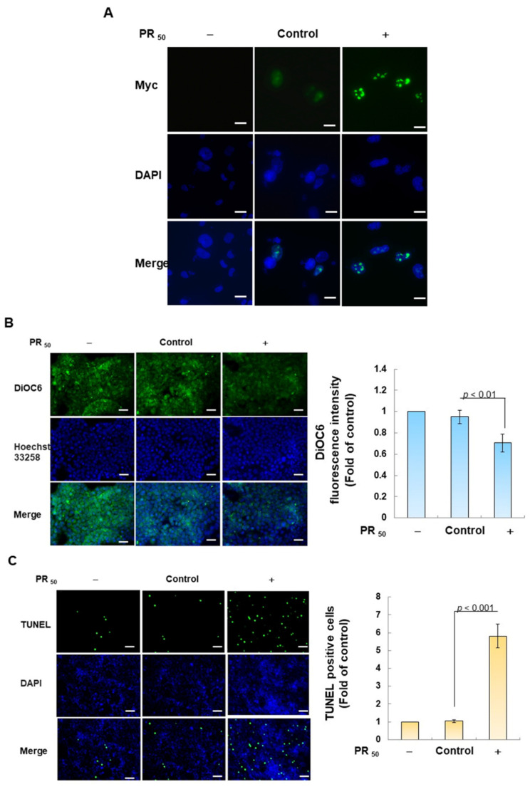
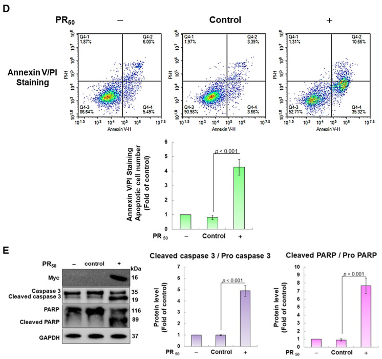
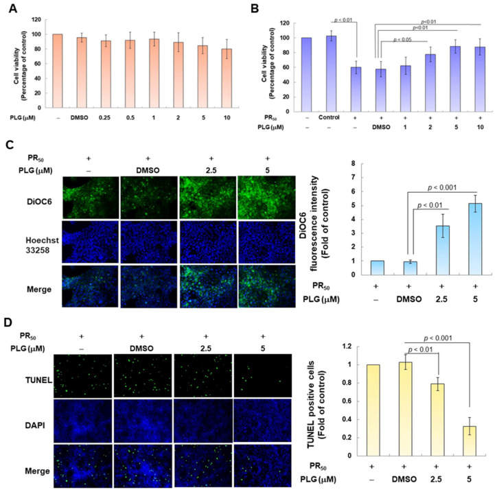
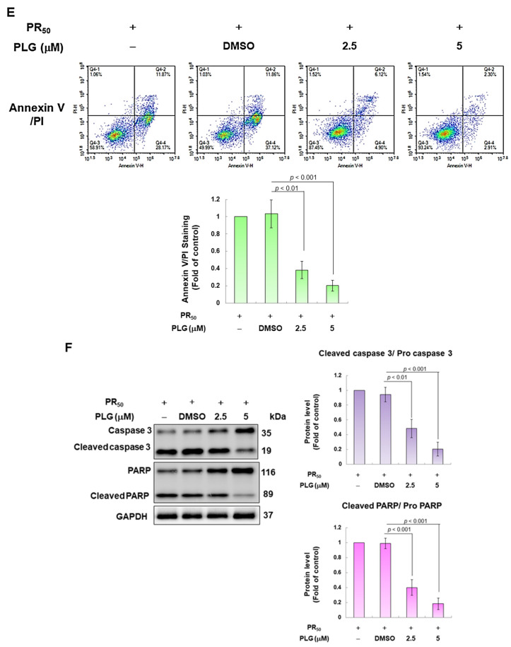
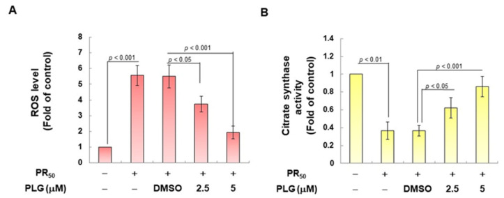
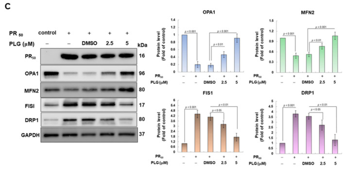
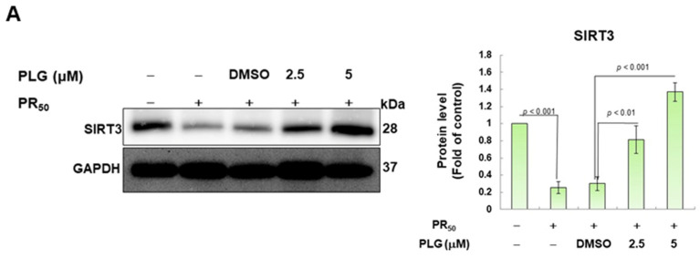
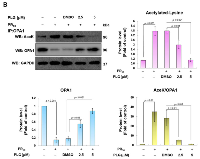
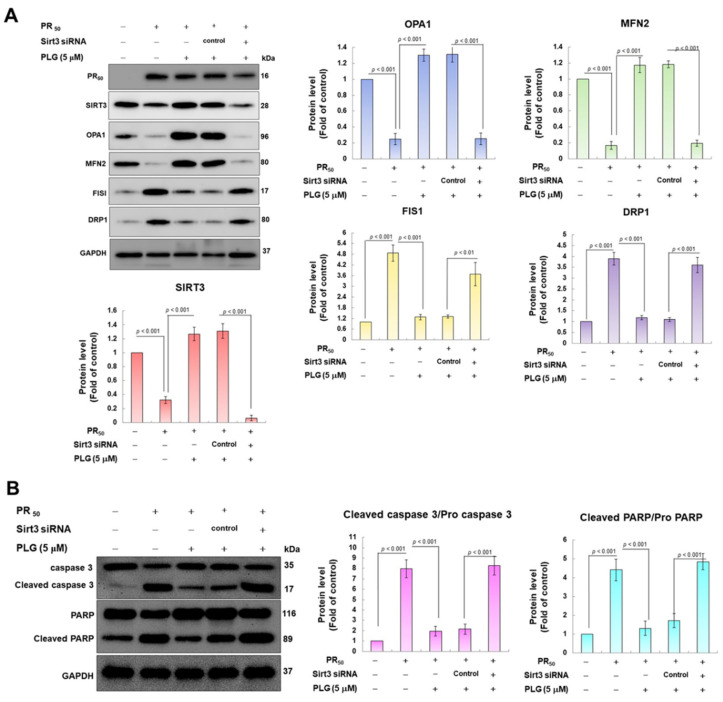
Amyotrophic lateral sclerosis (ALS) is considered a fatal progressive degeneration of motor neurons (MN) caused by oxidative stress and mitochondrial dysfunction. There are currently no treatments available. The most common inherited form of ALS is the C9orf72 mutation (C9-ALS). The proline-arginine dipeptide repeat protein (PR-DPR) produced by C9-ALS has been confirmed to be a functionally acquired pathogenic factor that can cause increased ROS, mitochondrial defects, and apoptosis in motor neurons. Pectolinarigenin (PLG) from the traditional medicinal herb Linaria vulgaris has antioxidant and anti-apoptotic properties. I established a mouse NSC-34 motor neuron cell line model expressing PR-DPR and confirmed the neuroprotective effect of PLG. The results showed that ROS production and apoptosis caused by PR-DPR could be improved by PLG treatment. In terms of mechanism research, PR-DPR inhibited the activity of the mitochondrial fusion proteins OPA1 and mitofusin 2. Conversely, the expression of fission protein fission 1 and dynamin-related protein 1 (DRP1) increased. However, PLG treatment reversed these effects. Furthermore, I found that PLG increased the expression and deacetylation of OPA1. Deacetylation of OPA1 enhances mitochondrial fusion and resistance to apoptosis. Finally, transfection with Sirt3 small interfering RNA abolished the neuroprotective effects of PLG. In summary, the mechanism by which PLG alleviates PR-DPR toxicity is mainly achieved by activating the SIRT3/OPA1 axis to regulate the balance of mitochondrial dynamics. Taken together, the potential of PLG in preclinical studies for C9-ALS drug development deserves further evaluation.

Keywords: C9orf72; OPA1; ROS; SIRT3; acetylation; amyotrophic lateral sclerosis (ALS); apoptosis; mitochondrial dynamics; pectolinarigenin; proline–arginine dipeptide repeat protein (PR-DPR).


Conflict of interest statement

The author declares no conflict of interest.

Figures

Figure 1
Figure 1
Molecular structure diagram of the flavonoid compound pectolinarigenin (PLG) derived from Linaria vulgaris.
Figure 2
Figure 2
PR50 expression causes apoptosis of mouse NSC-34 motor neuron cells. PR50 plasmids were transfected into NSC-34 cells for 24 h. (A) immunofluorescent staining using anti-myc-tag antibody (green). The position of the nucleus was confirmed by DAPI staining (blue). Microscope magnification is 400x. PR50 mainly forms punctates in the nucleus. (B) DiOC6 staining and fluorescence microscopy were used to detect changes in mitochondrial membrane potential (MMP). Microscope magnification is 100x. Quantitative values were obtained using ImageJ software (version 1.53). (C) the proportion of cells with chromatin fragmentation was assessed by fluorescence microscopy using the TUNEL assay. Microscope magnification is 100x. (D) cells co-stained with FITC-conjugated annexin V and propidium iodide (PI) were analyzed by flow cytometry to quantify the proportion of apoptotic cell populations. (E) the expression of apoptosis-related proteins was determined by Western blotting. GAPDH was used to normalize the total protein loading in each group. Quantitative values were obtained using ImageJ software (version 1.53). The results of Western blotting show the presence of the active form of the proteins since they are activated by proteolysis.
Figure 2
Figure 2
PR50 expression causes apoptosis of mouse NSC-34 motor neuron cells. PR50 plasmids were transfected into NSC-34 cells for 24 h. (A) immunofluorescent staining using anti-myc-tag antibody (green). The position of the nucleus was confirmed by DAPI staining (blue). Microscope magnification is 400x. PR50 mainly forms punctates in the nucleus. (B) DiOC6 staining and fluorescence microscopy were used to detect changes in mitochondrial membrane potential (MMP). Microscope magnification is 100x. Quantitative values were obtained using ImageJ software (version 1.53). (C) the proportion of cells with chromatin fragmentation was assessed by fluorescence microscopy using the TUNEL assay. Microscope magnification is 100x. (D) cells co-stained with FITC-conjugated annexin V and propidium iodide (PI) were analyzed by flow cytometry to quantify the proportion of apoptotic cell populations. (E) the expression of apoptosis-related proteins was determined by Western blotting. GAPDH was used to normalize the total protein loading in each group. Quantitative values were obtained using ImageJ software (version 1.53). The results of Western blotting show the presence of the active form of the proteins since they are activated by proteolysis.
Figure 3
Figure 3
PLG treatment can alleviate PR50-induced apoptosis of NSC-34 motor neuron cells. (A) NSC-34 cells were treated with different concentrations of PLG for 24 h, and cell viability was determined using the CellTiter Blue Cell Viability Assay. (B) PR50-expressing NSC-34 cells were treated with different concentrations of PLG for 24 h, and cell viability was measured. (CF) PR50-expressing NSC-34 cells were treated with 2.5 and 5 μM PLG for 24 h. (C) DiOC6 probe was used to detect changes in mitochondrial membrane potential (MMP) through fluorescence microscopy. Microscope magnification is 100x. The signal intensity was quantified using ImageJ software (version 1.53). (D) the number of cells with chromatin fragmentation was counted by fluorescence microscopy using the TUNEL assay. Microscope magnification is 100x. (E) FITC-conjugated Annexin V- and propidium iodide (PI) co-stained cells were used to count the number of apoptotic cell populations using flow cytometry. (F) the expression of apoptosis-related proteins was determined by Western blotting. GAPDH was used to normalize the total protein loading in each group. Quantitative values were obtained using ImageJ software (version 1.53). The results of Western blotting show the presence of the active form of the proteins since they are activated by proteolysis.
Figure 3
Figure 3
PLG treatment can alleviate PR50-induced apoptosis of NSC-34 motor neuron cells. (A) NSC-34 cells were treated with different concentrations of PLG for 24 h, and cell viability was determined using the CellTiter Blue Cell Viability Assay. (B) PR50-expressing NSC-34 cells were treated with different concentrations of PLG for 24 h, and cell viability was measured. (CF) PR50-expressing NSC-34 cells were treated with 2.5 and 5 μM PLG for 24 h. (C) DiOC6 probe was used to detect changes in mitochondrial membrane potential (MMP) through fluorescence microscopy. Microscope magnification is 100x. The signal intensity was quantified using ImageJ software (version 1.53). (D) the number of cells with chromatin fragmentation was counted by fluorescence microscopy using the TUNEL assay. Microscope magnification is 100x. (E) FITC-conjugated Annexin V- and propidium iodide (PI) co-stained cells were used to count the number of apoptotic cell populations using flow cytometry. (F) the expression of apoptosis-related proteins was determined by Western blotting. GAPDH was used to normalize the total protein loading in each group. Quantitative values were obtained using ImageJ software (version 1.53). The results of Western blotting show the presence of the active form of the proteins since they are activated by proteolysis.
Figure 4
Figure 4
PLG treatment reduced cellular ROS production and increased mitochondrial fusion in PR50-expressing NSC-34 cells. PR50-expressing NSC-34 cells were treated with PLG for 24 h. (A) the H2DCFDA probe was used to measure the ROS level in each group. (B) citrate synthase activity assay was used to determine intracellular mitochondrial activity in each group. (C) expression of fusion (OPA1 and MFN2) and fission (Fis1 and DRP1)-related proteins on mitochondrial dynamics was assessed using Western blotting. GAPDH was used as the control for total protein loading in each group. The signal intensity was quantitatively analyzed using ImageJ software (version 1.53).
Figure 4
Figure 4
PLG treatment reduced cellular ROS production and increased mitochondrial fusion in PR50-expressing NSC-34 cells. PR50-expressing NSC-34 cells were treated with PLG for 24 h. (A) the H2DCFDA probe was used to measure the ROS level in each group. (B) citrate synthase activity assay was used to determine intracellular mitochondrial activity in each group. (C) expression of fusion (OPA1 and MFN2) and fission (Fis1 and DRP1)-related proteins on mitochondrial dynamics was assessed using Western blotting. GAPDH was used as the control for total protein loading in each group. The signal intensity was quantitatively analyzed using ImageJ software (version 1.53).
Figure 5
Figure 5
PLG enhances SIRT3 expression and OPA1 deacetylation in PR50-expressing NSC-34 cells. PR50-expressing NSC-34 cells were treated with PLG for 24 h. (A) Western blotting was used to analyze expression. (B) determination of the degree of OPA1 acetylation using immunoprecipitation with an OPA1 antibody and Western blotting with a lysine acetylation-specific antibody. GAPDH was used to normalize the loading of all protein extracts from each group. The degree of OPA1 acetylation was corrected by normalizing OPA1 expression levels. The signal intensity was quantified using ImageJ software (version 1.53).
Figure 5
Figure 5
PLG enhances SIRT3 expression and OPA1 deacetylation in PR50-expressing NSC-34 cells. PR50-expressing NSC-34 cells were treated with PLG for 24 h. (A) Western blotting was used to analyze expression. (B) determination of the degree of OPA1 acetylation using immunoprecipitation with an OPA1 antibody and Western blotting with a lysine acetylation-specific antibody. GAPDH was used to normalize the loading of all protein extracts from each group. The degree of OPA1 acetylation was corrected by normalizing OPA1 expression levels. The signal intensity was quantified using ImageJ software (version 1.53).
Figure 6
Figure 6
Downregulation of SIRT3 expression eliminates the facilitation of mitochondrial dynamics fusion and the anti-apoptotic activities of PLG in PR50-expressing NSC-34 cells. Control or Sirt3 siRNA was delivered to PR50-expressing NSC34 cells for 16 h. This was followed by PLG (5 μM) treatment for 24 h. Finally, Western blotting was used to detect the expression of mitochondrial dynamics-related proteins (A) and the activity of the apoptotic core protein (B). GAPDH served as an internal control for loading total proteins in each group. The signal intensity was quantified using ImageJ software (version 1.53).

Similar articles

Cited by

References

    1. Goutman S.A., Savelieff M.G., Jang D.G., Hur J., Feldman E.L. The amyotrophic lateral sclerosis exposome: Recent advances and future directions. Nat. Rev. Neurol. 2023;19:617–634. doi: 10.1038/s41582-023-00867-2. - DOI - PMC - PubMed
    1. Kirola L., Mukherjee A., Mutsuddi M. Recent Updates on the Genetics of Amyotrophic Lateral Sclerosis and Frontotemporal Dementia. Mol. Neurobiol. 2022;59:5673–5694. doi: 10.1007/s12035-022-02934-z. - DOI - PubMed
    1. McGoldrick P., Robertson J. Unraveling the impact of disrupted nucleocytoplasmic transport systems in C9orf72-associated ALS. Front. Cell. Neurosci. 2023;17:1247297. doi: 10.3389/fncel.2023.1247297. - DOI - PMC - PubMed
    1. Mori K., Gotoh S., Ikeda M. Aspects of degradation and translation of the expanded C9orf72 hexanucleotide repeat RNA. J. Neurochem. 2023;166:156–171. doi: 10.1111/jnc.15847. - DOI - PubMed
    1. Fu R.H., Tsai C.W., Chiu S.C., Liu S.P., Chiang Y.T., Kuo Y.H., Shyu W.C., Lin S.Z. C9-ALS-Associated Proline-Arginine Dipeptide Repeat Protein Induces Activation of NLRP3 Inflammasome of HMC3 Microglia Cells by Binding of Complement Component 1 Q Subcomponent-Binding Protein (C1QBP), and Syringin Prevents This Effect. Cells. 2022;11:3128. doi: 10.3390/cells11193128. - DOI - PMC - PubMed