

2,4-Dichlorophenoxyacetic Acid Induces Degeneration of mDA Neurons In Vitro
- PMID: 38001883
- PMCID: PMC10669833
- DOI: 10.3390/biomedicines11112882
2,4-Dichlorophenoxyacetic Acid Induces Degeneration of mDA Neurons In Vitro
Abstract
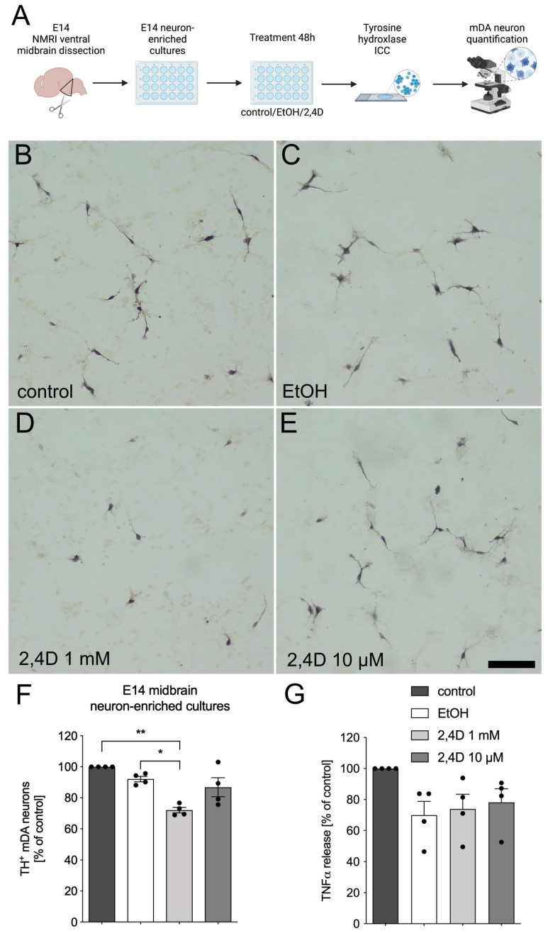
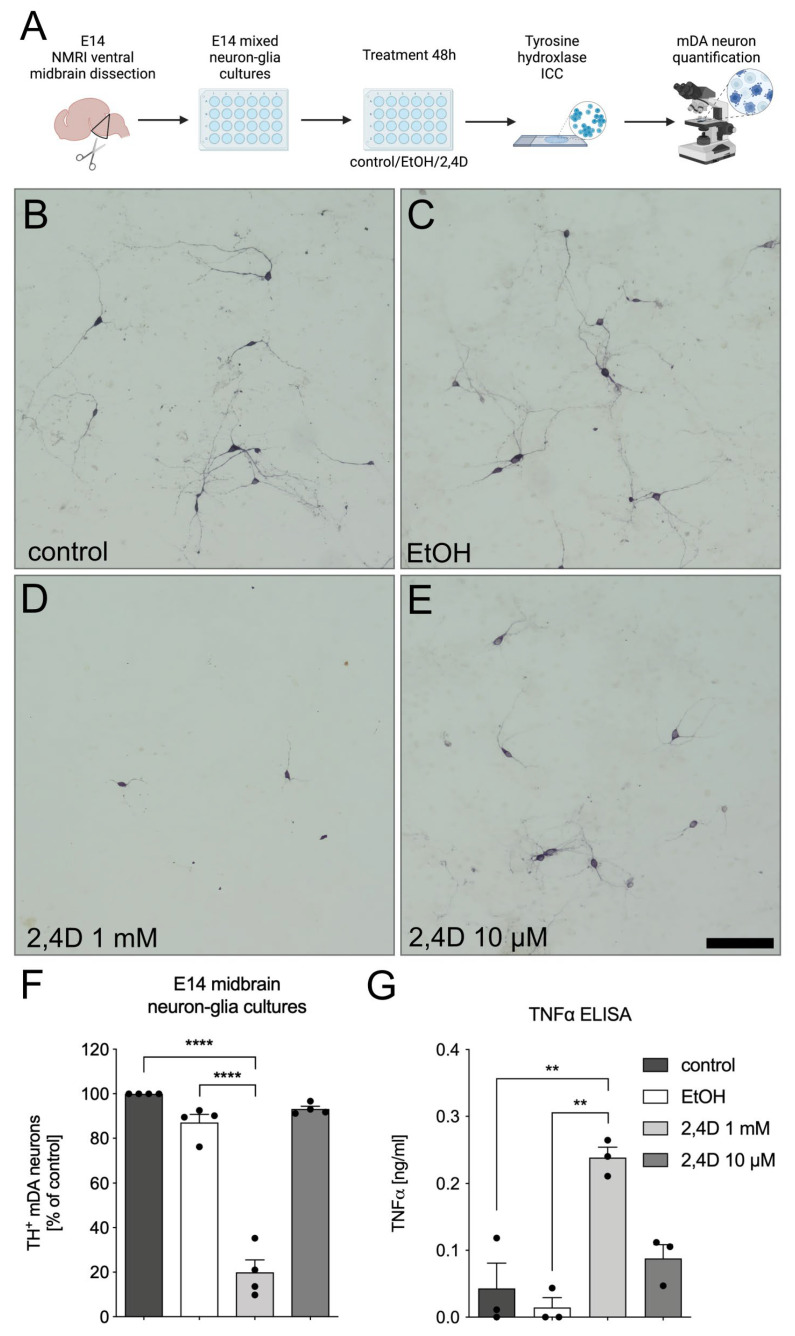
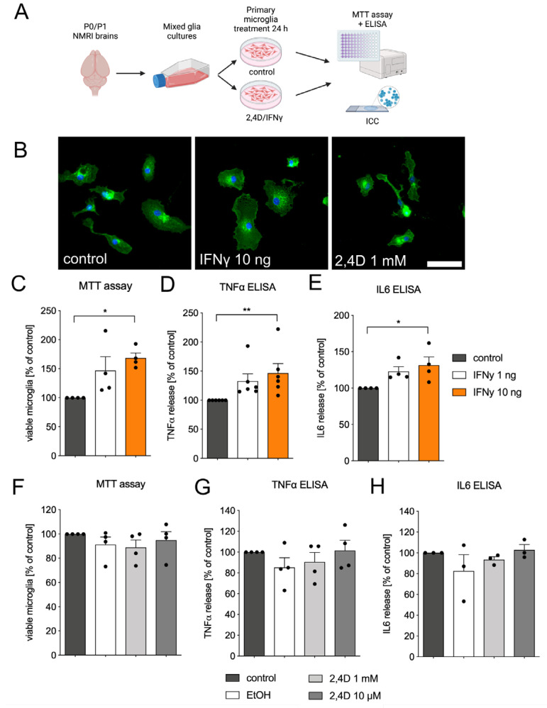
Background: Parkinson's disease (PD) affects 1-2% of the population over the age of 60 and the majority of PD cases are sporadic, without any family history of the disease. Neuroinflammation driven by microglia has been shown to promote the progression of midbrain dopaminergic (mDA) neuron loss through the release of neurotoxic factors. Interestingly, the risk of developing PD is significantly higher in distinct occupations, such as farming and agriculture, and is linked to the use of pesticides and herbicides. Methods: The neurotoxic features of 2,4-Dichlorophenoxyacetic acid (2,4D) at concentrations of 10 µM and 1 mM were analyzed in two distinct E14 midbrain neuron culture systems and in primary microglia. Results: The application of 1 mM 2,4D resulted in mDA neuron loss in neuron-enriched cultures. Notably, 2,4D-induced neurotoxicity significantly increased in the presence of microglia in neuron-glia cultures, suggesting that microglia-mediated neurotoxicity could be one mechanism for progressive neuron loss in this in vitro setup. However, 2,4D alone was unable to trigger microglia reactivity. Conclusions: Taken together, we demonstrate that 2,4D is neurotoxic for mDA neurons and that the presence of glia cells enhances 2,4D-induced neuron death. These data support the role of 2,4D as a risk factor for the development and progression of PD and further suggest the involvement of microglia during 2,4D-induced mDA neuron loss.
Keywords: 2,4-Dichlorophenoxyacetic acid; TNFα; mDA neurons; microglia; neurodegeneration.
Conflict of interest statement
The authors declare no conflict of interest. The funders had no role in the design of the study; in the collection, analyses, or interpretation of data.
Figures
Similar articles
-
TGFβ1 inhibits IFNγ-mediated microglia activation and protects mDA neurons from IFNγ-driven neurotoxicity.J Neurochem. 2015 Jul;134(1):125-34. doi: 10.1111/jnc.13111. Epub 2015 Apr 23. J Neurochem. 2015. PMID: 25827682
-
NADPH oxidase mediates lipopolysaccharide-induced neurotoxicity and proinflammatory gene expression in activated microglia.J Biol Chem. 2004 Jan 9;279(2):1415-21. doi: 10.1074/jbc.M307657200. Epub 2003 Oct 24. J Biol Chem. 2004. PMID: 14578353
-
3-hydroxymorphinan is neurotrophic to dopaminergic neurons and is also neuroprotective against LPS-induced neurotoxicity.FASEB J. 2005 Mar;19(3):395-7. doi: 10.1096/fj.04-1586fje. Epub 2004 Dec 13. FASEB J. 2005. PMID: 15596482
-
Microglia Polarization, Gene-Environment Interactions and Wnt/β-Catenin Signaling: Emerging Roles of Glia-Neuron and Glia-Stem/Neuroprogenitor Crosstalk for Dopaminergic Neurorestoration in Aged Parkinsonian Brain.Front Aging Neurosci. 2018 Feb 12;10:12. doi: 10.3389/fnagi.2018.00012. eCollection 2018. Front Aging Neurosci. 2018. PMID: 29483868 Free PMC article. Review.
-
Advances in NURR1-Regulated Neuroinflammation Associated with Parkinson's Disease.Int J Mol Sci. 2022 Dec 19;23(24):16184. doi: 10.3390/ijms232416184. Int J Mol Sci. 2022. PMID: 36555826 Free PMC article. Review.
Cited by
-
The Role of Dopamine in Neurological, Psychiatric, and Metabolic Disorders and Cancer: A Complex Web of Interactions.Biomedicines. 2025 Feb 17;13(2):492. doi: 10.3390/biomedicines13020492. Biomedicines. 2025. PMID: 40002905 Free PMC article.
-
Pesticide exposure and the development of Parkinson disease: a systematic review of Brazilian studies.Cad Saude Publica. 2025 Apr 11;41(4):e00011424. doi: 10.1590/0102-311XEN011424. eCollection 2025. Cad Saude Publica. 2025. PMID: 40243837 Free PMC article.
-
Soil to Synapse: Molecular Insights into the Neurotoxicity of Common Gardening Chemicals in Alzheimer's and Parkinson's Disease.Int J Mol Sci. 2025 Jul 4;26(13):6468. doi: 10.3390/ijms26136468. Int J Mol Sci. 2025. PMID: 40650243 Free PMC article. Review.
References
Grants and funding
LinkOut - more resources
Full Text Sources
