

Intermediate Monocytes and Circulating Endothelial Cells: Interplay with Severity of Atherosclerosis in Patients with Coronary Artery Disease and Type 2 Diabetes Mellitus
- PMID: 38001912
- PMCID: PMC10669450
- DOI: 10.3390/biomedicines11112911
Intermediate Monocytes and Circulating Endothelial Cells: Interplay with Severity of Atherosclerosis in Patients with Coronary Artery Disease and Type 2 Diabetes Mellitus
Abstract
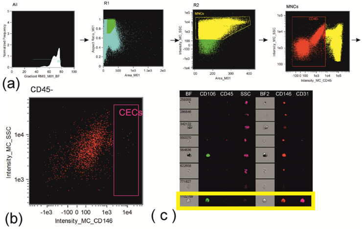
The aim was to investigate the association of monocyte heterogeneity and presence of circulating endothelial cells with the severity of coronary atherosclerosis in patients with coronary artery disease (CAD) and type 2 diabetes mellitus (T2DM). We recruited 62 patients with CAD, including 22 patients with DM2. The severity of atherosclerosis was evaluated using Gensini Score. Numbers of classical (CD14++CD16-), intermediate (CD14++CD16+), and non-classical (CD14+CD16++) monocyte subsets; circulating endothelial progenitor cells; and the presence of circulating endothelial cells were evaluated. Counts and frequencies of intermediate monocytes, but not glycaemia parameters, were associated with the severity of atherosclerosis in diabetic CAD patients (rs = 0.689; p = 0.001 and rs = 0.632; p = 0.002, respectively). Frequency of Tie2+ cells was lower in classical than in non-classical monocytes in CAD patients (p = 0.007), while in patients with association of CAD and T2DM, differences between Tie2+ monocytes subsets disappeared (p = 0.080). Circulating endothelial cells were determined in 100% of CAD+T2DM patients, and counts of CD14++CD16+ monocytes and concentration of TGF-β predicted the presence of circulating endothelial cells (sensitivity 92.3%; specificity 90.9%; AUC = 0.930). Thus, intermediate monocytes represent one of the key determinants of the appearance of circulating endothelial cells in all the patients with CAD, but are associated with the severity of atherosclerosis only in patients with association of CAD and T2DM.
Keywords: TGF-beta; atherosclerosis; circulating endothelial cells; coronary artery disease; endothelial progenitor cells; monocytes; type 2 diabetes mellitus.
Conflict of interest statement
The authors declare no conflict of interest.
Figures


Similar articles
-
Increased frequency of proangiogenic tunica intima endothelial kinase 2 (Tie2) expressing monocytes in individuals with type 2 diabetes mellitus.Cardiovasc Diabetol. 2022 May 12;21(1):72. doi: 10.1186/s12933-022-01497-6. Cardiovasc Diabetol. 2022. PMID: 35549955 Free PMC article.
-
Circulating CD14+CD16+ monocyte subsets as biomarkers of the severity of coronary artery disease in patients with stable angina pectoris.Circ J. 2012;76(10):2412-8. doi: 10.1253/circj.cj-12-0412. Epub 2012 Jun 29. Circ J. 2012. PMID: 22785372 Clinical Trial.
-
Frequency of monocyte subsets is linked to the severity of atherosclerosis in patients with ischemic heart disease: A case-control study.Biomedicine (Taipei). 2020 Jun 5;10(2):36-47. doi: 10.37796/2211-8039.1015. eCollection 2020. Biomedicine (Taipei). 2020. PMID: 33854919 Free PMC article.
-
Monocyte heterogeneity in human cardiovascular disease.Immunobiology. 2012 Dec;217(12):1273-84. doi: 10.1016/j.imbio.2012.07.001. Epub 2012 Jul 25. Immunobiology. 2012. PMID: 22898391 Review.
-
The Role of Different Monocyte Subsets in the Pathogenesis of Atherosclerosis and Acute Coronary Syndromes.Scand J Immunol. 2015 Sep;82(3):163-73. doi: 10.1111/sji.12314. Scand J Immunol. 2015. PMID: 25997925 Review.
Cited by
-
Immune cell contribution to vascular complications in diabetes.Front Endocrinol (Lausanne). 2025 May 21;16:1549945. doi: 10.3389/fendo.2025.1549945. eCollection 2025. Front Endocrinol (Lausanne). 2025. PMID: 40469434 Free PMC article. Review.
-
Immune Landscape Variation in Antineutrophil Cytoplasmic Antibody-Associated Vasculitis Circulation Before and After Plasmapheresis by Single-Cell Transcriptome.Mediators Inflamm. 2025 Apr 10;2025:5531382. doi: 10.1155/mi/5531382. eCollection 2025. Mediators Inflamm. 2025. PMID: 40256686 Free PMC article.
-
Editorial to the Special Issue "Molecular and Cellular Mechanisms of CVD: Focus on Atherosclerosis".Biomedicines. 2024 Sep 23;12(9):2148. doi: 10.3390/biomedicines12092148. Biomedicines. 2024. PMID: 39335661 Free PMC article.
References
-
- Cosentino F., Grant P.J., Aboyans V., Bailey C.J., Ceriello A., Delgado V., Federici M., Filippatos G., Grobbee D.E., Hansen T.B., et al. ESC Scientific Document Group. 2019 ESC Guidelines on Diabetes, Pre-Diabetes, and Cardiovascular Diseases Developed in Collaboration with the EASD. Eur. Heart J. 2020;41:255–323. doi: 10.1093/eurheartj/ehz486. - DOI - PubMed
Grants and funding
LinkOut - more resources
Full Text Sources
Research Materials
Miscellaneous
