. 2023 Oct 29;11(11):2924.
doi: 10.3390/biomedicines11112924.

Prolonged Door-to-Balloon Time Leads to Endothelial Glycocalyx Damage and Endothelial Dysfunction in Patients with ST-Elevation Myocardial Infarction

Affiliations

Prolonged Door-to-Balloon Time Leads to Endothelial Glycocalyx Damage and Endothelial Dysfunction in Patients with ST-Elevation Myocardial Infarction

Carl Vahldieck et al. Biomedicines. .

Abstract

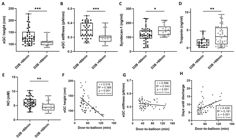
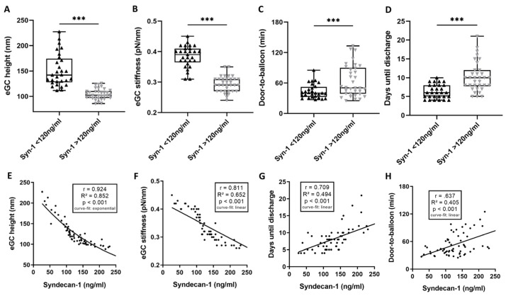
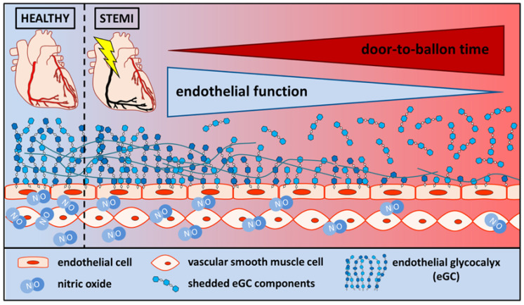
Damage to the endothelial glycocalyx (eGC) has been reported during acute ischemic events like ST-elevation myocardial infarction (STEMI). In STEMI, a door-to-balloon time (D2B) of <60 min was shown to reduce mortality and nonfatal complications. Here, we hypothesize that eGC condition is associated with D2B duration and endothelial function during STEMI. One hundred and twenty-six individuals were analyzed in this study (STEMI patients vs. age-/sex-matched healthy volunteers). After stimulating endothelial cells with patient/control sera, the eGC's nanomechanical properties (i.e., height/stiffness) were analyzed using the atomic force microscopy-based nanoindentation technique. eGC components were determined via ELISA, and measurements of nitric oxide levels (NO) were based on chemiluminescence. eGC height/stiffness (both p < 0.001), as well as NO concentration (p < 0.001), were reduced during STEMI. Notably, the D2B had a strong impact on the endothelial condition: a D2B > 60 min led to significantly higher serum concentrations of eGC components (syndecan-1: p < 0.001/heparan sulfate: p < 0.001/hyaluronic acid: p < 0.0001). A D2B > 60 min led to the pronounced loss of eGC height/stiffness (both, p < 0.001) with reduced NO concentrations (p < 0.01), activated the complement system (p < 0.001), and prolonged the hospital stay (p < 0.01). An increased D2B led to severe eGC shedding, with endothelial dysfunction in a temporal context. eGC components and pro-inflammatory mediators correlated with a prolonged D2B, indicating a time-dependent immune reaction during STEMI, with a decreased NO concentration. Thus, D2B is a crucial factor for eGC damage during STEMI. Clinical evaluation of the eGC condition might serve as an important predictor for the endothelial function of STEMI patients in the future.

Keywords: ST-elevation myocardial infarction (STEMI); door-to-balloon time; endothelial dysfunction; endothelial glycocalyx; nitric oxide.


Conflict of interest statement

The authors declare no conflict of interest. The funders had no role in the design of the study; in the collection, analyses, or interpretation of data; in the writing of the manuscript; or in the decision to publish the results.

Figures

Figure 1
Figure 1
ST-elevation myocardial infarction (STEMI) leads to endothelial glycocalyx (eGC) damage and endothelial dysfunction. Endothelial glycocalyx (eGC) height (A) and eGC stiffness (B) measured via AFM nanoindentation technique. Each dot represents one patient/healthy control (n = 126; each dot shows a mean of 50–60 cells per patient). Serum levels of syndecan-1 (C), heparan sulfate (D) and hyaluronic acid (hyaluronan) (E) were measured via ELISA. Nitric oxide (NO) products (F) were quantified via NO-Analyzer-280i. A-F: data shown as mean ± SD. Correlation of eGC height vs. eGC stiffness (G) and days until discharge from hospital (H). Correlation of eGC stiffness vs. days until discharge from hospital (I). Direct comparison of individual eGC height of STEMI patients and age- and sex-matched controls (J). Groups: CTR (control) stimulation with cell culture media + 10% serum of healthy controls; STEMI (ST-elevation myocardial infarction) stimulation with media + 10% serum of STEMI patients. p-values: ****: p < 0.0001; ***: p < 0.001. Rho (r), p-values (p), coefficient of determination (R2), and curve-fit model shown for correlations.
Figure 2
Figure 2
Door to balloon (D2B) time > 60 min leads to endothelial glycocalyx (eGC) damage and prolonged hospitalization. STEMI (ST-elevation myocardial infarction) patients were divided into cohorts: door-to-balloon time (D2B) ≤ 60 min and D2B > 60 min. Endothelial glycocalyx (eGC) height (A) and eGC stiffness (B) were measured via AFM nanoindentation technique. Each dot represents one patient/healthy control (n = 63; each dot shows a mean of 50–60 cells per patient). Quantification of syndecan-1 (C), troponin-t (D) and nitric oxide (E) serum levels. Correlation of D2B vs. eGC height (F), eGC stiffness (G) and days until discharge from hospital (H). p-values: *: p < 0.05; **: p < 0.01; ***: p < 0.001. Rho (r), p-values (p), coefficient of determination (R2) and curve-fit model shown for correlations.
Figure 3
Figure 3
High syndecan-1 is associated with unfavorable outcomes for endothelial glycocalyx (eGC) and patients. STEMI (ST-elevation myocardial infarction) patients were divided into cohorts: (a) syndecan-1 levels ≤ 120 ng/mL and (b) syndecan-1 levels > 120 ng/mL. Endothelial glycocalyx (eGC) height (A) and eGC stiffness (B) were measured via AFM nanoindentation technique. Each dot represents one patient/healthy control (n = 63; each dot shows a mean of 50–60 cells per patient). Group differences between (a) vs. (b) concerning door-to-balloon (D2B) time (C) and days until discharge from hospital (D). Correlation of syndecan-1 levels vs. eGC height (E), eGC stiffness (F), days until discharge from hospital (G), and door-to-balloon time (H). p-values: ***: p < 0.001. Rho (r), p-values (p), coefficient of determination (R2) and curve-fit model shown for correlations.
Figure 4
Figure 4
In healthy conditions, the vascular endothelial function is preserved, with adequate secretion of the endothelium-derived relaxing factor nitric oxide. Following ST-elevation myocardial infarction, endothelial function is impaired, with limited nitric oxide production. Endothelial function decreases as the door-to-balloon time increases. In addition, the eGC components are shed and become measurable in the blood stream.

Similar articles

Cited by

References

    1. Cosgun Z.C., Fels B., Kusche-Vihrog K. Nanomechanics of the Endothelial Glycocalyx: From Structure to Function. Am. J. Pathol. 2020;190:732–741. doi: 10.1016/j.ajpath.2019.07.021. - DOI - PubMed
    1. Abassi Z., Armaly Z., Heyman S.N. Glycocalyx Degradation in Ischemia-Reperfusion Injury. Am. J. Pathol. 2020;190:752–767. doi: 10.1016/j.ajpath.2019.08.019. - DOI - PubMed
    1. Ferdinandy P., Andreadou I., Baxter G.F., Bøtker H.E., Davidson S.M., Dobrev D., Gersh B.J., Heusch G., Lecour S., Ruiz-Meana M., et al. Interaction of Cardiovascular Nonmodifiable Risk Factors, Comorbidities and Comedications With Ischemia/Reperfusion Injury and Cardioprotection by Pharmacological Treatments and Ischemic Conditioning. Pharmacol. Rev. 2023;75:159–216. doi: 10.1124/pharmrev.121.000348. - DOI - PMC - PubMed
    1. Lee S.-J., Lee C.-K., Kang S., Park I., Kim Y.H., Kim S.K., Hong S.P., Bae H., He Y., Kubota Y., et al. Angiopoietin-2 exacerbates cardiac hypoxia and inflammation after myocardial infarction. J. Clin. Investig. 2018;128:5018–5033. doi: 10.1172/JCI99659. - DOI - PMC - PubMed
    1. Rehm M., Bruegger D., Christ F., Conzen P., Thiel M., Jacob M., Chappell D., Stoeckelhuber M., Welsch U., Reichart B., et al. Shedding of the endothelial glycocalyx in patients undergoing major vascular surgery with global and regional ischemia. Circulation. 2007;116:1896–1906. doi: 10.1161/CIRCULATIONAHA.106.684852. - DOI - PubMed

LinkOut - more resources