. 2023 Nov 1;11(11):2958.
doi: 10.3390/biomedicines11112958.

Cell Line-Based Human Bladder Organoids with Bladder-like Self-Organization-A New Standardized Approach in Bladder Cancer Research

Affiliations

Cell Line-Based Human Bladder Organoids with Bladder-like Self-Organization-A New Standardized Approach in Bladder Cancer Research

Mandy Berndt-Paetz et al. Biomedicines. .

Abstract

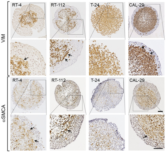
Three-dimensional tumor models have gained significant importance in bladder cancer (BCa) research. Organoids consisting of different cell types better mimic solid tumors in terms of 3D architecture, proliferation, cell-cell interaction and drug responses. We developed four organoids from human BCa cell lines with fibroblasts and smooth muscle cells of the bladder, aiming to find models for BCa research. The organoids were characterized in terms of cytokeratins, vimentin, α-actin and KI67 by immunoreactivity. Further, we studied ligand-dependent activation of the Wnt/β-catenin pathway and investigated the responses to anti-tumor therapies. The organoids mimicked the structure of an inverse bladder wall, with outside urothelial cells and a core of supportive cells. The cytokeratin staining patterns and proliferation rate were in conjunction with the origins of the BCa cells. RT-112 even showed stratification of the epithelium. Treatment with Wnt10B led to increased β-catenin (active) levels in high-grade organoids, but not in low-grade BCa cells. Doxorubicin treatment resulted in clearly reduced viability (10-30% vs. untreated). In contrast, the effectivity of radiotherapy depended on the proliferation status of BCa cells. In conclusion, cell-line-based organoids can form bladder-like structures and reproduce in vivo features such as urothelial differentiation and stratification. Thus, they can be useful tools for functional studies in BCa and anti-cancer drug development.

Keywords: Wnt/β-catenin activation; bladder cancer cell lines; drug response; organoids; self-organization; stratification.


Conflict of interest statement

The authors declare no conflict of interest.

Figures

Figure 1
Figure 1
Spontaneous formation of heterogeneous, compact BCa organoids. BCa cells (RT-4, RT-112, T-24, CAL-29) were co-cultured with hBF and hBSMC in an ULA plate. (a) Development of organoids over a period of up to 7 days. Live cell observation by transmitted light using a LSM800, exemplarily shown for RT-112 organoids. Spheroids reached sizes of up to 1 mm in diameter after 1 week. (b) Morphology of human BCa Orgs by HE stains after 4 days in culture. The morphology of BCa Orgs varied depending on the BCa cell line utilized (scale bar: 100 µm). (c) Organoid sizes ranged from 650 to 1000 µm in diameter, ** p < 0.01, *** p < 0.001, **** p < 0.0001. One-way ANOVA, mean + SD, n = 8. (d) Correlation analysis. Diameter of Orgs negatively correlated with the grading of BCa cell lines, Pearson r2 = 0.7875, p < 0.0001.
Figure 2
Figure 2
Bladder-like self-organization of BCa organoids. Distribution of BCa cells in urinary bladder organoids by analysis of panCK immunoreactivity (IR). (a) Representative images of BCa Orgs immunostained for panCK (brown); cell nuclei (blue). BCa cells showed different levels of cell separation, with the formation of an urothelium-like peripheral cell layer and tumor cell nests inside the organoid core. Scale bar: 100 µm. (b) Thickness of the BCa cell layer, normalized to the organoid size. The thickness of the RT-4 cell layer (RT-4: papillary, low-grade) was significantly lower than in BCa Orgs consisting of high-grade tumor cells, * p < 0.05, **** p < 0.0001. One-way ANOVA, mean + SD, n = 8. (c) There was a significant correlation of BCa layer thickness with the grading of the BCa cells, Pearson r2 = 0.1879, p < 0.0132. (d) Quantification of panCK-positive BCa cell nests in the inner core; stained area (%) was determined. RT-4 Orgs showed significantly higher amounts of panCK-positive cell nests within the inner cores than high-grade BCa Orgs, * p < 0.05, ** p < 0.01, *** p < 0.001, **** p < 0.0001. One-way ANOVA, mean + SD, n = 8. (e) The panCK-IR in the inner core was negatively correlated with the grading of the cell line which was used. Pearson r2 = 0.3686, p < 0.0002.
Figure 3
Figure 3
Bladder-like self-organization of BCa organoids. Distribution of hBF and hBSMC in urinary bladder organoids by analysis of VIM and αSMCA immunoreactivity. Representative images of BCa Orgs immunostained for VIM and αSMCA (brown), cell nuclei (blue). VIM- and αSMCA-IR were complementary to panCK staining and confirmed the presence of hBF and hBSMC in the organoid core. Arrows indicate the formation of VIM- and αSMCA-positive elongated cells underlying the urothelium. Scale bar: 100 µm.
Figure 4
Figure 4
Urothelial differentiation of BCa cells in organoids. Analysis of different CKs to detect poorly (CK7, basal), moderately (CK13, intermediate) or well-differentiated (CK20, superficial, umbrella cells) epithelial cells in BCa Orgs. (ac) Quantification of CK7 (a), CK13 (b) and CK20 (c) immunoreactivity in the BCa cell layer: * p < 0.05, *** p < 0.001, **** p < 0.0001. One-way ANOVA, mean + SD, n = 8. (d,e) Stratification of the urothelial-like peripheral cell layer. Distribution of CK7-, CK13- and CK20-positive cells in BCa organoids. (d) Immunohistochemical staining of CK7, CK13 and CK20 in the peripheral cell layer. Arrows refer to the labeling of superficial cells and the staining of intermediate and basal cells, respectively. Scale bar: 100 µm. (e) Schematic overview of CK staining patterns in BCa Orgs.
Figure 5
Figure 5
Proliferation status. Analysis of KI67 immunoreactivity in BCa cells. (a) Representative images of BCa Orgs immunostained for KI67 (brown), cell nuclei (blue). Scale bar: 100 µm. (b) Quantification of KI67-positive nuclei indicated a vigorous proliferation of tumor cells in the RT-112, T-24 and CAL-29 organoids, whereas low-grade RT-4 Orgs showed a proliferation index of <10%; **** p < 0.0001. One-way ANOVA, mean + SD, n = 8. (c) Proliferation indices were in conjunction with appropriate population doubling times of BCa cells in monolayer cultures.
Figure 6
Figure 6
Activation of the Wnt/β-catenin pathway in human BCa organoids by Wnt10B. (a,b) Translocation of active β-catenin into the nucleus in 2D cultured RT-112 cells. (a) Immunofluorescence staining of β-catenin (non-phospho, Ser33/37/Thr41). Membrane association was observed in control cells (vehicle control); Wnt10B-treated cells showed nuclear localization of β-catenin; arrows indicate nuclear β-catenin labeling. β-catenin (red), nuclei (blue). Scale bar: 75 µm. (b) Quantification of active β-catenin (FI; fluorescence intensity) in DAPI-positive nuclei revealed significantly increased nuclear β-catenin levels after 30 min of Wnt10B vs. controls. * Significance vs. vehicle-treated control (p ≤ 0.05, Mann–Whitney test), mean + SD. (cg) Increase in active β-catenin in the tumor cell layers of 3D cultured BCa cells. (c) Immunofluorescence staining of β-catenin (non-phospho, Ser33/37/Thr41) in paraffin-embedded BCa organoids, examplarily shown for RT-112 organoids. Localization of β-catenin was observed mainly in the submembranous cytoskeleton, but also in the nuclei of RT-112 tumor cells. β-catenin (orange), panCK (green), nuclei (blue). Scale bar: 75 µm. (d) Quantification of active β-catenin (FI; fluorescence intensity) in panCK-positive ROIs revealed significantly increased β-catenin levels after 5, 10 and 30 min of Wnt10B (vs. controls) in RT-112; (e) nuclear β-catenin levels were increased after 5, 10, 30 and 120 min. * Significance vs. vehicle-treated control (p ≤ 0.05, Mann–Whitney test), mean + SD. (f,g) Increased β-catenin (FI; fluorescence intensity; normalized to PBS vehicle control) was also detected in the BCa layer of T-24 and CAL-29 organoids (especially in panCK-positive ROIs), while RT-4 cells showed no response to Wnt10B.
Figure 7
Figure 7
Response to anticancer treatment. Metabolic activity was determined using the CellTiter-Glo® 3D Cell Viability Assay in BCa Orgs and the CellTiter-Blue® Cell Viability Assay in BCa monolayer cultures 72 h after treatment. (a) Drug response curves for BCa Orgs and monolayers treated with doxorubicin. IC50 values were determined by nonlinear regression of the dose response (inhibitor vs. normalized response). (b) Dose-dependent cytotoxic effects of radiotherapy on RT-4 and T-24 Orgs compared to monolayer cultures. * Significance vs. untreated control (UTC), p < 0.05, one-way ANOVA, mean + SD, n = 3. (c) Cell-line-dependent effects of radiotherapy in BCa Orgs. * Significance vs. untreated control (UTC), p < 0.05, Mann–Whitney test, mean + SD, n = 3.

References

    1. Babjuk M., Burger M., Capoun O., Cohen D., Compérat E.M., Dominguez Escrig J.L., Gontero P., Liedberg F., Masson-Lecomte A., Mostafid A.H., et al. European Association of Urology Guidelines on Non-muscle-invasive Bladder Cancer (Ta, T1, and Carcinoma in Situ) Eur. Urol. 2022;81:75–94. doi: 10.1016/j.eururo.2021.08.010. - DOI - PubMed
    1. Witjes J.A., Bruins H.M., Cathomas R., Compérat E.M., Cowan N.C., Gakis G., Hernández V., Linares Espinós E., Lorch A., Neuzillet Y., et al. European Association of Urology Guidelines on Muscle-invasive and Metastatic Bladder Cancer: Summary of the 2020 Guidelines. Eur. Urol. 2021;79:82–104. doi: 10.1016/j.eururo.2020.03.055. - DOI - PubMed
    1. Drost J., Clevers H. Organoids in cancer research. Nat. Rev. Cancer. 2018;18:407–418. doi: 10.1038/s41568-018-0007-6. - DOI - PubMed
    1. Schutgens F., Clevers H. Human Organoids: Tools for Understanding Biology and Treating Diseases. Annu. Rev. Pathol. 2020;15:211–234. doi: 10.1146/annurev-pathmechdis-012419-032611. - DOI - PubMed
    1. Doctor A., Seifert V., Ullrich M., Hauser S., Pietzsch J. Three-Dimensional Cell Culture Systems in Radiopharmaceutical Cancer Research. Cancers. 2020;12:2765. doi: 10.3390/cancers12102765. - DOI - PMC - PubMed

LinkOut - more resources