Review
. 2023 Nov 6;11(11):2985.
doi: 10.3390/biomedicines11112985.

The Prokineticin System in Inflammatory Bowel Diseases: A Clinical and Preclinical Overview

Affiliations
Review

The Prokineticin System in Inflammatory Bowel Diseases: A Clinical and Preclinical Overview

Giada Amodeo et al. Biomedicines. .

Abstract

Inflammatory bowel disease (IBD) includes Crohn's disease (CD) and ulcerative colitis (UC), which are characterized by chronic inflammation of the gastrointestinal (GI) tract. IBDs clinical manifestations are heterogeneous and characterized by a chronic relapsing-remitting course. Typical gastrointestinal signs and symptoms include diarrhea, GI bleeding, weight loss, and abdominal pain. Moreover, the presence of pain often manifests in the remitting disease phase. As a result, patients report a further reduction in life quality. Despite the scientific advances implemented in the last two decades and the therapies aimed at inducing or maintaining IBDs in a remissive condition, to date, their pathophysiology still remains unknown. In this scenario, the importance of identifying a common and effective therapeutic target for both digestive symptoms and pain remains a priority. Recent clinical and preclinical studies have reported the prokineticin system (PKS) as an emerging therapeutic target for IBDs. PKS alterations are likely to play a role in IBDs at multiple levels, such as in intestinal motility, local inflammation, ulceration processes, localized abdominal and visceral pain, as well as central nervous system sensitization, leading to the development of chronic and widespread pain. This narrative review summarized the evidence about the involvement of the PKS in IBD and discussed its potential as a druggable target.

Keywords: Crohn’s disease (CD); chronic pain; inflammatory bowel diseases (IBDs); prokineticin system; ulcerative colitis (UC).


Conflict of interest statement

The authors declare no conflict of interest.

Figures

Figure 5
Figure 5
PKRs and PK2 expression in the gastrointestinal tract. (A) Immunohistochemical analysis of PKR1 expression in the (a) myenteric and (b) submucosal plexus of the human gastrointestinal tract (in blue, DAPI; in red, PKR1). (B) PKR1 immunoreactivity in rat ileum. In detail (see white arrows), PKR1 was detected: (a) in the majority of submucosal neurons, (b) in some neurons and fibers in the myenteric plexus, (c) in the epithelial cells at the base of the crypts primarily, and (d) in the cell basolateral regions. (C) Immunohistochemical evaluations in the rat colon of (a) PKR2 and (b) PK2. Both PKR2- and PK2-positive expression (yellow stains, see red arrowheads) was mainly detected in the mucosal epithelium, proximal glands of the mucosal epithelium, interstitium, and colon muscle layer. These images were modified and republished, in accordance with specific editor copyright, by (A) Watson et al. [50]; (B) Wade et al. [51]; and (C) Zhou et al. [52].
Figure 7
Figure 7
PK1 and PKR1 in enteric neural crest cells. (A) Representative immunohistochemical images of PK1 ((a), in red) or PKR1 ((b), in red) and Tuj/1 ((a,b), in green) localization in mouse embryonic gut at 15.5 (a) and 13.5 (b) days, respectively. The enteric neural crest cells (NCCs), marked with Tuj/1, are localized in the gut mesenchyme and colocalized (arrowhead) with PKR1. PK1 is localized at the mucosa and mesenchyme. (B) NCCs cultured with vehicle or PK1 for 48 h. NCCs supplemented with PK1 showed a marked neuronal ((ad), in green; (e,f), in red) and glial expression ((gj), in red) in comparison with the vehicle. Neuronal markers: Tuj1 (class III beta-tubulin), TH (tyrosine hydroxylase), and NFL (neurofilament light); glial markers: GFAP (glial fibrillary acidic protein) and S100. These images have been edited and republished, in accordance with the specific editor copyright, by Ngan et al. [54].
Figure 8
Figure 8
PK1’s effect on endothelial cells in angiogenesis. (A) Capillary tube formation (stained tube-like structures in purple) was evaluated, using a specific angiogenesis kit (Kurabo Co), (a) in a standard cultured medium condition, (b) supplemented with PK1 (10 ng/mL), or (c) supplemented with both PK1 (10 ng/mL) and anti-PK1 mAb. (d) For each condition, capillary tube length was analyzed/quantified using the MacSCOPE program. The PK1-supplemented culture markedly increases the capillary tubes’ length, thereby stimulating angiogenesis, whereas, if the effect of PK1 is blocked, angiogenesis is also arrested. (B) Adrenal cortex-derived endothelial cells were cultured in (a) serum-free medium with/without (b) VEGF (0.13 nM) or (c) PK1 (50 nM). (d) Quantification of apoptotic cells using the fluorescence-activated cell sorting assay. After 24 h, starved endothelial cells showed 30% apoptosis, while VEGF or PK1 supplementation was able to rescue cells from this doom, indicating that PK1, similarly to VEGF, promotes endothelial cell survival. These images/graphs have been edited and republished, in accordance with the specific editor copyright, by Nakazawa et al. [65] and (B) Lin et al. [63].
Figure 1
Figure 1
IBDs: Ulcerative colitis vs. Crohn’s disease. (A) Colonic endoscopy. (B) Illustrative representation of the intestine; red indicates the possible inflammation site(s). (C) Illustrative representation of the colon longitudinal section and main pathophysiological features. (a) Healthy people, (b) patients with ulcerative colitis, and (c) patients with Crohn’s disease. Drawn by authors using BioRENDER online software.
Figure 2
Figure 2
PKS’s structure and signaling. (A) In mice, the PK1 gene maps to chromosome 3 and the PK2 gene maps to chromosome 6 (in humans, chromosomes 1p13.1 and 3p21.1, respectively). In both mouse and human, the PK1 gene has three exons encoding the mature protein (mouse, 105 amino acids (aa); human, 86 aa), while the PK2 gene has four exons, of which three exons encode the classical mature protein PK2 (mouse, 108 aa; human, 81 aa). Furthermore, in mice, it has been recently identified that via alternative splicing the PK2 gene can also originate other PK2 isoforms (PK2L or PK2beta, 129 aa, is encoded by all four exons; truncated PK2, 74 aa, is encoded by exons 1 and 2 and part of intron 2; and PK2C, 63 aa, is encoded by exons 1 and 4). (B) Both PKs can bind to their G protein-coupled receptors, PKR1 and PKR2, activating Gi (starting the MAPK/Akt cascade), Gs (promoting cAMP accumulation), or Gq (inducing calcium mobilization) and their downstream pathways. Drawn by the authors using BioRENDER online software.
Figure 3
Figure 3
Expression patterns of the prokineticin system. Illustrative representation of the expression of the (A) PKs [22] and (B) PKRs [27] in human tissues.
Figure 4
Figure 4
Neuronal connectome between the gut and the central nervous system (CNS). (A) A schematic representation of the CNS–gut connection. The intestine is connected to the CNS, both through interactions with the enteric nervous system (ENS) and independently through interactions with diverse gastrointestinal cells. Neural pathways that connect the gut to the CNS include the vagus nerve, which consists of the vagal afferent and efferent nerves (in blue, whose neurons reside in the nodose ganglion (NG) and in the brainstem, respectively); the spinal nociceptive nerves (whose neurons reside in the DRG, which are sensory neurons that innervate the viscera and the spinal cord neurons); the postsynaptic sympathetic nerves (whose neurons reside in celiac ganglia (CG) and in superior (SMG) and inferior mesenteric ganglia (IMG)); and the spinal sacral nerve, which directly connects the colon to spinal neurons (sympathetic and parasympathetic components). (B) A detailed transversal section of the colon. The gut has its own nervous system (ENS), whose neurons are located in two plexuses, namely the myenteric plexus and submucosal plexus. Neurons of both plexuses innervate different tissue regions, performing several functions; neural pathways of the submucosal plexus mainly regulate fluid exchange across the intestinal mucosa, while those of the myenteric plexus coordinate intestine contractile activity. Additionally, in these intestinal layers, several enteric glial cells are present. (C) Illustrative magnification of the intestinal layers, showing neurons and glial cell distribution. Drawn by the authors using BioRENDER online software.
Figure 6
Figure 6
The PKS and nNOS. (A) Immunofluorescence investigations show that (ac) PKR1 co-localizes with neuronal nitric oxide synthase (nNOS) in the mouse musculo-myenteric plexus at the colonic level, and that (B) NCCs cultured for 1 week in the presence of (b) PK1 display upregulated nNOS expression compared to those cultured with (a) vehicle alone. These images have been edited and republished, in accordance with the specific editor copyright, by (A) Hoogerwerf [53] and (B) Ngan et al. [54].
Figure 9
Figure 9
PK1 expressions in colorectal cancer patients and their prognosis. (A) Plasma PK1 concentration was evaluated in 130 CRC patients; the graph shows an increasing trend of PK1 with the cancer stage progression. (B) In CRC patients’ stage III, PK1 levels were evaluated in both plasma and primary tumor lesions (every 10 months, 5-year follow-up). The results were then correlated to the CRC patient mortality rate. From these evaluations, it emerged that in patients who did not express PK1 either in the plasma or at the level of the primary lesion, the mortality was 15.5%, in patients who expressed plasma PK1 the mortality rate was 25.7% (increase of 10.2%), while patients expressing PK1 in both plasma and primary tumors had a mortality of 40.7% (25.2% and 15% increase, respectively). These data have been edited and republished, in accordance with the specific editor copyright, by Tagai et al. [80].
Figure 10
Figure 10
PKR1 expression in rat DRGs. Immunostaining in naïve rat DRG shows that PKR1 is not expressed in large-diameter neurons but co-localizes with TRPV1 in the cell bodies of small/medium-diameter nociceptive sensory neurons. These images have been edited and republished, in accordance with the specific editor copyright, by Watson et al. [50].
Figure 11
Figure 11
PK2 increase and visceral pain. During intestinal inflammation, PK2 is released by immune cells. Its increase at the inflammatory site could modulate/drive visceral pain activating PKRs, which are largely expressed in the gut and enteric nervous system. PK2 can act by: (i) attracting monocytes/macrophages to the site of inflammation (chemotactic action); (ii) stimulating the release of pro-inflammatory cytokines by immune cells (helping to create a vicious cycle for its production); and (iii) by sensitizing TRPV1 localized on the afferents of sensory neurons that innervate the gastrointestinal tract. Drawn by the authors using BioRENDER online software.
Figure 12
Figure 12
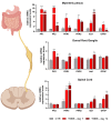
PKS characterization in nervous tissue of TNBS mice. PKS and glial marker expression levels in a TNBS murine model. The evaluations were performed in important stations involved in IBD pain transmission, i.e., the myenteric plexus, dorsal root ganglia, and spinal cord, at two different time points (day 7 and day 14 post-TNBS). *** p < 0.001, ** p < 0.01, and * p < 0.05 vs. respective CTR; $$$ p < 0.001, $$ p < 0.01, and $ < 0.05 vs. TNBS d7. These images have been edited and republished, in accordance with the specific editor copyright, by Amodeo et al. [39].

Similar articles

Cited by

References

    1. Zhang Y.-Z., Li Y.-Y. Inflammatory bowel disease: Pathogenesis. World J. Gastroenterol. 2014;20:91–99. doi: 10.3748/wjg.v20.i1.91. - DOI - PMC - PubMed
    1. Kaplan G.G. The global burden of IBD: From 2015 to 2025. Nat. Rev. Gastroenterol. Hepatol. 2015;12:720–727. doi: 10.1038/nrgastro.2015.150. - DOI - PubMed
    1. Jairath V., Feagan B.G. Global burden of inflammatory bowel disease. Lancet Gastroenterol. Hepatol. 2020;5:2–3. doi: 10.1016/S2468-1253(19)30358-9. - DOI - PubMed
    1. Lemmens B., De Hertogh G., Sagaert X. Inflammatory Bowel Diseases. Pathobiol. Hum. Dis. 2014;2014:1297–1304. doi: 10.1016/B978-0-12-386456-7.03806-5. - DOI
    1. Liverani E., Scaioli E., Digby R.J., Bellanova M., Belluzzi A. How to predict clinical relapse in inflammatory bowel disease patients. World J. Gastroenterol. 2016;22:1017–1033. doi: 10.3748/wjg.v22.i3.1017. - DOI - PMC - PubMed