. 2023 Nov 10;11(11):3017.
doi: 10.3390/biomedicines11113017.

The Ultraviolet Irradiation of Keratinocytes Induces Ectopic Expression of LINE-1 Retrotransposon Machinery and Leads to Cellular Senescence

Affiliations

The Ultraviolet Irradiation of Keratinocytes Induces Ectopic Expression of LINE-1 Retrotransposon Machinery and Leads to Cellular Senescence

Fadi Touma et al. Biomedicines. .

Abstract

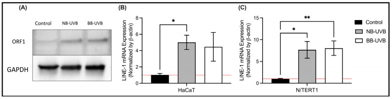
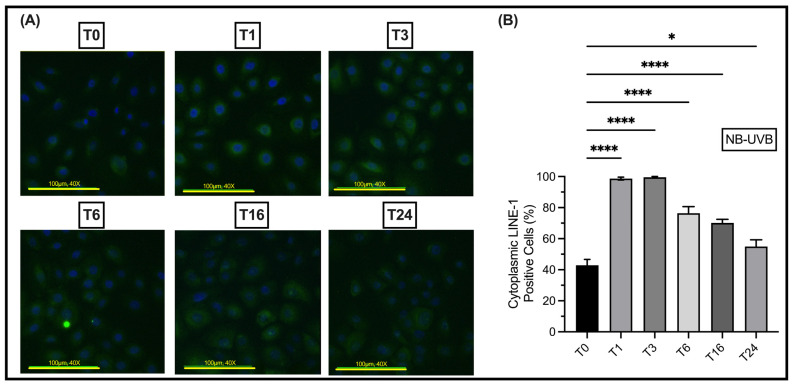
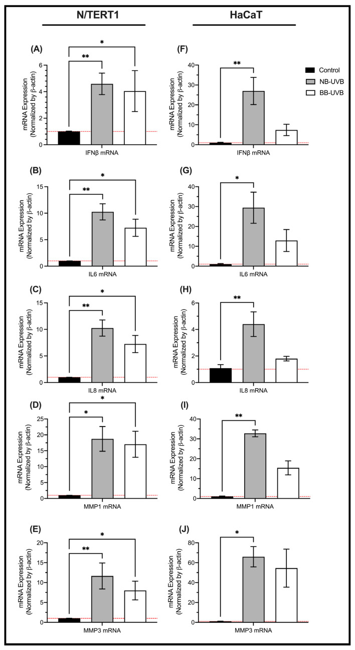
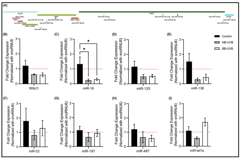
Retrotransposons have played an important role in evolution through their transposable activity. The largest and the only currently active human group of mobile DNAs are the LINE-1 retrotransposons. The ectopic expression of LINE-1 has been correlated with genomic instability. Narrow-band ultraviolet B (NB-UVB) and broad-band ultraviolet B (BB-UVB) phototherapy is commonly used for the treatment of dermatological diseases. UVB exposure is carcinogenic and can lead, in keratinocytes, to genomic instability. We hypothesize that LINE-1 reactivation occurs at a high rate in response to UVB exposure on the skin, which significantly contributes to genomic instability and DNA damage leading to cellular senescence and photoaging. Immortalized N/TERT1 and HaCaT human keratinocyte cell lines were irradiated in vitro with either NB-UVB or BB-UVB. Using immunofluorescence and Western blotting, we confirmed UVB-induced protein expression of LINE-1. Using RT-qPCR, we measured the mRNA expression of LINE-1 and senescence markers that were upregulated after several NB-UVB exposures. Selected miRNAs that are known to bind LINE-1 mRNA were measured using RT-qPCR, and the expression of miR-16 was downregulated with UVB exposure. Our findings demonstrate that UVB irradiation induces LINE-1 reactivation and DNA damage in normal keratinocytes along with the associated upregulation of cellular senescence markers and change in miR-16 expression.

Keywords: BB-UVB; HaCaT; N/TERT1; NB-UVB; cellular senescence; genomic instability; keratinocytes; long interspersed nucleotide element-1 (LINE-1); miR-16; microRNAs; phototherapy.


Conflict of interest statement

The authors declare no conflict of interest. The funder had no role in the design of the study; in the collection, analyses, or interpretation of data; in the writing of the manuscript; or in the decision to publish the results.

Figures

Figure A1
Figure A1
UV irradiation decreases cell proliferation and increases cell diameter. (A) The proliferation curve of N/TERT1 cells at 1, 2, 3, and 4 days following 6 UV irradiations with either NB-UVB or BB-UVB as compared to an unirradiated control sample. (B) Cell diameter of N/TERT1 or cells after 6 UV irradiations (50–70 cells/condition). The photos were taken on an Etaluma Lumascope LS720 microscope with a 60X objective (Meiji MA969) (scale bar 10 μm). Significance for N/TERT1 samples was calculated by a two-way ANOVA test and corrected for multiple comparisons using Dunnett’s test (both the day of measurement and the UV radiation were significant as compared with Day 1 and the control, respectively, p < 0.05).
Figure 1
Figure 1
UV irradiation induces DNA damage. (A) The expression of γH2AX (green) in N/TERT1 keratinocytes as shown by immunofluorescence staining following 24 h of UV irradiation with either NB-UVB or BB-UVB as compared to unirradiated control samples (n = 3 with 500 cells/condition). The three patterns of γH2AX staining correspond to the number of double-stranded DNA breaks, i.e., type 1 expression <10 nuclear foci (low-level DNA damage), type 2 expression >10 nuclear foci (high-level DNA damage), and type 3 pan-nuclear expression (pre-apoptotic state). The photos were taken on an Etaluma Lumascope LS720 microscope with a 60X objective (Meiji MA969) (scale bar 10 μm). Significance was calculated using a mixed-effects model to analyze the data, and for multiple comparisons correction, Dunnett’s test was applied. (B) Nuclear size counts of NTERT cells after 6 UV irradiations (n = 3 with 50–69 cells/condition). Significance was calculated using the one-way ANOVA test and was corrected for multiple comparisons using Dunnett’s test. (**** p value < 0.0001, ** p value < 0.0021, * p value < 0.05).
Figure 2
Figure 2
UV irradiation decreases cell proliferation and increases cell diameter. (A) The proliferation curve of HaCaT (n = 3) cells at 1, 2, 3, and 4 days following 6 UV irradiations with either NB-UVB or BB-UVB as compared to unirradiated control samples. (B) Cell diameter of HaCaT (n = 3) cells after 6 UV irradiations (50–70 cells/condition). The photos were taken on an Etaluma Lumascope LS720 microscope with a 60X objective (Meiji MA969) (scale bar 10 μm). Significance was calculated using a mixed-effects model to analyze the data, and for multiple comparisons correction, Tukey’s test was performed. (**** p value < 0.0001, ** p value < 0.0021, * p value < 0.05).
Figure 3
Figure 3
LINE-1 protein expression on HaCaT and N/TERT1 cells after 6 UV exposures with immunofluorescence. The expression of ORF1 proteins of LINE1 elements (red) as counterstained by DAPI (blue) in HaCaT (A,B) and N/TERT1 (C,D) keratinocytes as shown by immunofluorescence staining after 24 h of 6 UV irradiations with either NB-UVB or BB-UVB as compared to unirradiated control samples. The photos were taken on an Etaluma Lumascope LS720 microscope with a 60X objective (Meiji MA969) (scale bar 20 μm). Significance was calculated by a one-way ANOVA test and was corrected for multiple comparisons using Dunnett’s test. (*** p value < 0.0002, ** p value < 0.0021).
Figure 4
Figure 4
LINE-1 protein and mRNA expression in HaCaT and N/TERT1 cells after 6 UV exposures. (A) Measurement of ORF1 protein of LINE-1 elements using Western blotting in N/TERT1 cells. The mRNA expression of ORF2 protein of LINE-1 elements in N/TERT1 cells (n = 6) (B) and HaCaT cells (n = 3) (C) as normalized by the mRNA expression of GAPDH. Kruskal–Wallis nonparametric test was performed, and the statistical significance was corrected for multiple comparisons using Dunn’s test (** p value < 0.0021, * p value < 0.05).
Figure 5
Figure 5
Time course of LINE-1 protein expression in N/TERT1 cells following 6 UV exposures with NB-UVB. (A) Immunofluorescence visualization of the expression of ORF1 protein (green) counterstained with DAPI (blue) in N/TERT1 cells (n = 5) after 6 irradiations with NB-UVB at different time points (0, 1, 3, 6, 16, and 24 h) (B) Quantification of LINE-1 expression at different time points compared with the expression immediately after NB-UVB exposure. The photos were taken on an Etaluma Lumascope LS720 microscope with a 60X objective (Meiji MA969) (scale bar 20 μm). Significance was calculated by a one-way ANOVA test corrected for multiple comparisons using Dunnett’s test. (**** p value < 0.0001, * p value < 0.05).
Figure 6
Figure 6
The mRNA expression of senescence markers following multiple UV exposures. The mRNA expression of senescence markers in N/TERT1 cells (n = 4) (AE) and HaCaT cells (n = 3) (FJ) following 6 UV irradiations. Significance was calculated by the Kruskal–Wallis nonparametric test using the uncorrected Dunn’s test (** p value < 0.0021, * p value < 0.05).
Figure 7
Figure 7
The expression of regulatory RNAs of LINE-1 reactivation. (A) LINE-1 mRNA sequence and its microRNAs binding sites. The microRNAs selected are those from the cross-search between microRNAs dysregulated upon UV irradiation and the ones that are able to target LINE-1 mRNA. (BI) The expression of selected miRNAs in N/TERT1 cells following 6 repeated UV exposures (n = 4). Significance was calculated by one-way ANOVA using Fisher’s LSD test (* p value < 0.05).

Similar articles

Cited by

References

    1. Solano F. Photoprotection and Skin Pigmentation: Melanin-Related Molecules and Some Other New Agents Obtained from Natural Sources. Molecules. 2020;25:1537. doi: 10.3390/molecules25071537. - DOI - PMC - PubMed
    1. De Gruijl F.R. Methods in Enzymology. Volume 319. Elsevier; Amsterdam, The Netherlands: 2000. [33] Photocarcinogenesis: UVA vs. UVB; pp. 359–366. - PubMed
    1. Myers E., Kheradmand S., Miller R. An Update on Narrowband Ultraviolet B Therapy for the Treatment of Skin Diseases. Cureus. 2021;13:e19182. doi: 10.7759/cureus.19182. - DOI - PMC - PubMed
    1. Enninga I.C., Groenendijk R.T., Filon A.R., van Zeeland A.A., Simons J.W. The Wavelength Dependence of u.v.-Induced Pyrimidine Dimer Formation, Cell Killing and Mutation Induction in Human Diploid Skin Fibroblasts. Carcinogenesis. 1986;7:1829–1836. doi: 10.1093/carcin/7.11.1829. - DOI - PubMed
    1. Kielbassa C. Wavelength Dependence of Oxidative DNA Damage Induced by UV and Visible Light. Carcinogenesis. 1997;18:811–816. doi: 10.1093/carcin/18.4.811. - DOI - PubMed

Grants and funding

LinkOut - more resources