. 2023 Nov 14;11(11):3050.
doi: 10.3390/biomedicines11113050.

Transient Expression in HEK-293 Cells in Suspension Culture as a Rapid and Powerful Tool: SARS-CoV-2 N and Chimeric SARS-CoV-2N-CD154 Proteins as a Case Study

Affiliations

Transient Expression in HEK-293 Cells in Suspension Culture as a Rapid and Powerful Tool: SARS-CoV-2 N and Chimeric SARS-CoV-2N-CD154 Proteins as a Case Study

Thailin Lao et al. Biomedicines. .

Abstract

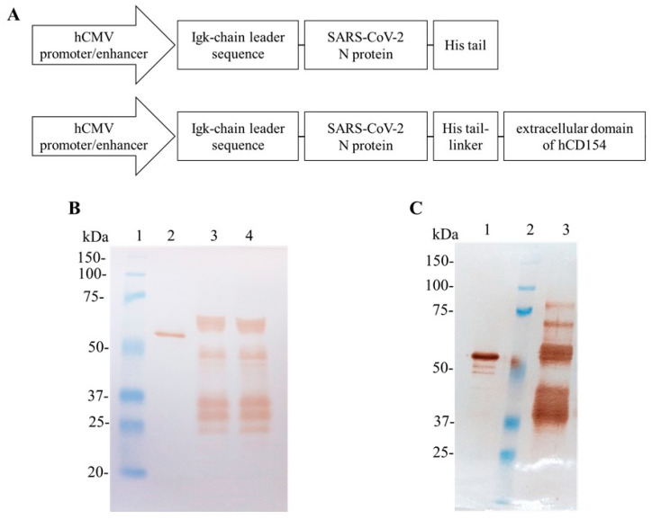
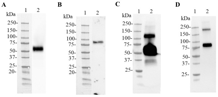
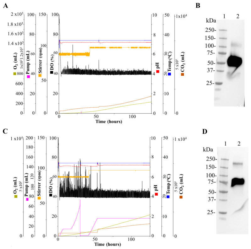
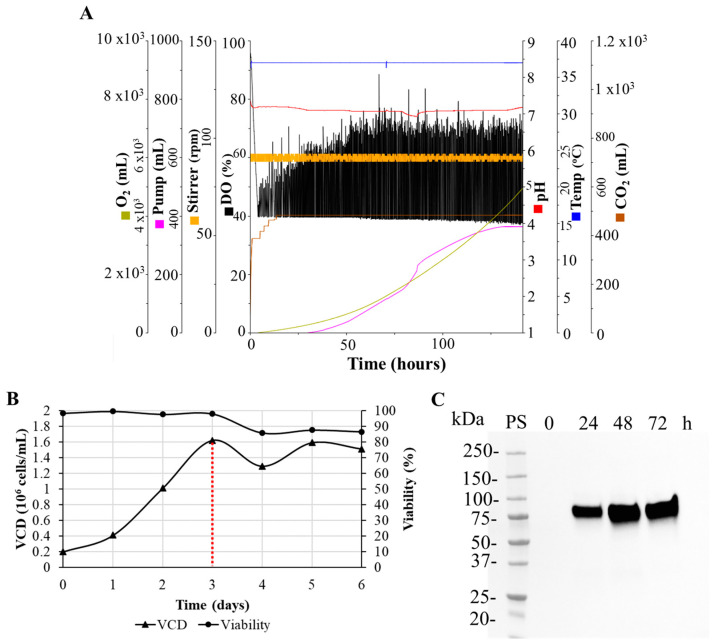
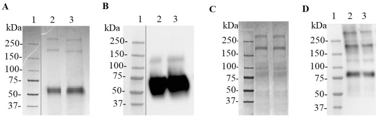
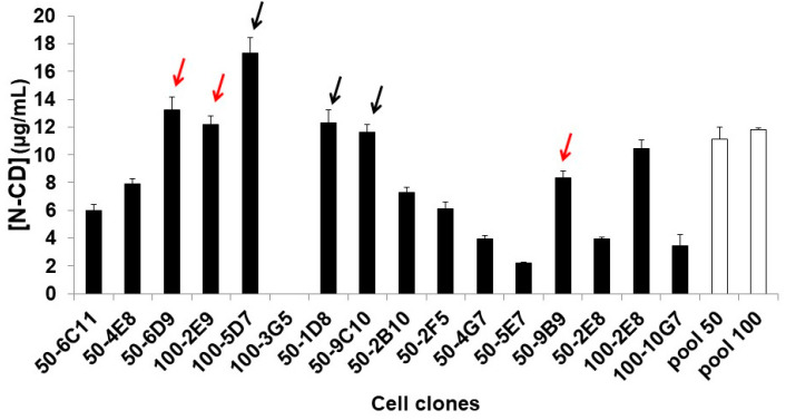
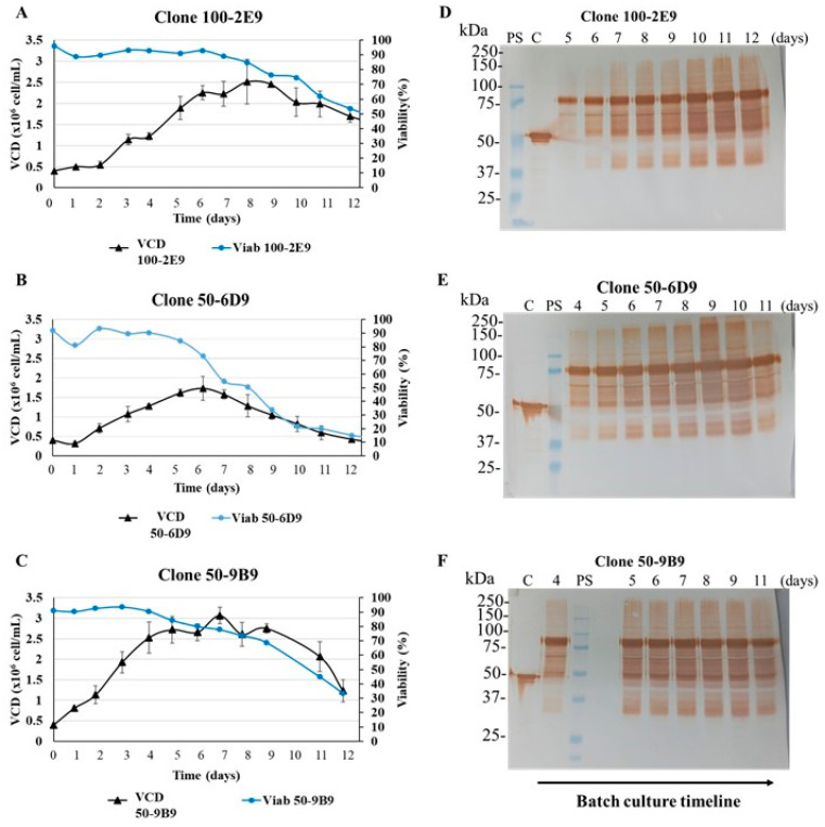
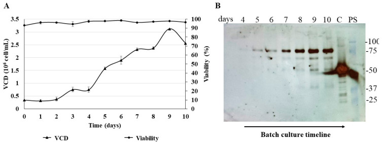
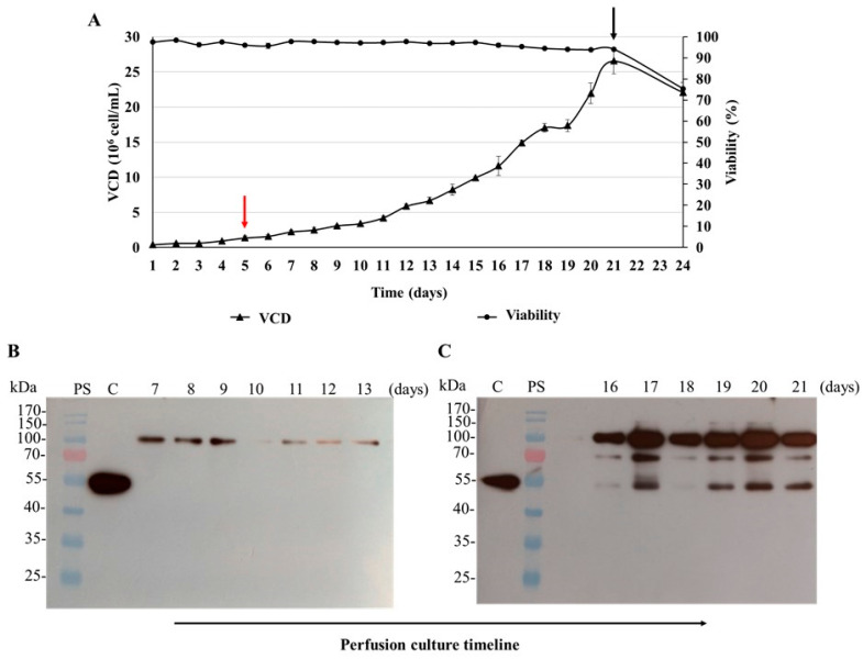
In a previous work, we proposed a vaccine chimeric antigen based on the fusion of the SARS-CoV-2 N protein to the extracellular domain of the human CD40 ligand (CD154). This vaccine antigen was named N-CD protein and its expression was carried out in HEK-293 stably transfected cells, grown in adherent conditions and serum-supplemented medium. The chimeric protein obtained in these conditions presented a consistent pattern of degradation. The immunization of mice and monkeys with this chimeric protein was able to induce a high N-specific IgG response with only two doses in pre-clinical experiments. In order to explore ways to diminish protein degradation, in the present work, the N and N-CD proteins were produced in suspension cultures and serum-free media following transient transfection of the HEK-293 clone 3F6, at different scales, including stirred-tank controlled bioreactors. The results showed negligible or no degradation of the target proteins. Further, clones stably expressing N-CD were obtained and adapted to suspension culture, obtaining similar results to those observed in the transient expression experiments in HEK-293-3F6. The evidence supports transient protein expression in suspension cultures and serum-free media as a powerful tool to produce in a short period of time high levels of complex proteins susceptible to degradation, such as the SARS-CoV-2 N protein.

Keywords: HEK-293; N protein; SARS-CoV-2; serum-free; suspension culture; transient expression.


Conflict of interest statement

The authors declare no conflict of interest. The funders had no role in the design of the study; in the collection, analyses or interpretation of data; in the writing of the manuscript; or in the decision to publish the results.

Figures

Figure 1
Figure 1
SARS-CoV-2 N and N-CD proteins produced in adherent conditions by recombinant HEK-293 cells. (A) Schematic diagram of the expression cassettes pl6twblast-CMV-N-His and pl6twblast-CMV-N-CD. (B,C): Western blot analysis under reducing conditions. SARS-CoV-2 N protein produced in E. coli was used as positive control. The antibody CBSSNCoV-1-HRP was used for protein detection. (B) Cell culture supernatant harvested after 5 days of culture in DMEM/F12 medium. Recombinant cells were seeded in 24-well plates at 0.3 × 10⁶ cells/well in 1 mL of DMEM/F12-FBS culture medium. Twenty-four hours later, the cell monolayer was carefully washed with PBS and 1 mL of DMEM/F12 medium was added per well. Lane 1: Precision Plus ProteinTM All Blue Prestained Standard; lane 2: positive control; lane 3: cell culture supernatant of HEK-293-N-100 cells; lane 4: cell culture supernatant of HEK-293-N-200 cells. (C) N-CD protein purified previously in the laboratory with IMAC. Lane 1: positive control; lane 2: Precision Plus ProteinTM All Blue Prestained Standard; lane 3: purified N-CD protein. Protein visualization was carried out with DAB.
Figure 2
Figure 2
Analysis of the SARS-CoV-2 N and N-CD proteins produced in HEK-293SF-3F6 cells using shake flasks. Western blot of culture supernatant of cells transfected to express the N protein (A,C) or the N-CD protein (B,D). HEK-293SF cells were transfected in 1 L shake flasks using HyCell TransFx-H medium. Seventy-two hours post transfection, the cell culture supernatant was collected. Equal volumes of 5-fold (A,C,D) or 11-fold (B) concentrated cell culture supernatant were loaded onto the SDS-PAGE gel under reducing conditions. Amicon® Ultra-2 mL centrifugal filters were used for concentration. For (A,B), a 6x-His Tag monoclonal antibody was used for detection. For (C,D), a SARS-CoV-2 N protein mouse monoclonal antibody was used as primary antibody. (A,C), Lane 1: Precision Plus ProteinTM All Blue Prestained Standard, lane 2: cell culture supernatant of cells transfected to express the N protein. (B,D) Lane 1: Precision Plus ProteinTM All Blue Prestained Standard, lane 2: cell culture supernatant of cells transfected to express the N-CD protein. Protein visualization was carried out with ECL detection system.
Figure 3
Figure 3
Production of the SARS-CoV-2 N and N-CD proteins in 1 L bioreactors. HEK-293SF-3F6 cells were transfected with linear PEI (MW 25,000) in HyCell TransFx-H medium with the plasmid pl6twblast-N-His or pl6twblast-N-CD. Injected O2 and CO2, base pumping (pump), agitation rate (stirrer), dissolved oxygen (DO), pH and temperature were monitored during the run before and after the transfection to produce the N protein (A) or the N-CD protein (C). Western blot of culture supernatant of cells transfected to express the N protein (B) or the N-CD protein (D). Seventy-two hours post transfection, the cell culture supernatant was collected and concentrated with Amicon® Ultra-2 mL centrifugal filters. Equal volumes of 5-fold concentrated cell culture supernatant were loaded onto the SDS-PAGE gel under reducing conditions. SARS-CoV-2 N protein monoclonal antibody was used as primary antibody. (B) Lane 1: Precision Plus ProteinTM All Blue Prestained Standard, lane 2: cell culture supernatant of cells transfected to express the N protein. (D) Lane 1: Precision Plus ProteinTM All Blue Prestained Standard, lane 2: cell culture supernatant of cells transfected to express the N-CD protein. Protein visualization was carried out with an ECL detection system.
Figure 4
Figure 4
Analysis of growth and expression kinetics of the N-CD protein produced in the 3 L bioreactor. (A) Graphic representation of injected O2 and CO2, base pumping (pump), agitation rate (stirrer), dissolved oxygen (DO), pH and temperature monitored during the run. (B) Growth curve of HEK-293SF-3F6 cells cultured in HyCell TransFx-H medium in batch mode. Every 24 h, samples of cell culture were collected and viable cell density (VCD) and cell viability were measured. Cells were transfected with the plasmid pl6twblast-N-CD using linear PEI (MW 25,000). The transfection day is indicated with a red dotted line. (C) Samples of cell culture supernatant were collected before transfection (time 0 h) and every 24 h after transfection. Sample concentration was carried out in Amicon® Ultra-2 mL centrifugal filters. Equal volumes of 5-fold concentrated cell culture supernatant were loaded onto the SDS-PAGE gel under reducing conditions. A SARS-CoV-2 N protein mouse monoclonal antibody was used as primary antibody. Precision Plus ProteinTM All Blue Prestained Standard was used as protein standard (PS). Protein visualization was carried out with an ECL detection system.
Figure 5
Figure 5
Analysis of the downstream processing of the SARS-CoV-2 N and N-CD proteins produced in suspension culture and protein-free medium. Equal volumes of samples were analyzed under reducing conditions. For protein detection in Western Blot, an anti- SARS-CoV-2 N monoclonal antibody was used as primary antibody. A and B Analysis with Coomassie-stained SDS-PAGE and Western blot of the N protein in formulation buffer, respectively. Lane 1: Precision Plus ProteinTM Unstained Standard (A) or Precision Plus ProteinTM All Blue Prestained Standard (B), lane 2: N protein after buffer exchange to formulation buffer, lane 3: Main peak of elution fraction at 250 mM imidazole. (C,D) Analysis with Coomassie-stained SDS-PAGE and Western blot of the N-CD protein in formulation buffer, respectively. Lane 1: Precision Plus ProteinTM Unstained Standard (C) or Precision Plus ProteinTM All Blue Prestained Standard (D), lane 2: Main peak of elution fraction at 250 mM imidazole, lane 3: N-CD protein after buffer exchange to formulation buffer. For Western blot, protein visualization was carried out with an ECL detection system.
Figure 6
Figure 6
Evaluation of protein expression in N-CD-expressing cell clones generated via limiting dilution cloning from N-CD-expressing HEK-293 cell pools (MOI 50 and 100). The cells were seeded in 24-well plates in DMEM/F12-FBS at 0.3 × 10⁶ cells/well in triplicate. After 7 days, cell culture was harvested and N protein was measured with ELISA. The data correspond to mean ± standard deviation. The white bars correspond to the two different N-CD-expressing HEK-293 cell pools and the black bars to the cell clones derived from these cell pools. The six arrows point out the cell clones selected for adaptation to protein-free and suspension culture conditions in CDM4HEK293 medium. The red arrows indicate the cell clones that survived to the adaptation process.
Figure 7
Figure 7
Batch culture of N-CD-expressing cell clones adapted to protein-free medium and suspension culture conditions. (AC) Growth profile of cell clones 100-2E9, 50-6D9 and 50-9B9, respectively. The cells were seeded in 250 mL shake flasks at 0.4 × 10⁶ cells/mL in 90 mL of CDM4HEK293 medium. The shake flasks were set in a shaker (130 rpm) and incubated in a warm chamber (37 °C) without CO2. The experiment was performed in duplicate. Every 24 h, samples of cell culture were collected and viable cell density (VCD) and cell viability were measured. After sampling, the flasks were incubated at 37 °C with 5% CO2 in static conditions for 2 or 3 h, and, later, they were incubated again in the conditions described above. (DF) Analysis with Western blot of cell culture supernatant of cell clones 100-2E9, 50-6D9 and 50-9B9, respectively. Total proteins from equal volumes of cell culture supernatant, precipitated by the methods of TCA and sodium deoxycholate, were loaded onto the SDS-PAGE gel under reducing conditions. The supernatant samples analyzed corresponded to the exponential and stationary growth phases. The specific days analyzed are pointed out in the figure. For protein detection, the membrane was exposed to an HRP-conjugated anti-SARS-CoV-2 N-protein monoclonal antibody. PS: Precision Plus ProteinTM All Blue Prestained Standard was used as protein standard. C: SARS-CoV-2 N protein expressed in E. coli was used as positive control. Protein visualization was carried out with DAB.
Figure 8
Figure 8
Batch culture of the N-CD-expressing cell clone 100-2E9 in 2 L roller bottles. (A) Growth profile of cell clone 100-2E9 in protein-free medium and suspension culture conditions. The 2 L roller bottles were seeded at 0.35 × 10⁶ cells/mL in 500 mL of CDM4HEK293 medium and placed vertically in an incubator (Infors HT, Switzerland) at 37 °C with 5% CO2 in shaking conditions (120 rpm). The experiment was performed in duplicate. Every 24 h, samples of cell culture were collected and viable cell density (VCD) and cell viability were measured. (B) Analysis with Western blot of cell culture supernatant. Total proteins from equal volumes of cell culture supernatant, precipitated by the methods of TCA and sodium deoxycholate, were loaded onto the SDS-PAGE gel under reducing conditions. The analyzed supernatant samples correspond to the exponential growth phase. For protein detection, the membrane was exposed to an HRP-conjugated anti-SARS-CoV-2 N-protein monoclonal antibody. C: SARS-CoV-2 N protein expressed in E. coli (45 kDa) was used as positive control. PS: Precision Plus ProteinTM All Blue Prestained Standard was used as protein standard. Protein visualization was carried out with an ECL detection system.
Figure 9
Figure 9
Simulation of perfusion culture of the N-CD-expressing cell clone 100-2E9 in shake flasks. (A) Growth profile of cell clone 100-2E9 in protein-free medium and suspension culture conditions. The 250 mL shake flasks were seeded at 0.4 × 10⁶ cells/mL in 90 mL of CDM4HEK293 medium and incubated at 37 °C with 5% CO2 in shaking conditions (120 rpm). The experiment was performed in duplicate. The cells were maintained in batch culture conditions until the exponential phase and maximum growth rate were reached. The CSPR was fixed to 0.15 nL/cell/day. Every 24 h, samples of cell culture were collected. Viable cell density (VCD) and cell viability were measured. At the end of perfusion culture, the cells were grown in batch culture for 3 days. The beginning and the end of the perfusion culture are indicated with red and black arrows, respectively. (B) Analysis with Western blot of cell culture supernatant. Total proteins from equal volumes of cell culture supernatant, precipitated by the methods of TCA and sodium deoxycholate, were loaded onto the SDS-PAGE gel under reducing conditions. The analyzed supernatant samples correspond to the exponential growth phase; the specific analyzed days are pointed out in the figure. For protein detection, the membrane was exposed to an HRP-conjugated anti-SARS-CoV-2 N-protein monoclonal antibody. PS: Page RulerTM Prestained Protein Ladder was used as protein standard. (C) SARS-CoV-2 N protein expressed in E. coli (45 kDa) was used as positive control. Protein visualization was carried out with an ECL detection system.

Similar articles

References

    1. McLean G., Kamil J., Lee B., Moore P., Schulz T.F., Muik A., Sahin U., Türeci Ö., Pather S. The impact of evolving SARS-CoV-2 mutations and variants on COVID-19 vaccines. MBio. 2022;13:e02979-21. doi: 10.1128/mbio.02979-21. - DOI - PMC - PubMed
    1. Guan X., Yang Y., Du L. Advances in SARS-CoV-2 receptor-binding domain-based COVID-19 vaccines. Expert Rev. Vaccines. 2023;22:422–439. doi: 10.1080/14760584.2023.2211153. - DOI - PMC - PubMed
    1. Ahlén G., Frelin L., Nikouyan N., Weber F., Höglund U., Larsson O., Westman M., Tuvesson O., Gidlund E.-K., Cadossi M. The SARS-CoV-2 N protein is a good component in a vaccine. J. Virol. 2020;94:10–1128. doi: 10.1128/JVI.01279-20. - DOI - PMC - PubMed
    1. Dutta N.K., Mazumdar K., Gordy J.T. The nucleocapsid protein of SARS–CoV-2: A target for vaccine development. J. Virol. 2020;94:10–1128. doi: 10.1128/JVI.00647-20. - DOI - PMC - PubMed
    1. Bai Z., Cao Y., Liu W., Li J. The SARS-CoV-2 nucleocapsid protein and its role in viral structure, biological functions, and a potential target for drug or vaccine mitigation. Viruses. 2021;13:1115. doi: 10.3390/v13061115. - DOI - PMC - PubMed