. 2023 Oct 24;13(11):1567.
doi: 10.3390/biom13111567.

Optical Genome Mapping for the Molecular Diagnosis of Facioscapulohumeral Muscular Dystrophy: Advancement and Challenges

Affiliations

Optical Genome Mapping for the Molecular Diagnosis of Facioscapulohumeral Muscular Dystrophy: Advancement and Challenges

Stephanie Efthymiou et al. Biomolecules. .

Abstract

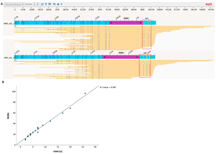
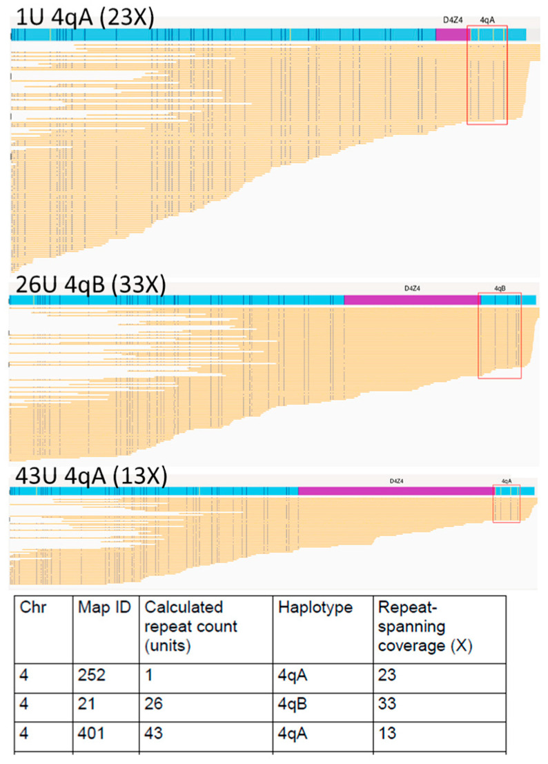
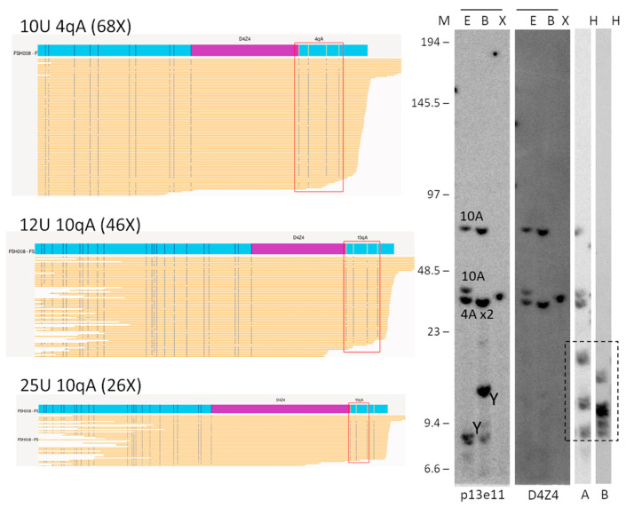
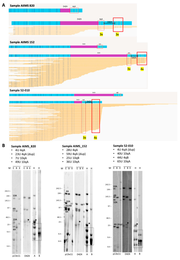
Facioscapulohumeral muscular dystrophy (FSHD) is the second most common muscular dystrophy in adults, and it is associated with local D4Z4 chromatin relaxation, mostly via the contraction of the D4Z4 macrosatellite repeat array on chromosome 4q35. In this study, we aimed to investigate the use of Optical Genome Mapping (OGM) as a diagnostic tool for testing FSHD cases from the UK and India and to compare OGM performance with that of traditional techniques such as linear gel (LGE) and Pulsed-field gel electrophoresis (PFGE) Southern blotting (SB). A total of 6 confirmed and 19 suspected FSHD samples were processed with LGE and PFGE, respectively. The same samples were run using a Saphyr Genome-Imaging Instrument (1-color), and the data were analysed using custom EnFocus FSHD analysis. OGM was able to confirm the diagnosis of FSHD1 in all FSHD1 cases positive for SB (n = 17), and D4Z4 sizing highly correlated with PFGE-SB (p < 0.001). OGM correctly identified cases with mosaicism for the repeat array contraction (n = 2) and with a duplication of the D4Z4 repeat array. OGM is a promising new technology able to unravel structural variants in the genome and seems to be a valid tool for diagnosing FSHD1.

Keywords: Bionano Genomics; D4Z4 contraction; FSHD; optical genome mapping.


Conflict of interest statement

The authors declare no conflict of interest.

Figures

Figure 1
Figure 1
(A) Case example processed with OGM. Blue bar is the sample consensus map; yellow bars are individual sample molecules; D4Z4 region is highlighted in purple. The blue vertical lines represent the positions of the fluorescent labels (OGM markers). The number of D4Z4 repeats was calculated by measuring the distance between the most proximal and distal OGM markers. Label patterns are also used to differentiate the haplotypes A and B (red square). (B) OGM detected a 4q35 D4Z4 macrosatellite contraction ranging from 1 to 26 D4Z4 repeats units and showed a strong correlation with SB allele sizing and haplotyping (100% haplotype match). Blue dots represent individual patients D4Z4 sizing. Linear regression showed that repeat array allele size according to OGM correlates well with the size determined via SB analysis.
Figure 2
Figure 2
Optical genome mapping (EnFocus FSHD analysis (version 1.0; Bionano Access Software v1.7) for the analysis of proband NHNN_FSH006 with somatic mosaicism. The output files show all reads aligned to the two D4Z4 alleles on chromosome 4. Specific fluorescence tags proximal and distal to the D4Z4 arrays enabled chromosomal assignment and A-B haplotyping. The fluorescence tags are absent in the D4Z4 region; here, the number of D4Z4 units was derived from the size of this unlabelled region.
Figure 3
Figure 3
Comparison of OGM (EnFocus FSHD analysis (version 1.0; Bionano Access Software v1.7)) and SB hybridisation and SSLP haplotyping for the analysis of proband FSH008 with homozygous 10 U alleles. The OGM output files (left panel) show one Chr 4 map with a higher number of reads, suggesting the presence of a homozygous D4Z4 region. SB analysis (right panel) showed hybridisations with probes p13E-11 and D4Z4 on genomic DNA digested using the enzymes EcoRI/HindIII €, EcoRI/BlnI (B), and XapI (X) and hybridisations with probes A and B on genomic DNA digested with HindIII (H). The alleles based on the different blots are indicated in the p13E-11 blot. The chromosome Y fragment is indicated, and the cross-hybridising fragments in the A/B hybridisations are marked with a dotted box. The size of the molecular weight marker is indicated on the left. Original blot images can be found in Figure S1.
Figure 4
Figure 4
(A) Duplicated 4qA signatures identified via visual inspection of OGM data using EnFocus FSHD analysis browser view (version 1.0; Bionano Access Software v1.7) for the analysis of probands AIIMS 820, AIIMS 152, and 52-010. The OGM output files show that all reads aligned to the D4Z4 alleles on chromosome 4. Nick sites are denoted by the red box, and the number of sites is given in yellow. (B) Comparison with SB analysis shows hybridisations with probes p13E-11 and D4Z4 on genomic DNA digested using the enzymes EcoRI/HindIII (E), EcoRI/BlnI (B), and XapI (X) and hybridisations with probes A and B on genomic DNA digested with HindIII (H). The alleles based on the different blots are indicated in the p13E-11 blot. The chromosome Y fragment is indicated, and the cross-hybridising fragments in the A/B hybridisations are marked with a dotted box. The size of the molecular weight marker is indicated on the left, and the asterisk (*) indicates the extra allele that appears upon D4Z4 and 4qA hybridisation and depicts the duplication allele. Original blot images can be found in Figure S1.

References

    1. Deenen J.C., Arnts H., van der Maarel S.M., Padberg G.W., Verschuuren J.J., Bakker E., Weinreich S.S., Verbeek A.L., van Engelen B.G. Population-based incidence and prevalence of facioscapulohumeral dystrophy. Neurology. 2014;83:1056–1059. doi: 10.1212/WNL.0000000000000797. - DOI - PMC - PubMed
    1. van Deutekom J.C., Wijmenga C., van Tienhoven E.A., Gruter A.M., Hewitt J.E., Padberg G.W., van Ommen G.J., Hofker M.H., Frants R.R. FSHD associated DNA rearrangements are due to deletions of integral copies of a 3.2 kb tandemly repeated unit. Hum. Mol. Genet. 1993;2:2037–2042. doi: 10.1093/hmg/2.12.2037. - DOI - PubMed
    1. Wijmenga C., Hewitt J.E., Sandkuijl L.A., Clark L.N., Wright T.J., Dauwerse H.G., Gruter A.M., Hofker M.H., Moerer P., Williamson R., et al. Chromosome 4q DNA rearrangements associated with facioscapulohumeral muscular dystrophy. Nat. Genet. 1992;2:26–30. doi: 10.1038/ng0992-26. - DOI - PubMed
    1. Lemmers R.J., van der Vliet P.J., Klooster R., Sacconi S., Camano P., Dauwerse J.G., Snider L., Straasheijm K.R., van Ommen G.J., Padberg G.W., et al. A unifying genetic model for facioscapulohumeral muscular dystrophy. Science. 2010;329:1650–1653. doi: 10.1126/science.1189044. - DOI - PMC - PubMed
    1. van Deutekom J.C., Bakker E., Lemmers R.J., van der Wielen M.J., Bik E., Hofker M.H., Padberg G.W., Frants R.R. Evidence for subtelomeric exchange of 3.3 kb tandemly repeated units between chromosomes 4q35 and 10q26: Implications for genetic counselling and etiology of FSHD1. Hum. Mol. Genet. 1996;5:1997–2003. doi: 10.1093/hmg/5.12.1997. - DOI - PubMed

Publication types

Supplementary concepts

LinkOut - more resources