. 2023 Nov 6;13(11):1623.
doi: 10.3390/biom13111623.

Identification of a Novel Acid Sphingomyelinase Activity Associated with Recombinant Human Acid Ceramidase

Affiliations

Identification of a Novel Acid Sphingomyelinase Activity Associated with Recombinant Human Acid Ceramidase

Xingxuan He et al. Biomolecules. .

Abstract

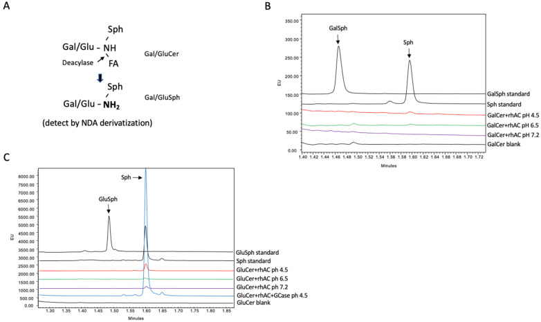
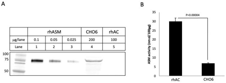
Acid ceramidase (AC) is a lysosomal enzyme required to hydrolyze ceramide to sphingosine by the removal of the fatty acid moiety. An inherited deficiency in this activity results in two disorders, Farber Lipogranulomatosis and spinal muscular atrophy with myoclonic epilepsy, leading to the accumulation of ceramides and other sphingolipids in various cells and tissues. In addition to ceramide hydrolysis, several other activities have been attributed to AC, including a reverse reaction that synthesizes ceramide from free fatty acids and sphingosine, and a deacylase activity that removes fatty acids from complex lipids such as sphingomyelin and glycosphingolipids. A close association of AC with another important enzyme of sphingolipid metabolism, acid sphingomyelinase (ASM), has also been observed. Herein, we used a highly purified recombinant human AC (rhAC) and novel UPLC-based assay methods to investigate the recently described deacylase activity of rhAC against three sphingolipid substrates, sphingomyelin, galactosyl- and glucosylceramide. No deacylase activities were detected using this method, although we did unexpectedly identify a significant ASM activity using natural (C-18) and artificial (Bodipy-C12) sphingomyelin substrates as well as the ASM-specific fluorogenic substrate, hexadecanoylamino-4-methylumbelliferyl phosphorylcholine (HMU-PC). We showed that this ASM activity was not due to contaminating, hamster-derived ASM in the rhAC preparation, and that the treatment of ASM-knockout mice with rhAC significantly reduced sphingomyelin storage in the liver. However, unlike the treatment with rhASM, this did not lead to elevated ceramide or sphingosine levels.

Keywords: Farber disease; ceramidase; deacylase; sphingomyelinase.


Conflict of interest statement

The authors declare no conflict of interest.

Figures

Figure 1
Figure 1
rhAC was purified from the media of overexpressing CHO cells and run on SDS-PAGE-reducing gels as described in the Methods. Three AC polypeptides (precursor, pre; beta, β and alpha, α) were identified by (A) Coomassie blue staining, (B) silver staining and (C) Western blotting using an anti-human AC polyclonal antibody. A total of 100 μg of rhAC was loaded per lane. (D) Ceramidase activity was determined using C12-Cer as the substrate at three different pH values. The Sph product was derivatized with NDA as described in the Methods, and the fluorescent Sph was identified and quantified by UPLC. The experiments were repeated three times.
Figure 2
Figure 2
The deacylase activity of rhAC was analyzed using either GalCer or GluCer as substrates. (A) Schematic depiction of the deacylation reaction. The GalSph or GluSph products were derivatized with NDA and quantified by UPLC as described in the Methods. Reactions were incubated for up to 24 h at 37 °C prior to analysis. Note that no GalSph (B) or GluSph (C) products were produced in these reactions under three different pH conditions. For GluCer, an additional control reaction was included, in which GluCer was first incubated with the enzyme beta glucocerebrosidase (GCase) followed by rhAC. In this combined reaction, GluCer was first hydrolyzed to Cer by GCase, and then Cer was hydrolyzed to Sph by rhAC. The Sph product was derivatized with NDA and quantified by UPLC. Note the significant Sph product produced by the dual enzyme reaction at pH 4.5, confirming the high AC activity associated with rhAC. The experiments were repeated three times.
Figure 3
Figure 3
Evaluation of the Spm deacylation reaction using rhAC and identification of a novel rhAC-associated ASM activity using three different substrates. (A) Schematic depiction showing the potential deacylase reaction using C18-Spm. Detection of the Spc product after derivatization with NDA and quantification by UPLC was performed as described in the Methods. (B) Schematic depiction showing that a potential combined reaction of sphingomyelinase activity followed by ceramidase activity of C18-Spm would result in Sph, which could be detected and quantified using NDA. (C) Deacylation reactions were carried out for 24 h with C18-Spm as the substrate, but no Spc product was detected under three different pH conditions. However, a significant Sph product was detected at pH 4.5, indicating the presence of an ASM activity. (D) To ensure that Spc could not be directly hydrolyzed to Sph by rhAC, we incubated rhAC with Spc, but no Sph product was found. To further confirm the rhAC-associated ASM activity, we incubated rhAC with Bodipy-conjugated C12-Spm (Bp-Spm) at pH 4.5 and found a high ASM activity (Bp-FA product) that was sensitive to denaturation using urea and DTT (E). (F) As an additional confirmation of the rhAC-associated ASM activity, we used the ASM-specific fluorogenic substrate HMU-PC and found significant ASM activity in rhAC. The experiments were repeated three times. p values are shown in (F) comparing the activities with varying amounts of rhAC added to the reaction mixtures to the buffer controls. The mean and standard deviation values were 52.5 +/− 9.92, 297.35 +/− 16.1 and 617 +/− 27.1 for buffer alone (0 rhAC added), 0.5 and 1.0 μg/rhAC, respectively.
Figure 4
Figure 4
To further rule out the possibility that the rhAC-associated ASM activity could be derived from contaminating CHO ASM, we performed Western blot analysis using an anti-ASM antibody that cross-reacted with CHO ASM (A). Note that CHO6 cell extracts overexpressing rhAC had a cross-reacting CHO ASM band that was readily visible. In contrast, the highly purified rhAC did not. We then compared the ASM activity in purified rhAC vs. CHO6 cells, and despite the fact that no cross-reacting CHO ASM was detected by Western blot in rhAC, the ASM activity was >5 fold greater than that in CHO6 cell extracts (B). This demonstrated that the rhAC-associated ASM activity could not be derived from CHO ASM. The experiments were repeated three times. The mean ASM activity value for rhAC in (B) was 29.9, with a standard deviation of +/−2.0, while for the CHO6 cells, it was 7.0 +/− 0.136. p values are shown.
Figure 5
Figure 5
Four-month-old ASMKO mice accumulating high levels of Spm in their livers were treated with 6 injections of rhASM (1 mg/kg) or rhAC (10 mg/kg). A total of 24 h after the last injection, the mice were euthanized, and the livers were collected for Spm, Cer or Sph analysis as described in the Methods. Previous studies have shown that treatment of ASMKO mice with 10 mg/kg rhASM leads to acute toxicity [29], including lethargy and death. No toxicity was observed using 10 mg/kg rhAC, despite the equivalent level of Spm reduction by the two enzyme treatments (A). As expected, treatment with rhASM led to Cer and Sph elevations (B,C). However, despite the equivalent reduction in Spm in the ASMKO mouse livers, no Cer or Sph elevation was observed after rhAC treatment, consistent with the fact that dosing of these mice with 10 mg/kg of rhAC did not lead to toxicity. The experiments were repeated three times. In (A), the mean SPM values and standard deviations are 9.34 +/− 2.22, 5.50 +/− 0.482 and 5.64 +/− 0.910 for saline, rhAC and rhAC, respectively; in (B), the mean Cer values and standard deviations are 0.093 +/− 0.011, 0.181 +/− 0.033 and 0.074 +/− 0.016, respectively; and in (C), the mean Sph values are 0.014 +/− 0.004, 0.148 +/− 0.044 and 0.015 +/− 0.009, respectively. p values are shown.

Similar articles

References

    1. Mao C., Obeid L.M. Ceramidases: Regulators of cellular responses mediated by ceramide, sphingosine, and sphingosine-1-phosphate. Biochim. Biophys. Acta. 2008;1781:424. doi: 10.1016/j.bbalip.2008.06.002. - DOI - PMC - PubMed
    1. Coant N., Sakamoto W., Mao C., Hannun Y.A. Ceramidases, roles in sphingolipid metabolism and in health and disease. Adv. Biol. Regul. 2017;63:122. doi: 10.1016/j.jbior.2016.10.002. - DOI - PMC - PubMed
    1. Frohbergh M., He X., Schuchman E.H. The molecular medicine of acid ceramidase. Biol. Chem. 2015;396:759. doi: 10.1515/hsz-2014-0290. - DOI - PubMed
    1. Ueda N. A rheostat of ceramide and sphingosine-1-phosphate as a determinant of oxidative stress-mediated kidney injury. Int. J. Mol. Sci. 2022;23:4010. doi: 10.3390/ijms23074010. - DOI - PMC - PubMed
    1. Piccoli M., Cirillo F., Ghiroldi A., Rota P., Coviello S., Tarantino A., La Rocca P., Lavota I., Creo P., Signorelli P., et al. Sphingolipids and atherosclerosis: The dual role of ceramide and sphingosine-1-phosphate. Antioxidants. 2023;12:143. doi: 10.3390/antiox12010143. - DOI - PMC - PubMed

Publication types

LinkOut - more resources