

Controlling Trophoblast Cell Fusion in the Human Placenta-Transcriptional Regulation of Suppressyn, an Endogenous Inhibitor of Syncytin-1
- PMID: 38002309
- PMCID: PMC10668956
- DOI: 10.3390/biom13111627
Controlling Trophoblast Cell Fusion in the Human Placenta-Transcriptional Regulation of Suppressyn, an Endogenous Inhibitor of Syncytin-1
Abstract
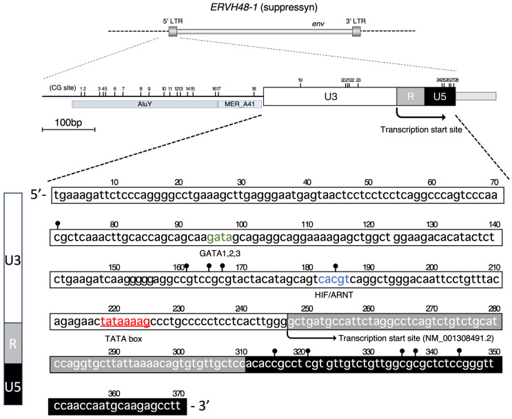
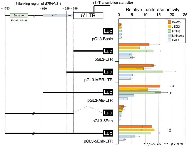
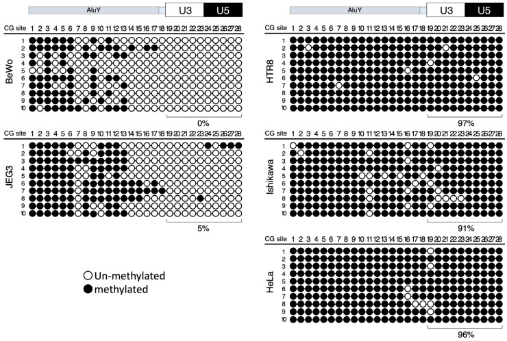
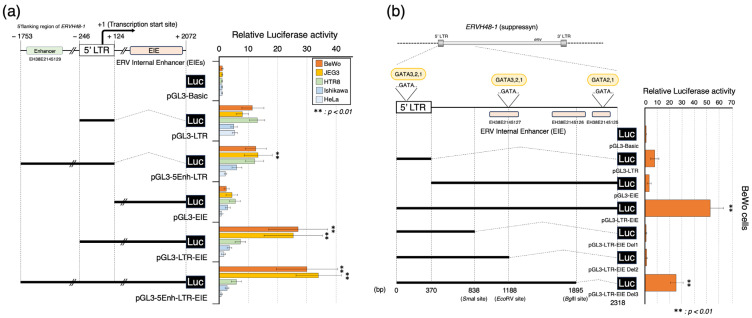
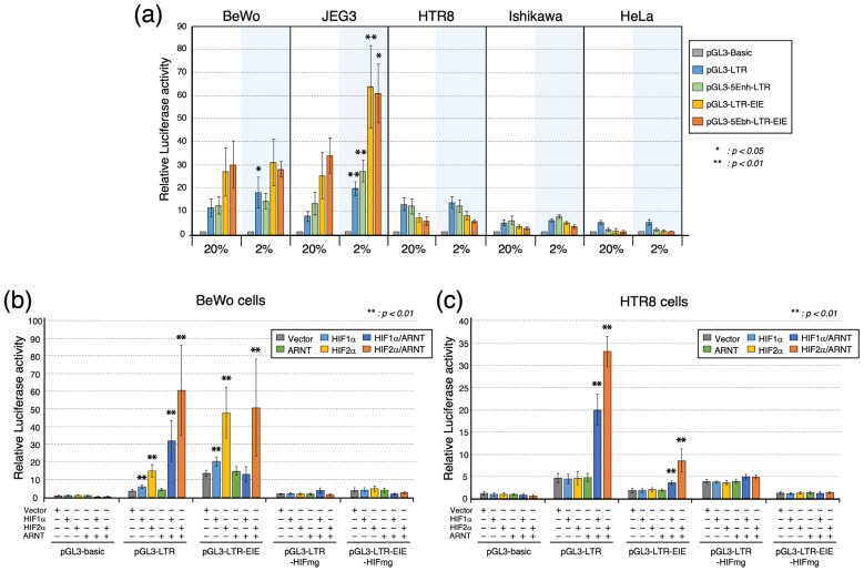
Cell fusion in the placenta is tightly regulated. Suppressyn is a human placental endogenous retroviral protein that inhibits the profusogenic activities of another well-described endogenous retroviral protein, syncytin-1. In this study, we aimed to elucidate the mechanisms underlying suppressyn's placenta-specific expression. We identified the promoter region and a novel enhancer region for the gene encoding suppressyn, ERVH48-1, and examined their regulation via DNA methylation and their responses to changes in the oxygen concentration. Like other endogenous retroviral genes, the ERVH48-1 promoter sequence is found within a characteristic retroviral 5' LTR sequence. The novel enhancer sequence we describe here is downstream of this LTR sequence (designated EIEs: ERV internal enhancer sequence) and governs placental expression. The placenta-specific expression of ERVH48-1 is tightly controlled by DNA methylation and further regulated by oxygen concentration-dependent, hypoxia-induced transcription factors (HIF1α and HIF2α). Our findings highlight the involvement of (1) tissue specificity through DNA methylation, (2) expression specificity through placenta-specific enhancer regions, and (3) the regulation of suppressyn expression in differing oxygen conditions by HIF1α and HIF2α. We suggest that these regulatory mechanisms are central to normal and abnormal placental development, including the development of disorders of pregnancy involving altered oxygenation, such as preeclampsia, pregnancy-induced hypertension, and fetal growth restriction.
Keywords: DNA methylation; HERV; cell fusion; hypoxia inducible factor (HIF); placenta; promoter; suppressyn; syncytin.
Conflict of interest statement
The authors declare no conflict of interest.
Figures
Similar articles
-
Could the Human Endogenous Retrovirus-Derived Syncytialization Inhibitor, Suppressyn, Limit Heterotypic Cell Fusion Events in the Decidua?Int J Mol Sci. 2021 Sep 23;22(19):10259. doi: 10.3390/ijms221910259. Int J Mol Sci. 2021. PMID: 34638599 Free PMC article.
-
Syncytins expressed in human placental trophoblast.Placenta. 2021 Sep 15;113:8-14. doi: 10.1016/j.placenta.2021.01.006. Epub 2021 Jan 15. Placenta. 2021. PMID: 33504453 Free PMC article. Review.
-
A novel human endogenous retroviral protein inhibits cell-cell fusion.Sci Rep. 2013;3:1462. doi: 10.1038/srep01462. Sci Rep. 2013. PMID: 23492904 Free PMC article.
-
Acquisition and Exaptation of Endogenous Retroviruses in Mammalian Placenta.Biomolecules. 2023 Oct 4;13(10):1482. doi: 10.3390/biom13101482. Biomolecules. 2023. PMID: 37892164 Free PMC article. Review.
-
Epigenetic and non-epigenetic regulation of syncytin-1 expression in human placenta and cancer tissues.Cell Signal. 2014 Mar;26(3):648-56. doi: 10.1016/j.cellsig.2013.11.002. Epub 2013 Nov 9. Cell Signal. 2014. PMID: 24216608 Review.
Cited by
-
UNet with Attention Networks: A Novel Deep Learning Approach for DNA Methylation Prediction in HeLa Cells.Genes (Basel). 2025 May 28;16(6):655. doi: 10.3390/genes16060655. Genes (Basel). 2025. PMID: 40565547 Free PMC article.
-
Trophoblast Fusion in Hypertensive Disorders of Pregnancy and Preeclampsia.Int J Mol Sci. 2025 Mar 21;26(7):2859. doi: 10.3390/ijms26072859. Int J Mol Sci. 2025. PMID: 40243430 Free PMC article. Review.
-
Persistent type I interferon signaling within the brain of people with HIV on ART with cognitive impairment.PLoS Pathog. 2025 Aug 20;21(8):e1013411. doi: 10.1371/journal.ppat.1013411. eCollection 2025 Aug. PLoS Pathog. 2025. PMID: 40833988 Free PMC article.
-
Endogenous retroviral ERVH48-1 promotes human urine cell reprogramming.Cell Regen. 2024 Sep 13;13(1):17. doi: 10.1186/s13619-024-00200-2. Cell Regen. 2024. PMID: 39269631 Free PMC article.
-
Genetic Diversity in the Suppressyn Gene Sequence: From Polymorphisms to Loss-of-Function Mutations.Biomolecules. 2025 Jul 21;15(7):1051. doi: 10.3390/biom15071051. Biomolecules. 2025. PMID: 40723922 Free PMC article.
References
Publication types
MeSH terms
Substances
Grants and funding
LinkOut - more resources
Full Text Sources
Molecular Biology Databases
