. 2023 Nov 7;13(11):1628.
doi: 10.3390/biom13111628.

Low-Entropy Hydration Shells at the Spike RBD's Binding Site May Reveal the Contagiousness of SARS-CoV-2 Variants

Affiliations

Low-Entropy Hydration Shells at the Spike RBD's Binding Site May Reveal the Contagiousness of SARS-CoV-2 Variants

Lin Yang et al. Biomolecules. .

Abstract

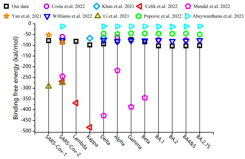
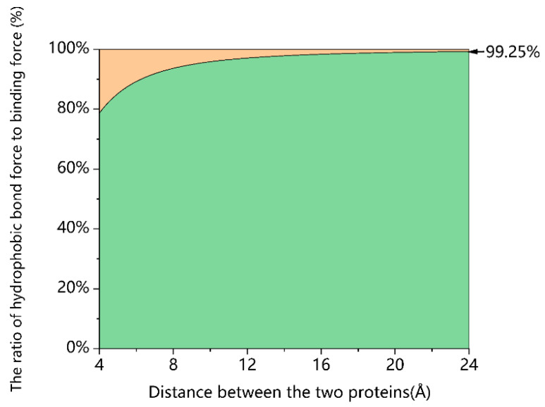
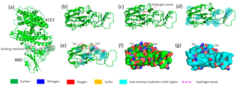
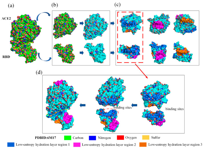
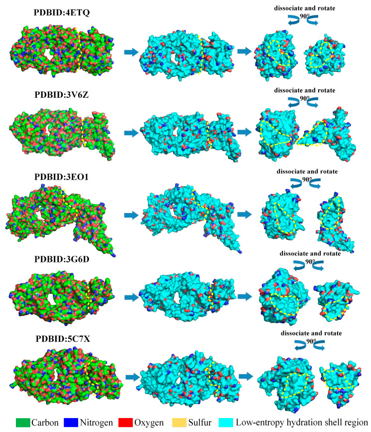
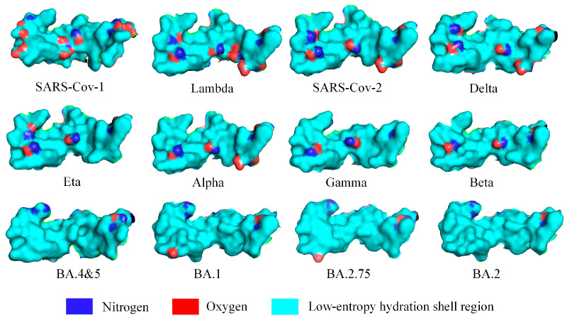
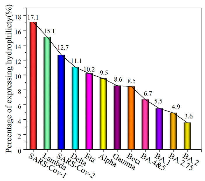
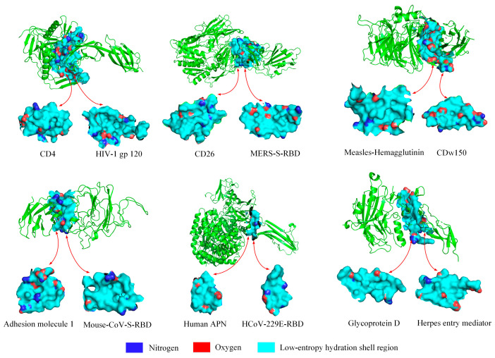
The infectivity of the severe acute respiratory syndrome coronavirus 2 (SARS-CoV-2) is primarily determined by the binding affinity between the receptor-binding domain (RBD) of the spike protein and the angiotensin-converting enzyme 2 (ACE2) receptor. Here, through screening off pseudo hydrophilic groups on protein surfaces, the distribution of low-entropy regions on hydration shells of the ACE2 receptor and the RBDs of multiple SARS-CoV-2 variants was demonstrated. Shape matching between the low-entropy hydration shells of multiple SARS-CoV-2 variants and the ACE2 receptor has been identified as a mechanism that drives hydrophobic attraction between the RBDs and the ACE2 receptor, which estimates the binding affinity. Low-entropy regions of the hydration shells, which play important roles in determining the binding of other viruses and their receptors, are demonstrated. The RBD-ACE2 binding is thus found to be guided by hydrophobic collapse between the shape-matched low-entropy regions of the hydration shells of the proteins. A measure of the low-entropy status of the hydration shells can be estimated by calculating genuine hydrophilic groups within the binding sites. An important indicator of the contagiousness of SARS-CoV-2 variants is the low-entropy level of its hydration shells at the spike protein binding site.

Keywords: SARS-CoV-2 variants; contagiousness; low-entropy hydration shell; protein–protein interaction.


Conflict of interest statement

The authors declare no conflict of interest.

Figures

Figure 1
Figure 1
The RBD–ACE2 binding free energy for SARS-CoV-1 and SARS-CoV-2 variants, calculated by considering electrostatic interaction, van der Waals interaction, polar solvation energy, and non-polar solvation energy, compared with the binding energies reported in other references [12,13,14,15,16,17,18,19,20].
Figure 2
Figure 2
Percentage share of hydrophobic interaction force in the overall attractive force between the ACE2 receptor and the SARS-CoV-2 RBD during the binding process.
Figure 3
Figure 3
Schematic workflow of screening off pseudo hydrophilic groups on the binding site of the SARS-CoV-2 S RBD. (a) Molecular structure of SARS-CoV-2 RBD and ACE2; the following figures exhibit only the RBD binding interface. (b) Amino acids adjacent to ACE2 on the RBD binding interface. (c) Intramolecular hydrogen bonding on the RBD binding interface (highlighted by red dashed lines). (d) Hydrophobic sidechains protruding on the surface of the protein (carbon atoms are highlighted by cyan dots). (e) The distribution of tyrosine and lysine on the RBD binding interface (oxygen, nitrogen, and carbon atoms are highlighted by red, blue, and cyan dots, respectively). (f) The topography distribution of hydrophilic nitrogen and oxygen atoms on the normal RBD surface. (g) The topography distribution of the low-entropy hydration shell on RBD surface (highlighted in cyan).
Figure 4
Figure 4
Low-entropy hydration shells on the binding sites of SARS-CoV-2 RBD and ACE2. (a) The normal surface of ACE2 and SARS-CoV-2 RBD. (b) The low-entropy hydration shell regions of RBD and ACE2 (highlighted in cyan). (c) Three larger low-entropy hydration shell regions of RBD and ACE2 are depicted in marine (region 1), magenta (region 2), and orange (region 3). (d) The larger low-entropy regions of hydration shells covering the binding sites of RBD and ACE2.
Figure 5
Figure 5
Low-entropy hydration shells at binding sites guide protein binding using five antibody–antigen test cases for Docking Benchmark 5.5. The low-entropy hydration shell regions are shown in cyan, and the curves and irregular circles marked with yellow dashed lines represent the binding surface and binding sites, respectively.
Figure 6
Figure 6
Distribution of the surface area of expressed hydrophilic groups on the low-entropy hydration shells at the RBD binding sites of SARS-CoV-1, SARS-CoV-2, and SARS-CoV-2 variants with the ACE2 receptor. The low-entropy region of the hydration shells is shown in cyan. PDBIDs include: 2AJF, 7FEM, 6M17, 7W9I, 7NXC, 7R11, 7WBL, 7XWA, 8ASY, etc.
Figure 7
Figure 7
The proportion of the surface area of expressed hydrophilic groups on the low-entropy hydration shells at the RBD binding sites of SARS-CoV-1, SARS-CoV-2, and 10 SARS-CoV-2 variants.
Figure 8
Figure 8
Expressed hydrophilic groups in the low-entropy regions of hydration shells at the binding sites of six other viruses. The six viruses are HIV (PDBID: 4RQS), MERS (PDBID: 4KR0), mouse coronavirus (PDBID: 6VSJ), measles virus hemagglutinin (PDBID: 3ALZ), human coronavirus HCoV-229E (PDBID: 6U7G), and herpes (PDBID: 1JMA), respectively.
Figure 9
Figure 9
Percentage surface area occupied by hydrophilic groups expressed in the low-entropy region at the binding sites of six viruses.
Figure 10
Figure 10
(a) A schematic plot of the eight representative antibodies bound to five low-entropy regions of hydration shells on the surface of the SARS-CoV-2 spike protein RBD; The contour of the binding site is a yellow dashed circle.: marine (region 1), magenta (region 2), orange (region 3), chartreuse (region 4), and pink (region 5). (b) Schematic representation of eight representative antibodies bound to SARS-CoV-2 spike protein RBD; the antibodies are shown in transparent burgundy.
Figure 11
Figure 11
Schematic illustration of the proportions of the expressed hydrophilic groups in the low-entropy region of Ab709, H104, 10G4, and S309 antibodies and RBD at binding sites. The low-entropy hydration shell regions are shown in cyan; the curves and irregular circles marked with yellow dashed lines represent the binding surface and binding sites, respectively.

Similar articles

References

    1. Lan J., Ge J., Yu J., Shan S., Zhou H., Fan S., Zhang Q., Shi X., Wang Q., Zhang L. Structure of the SARS-CoV-2 spike receptor-binding domain bound to the ACE2 receptor. Nature. 2020;581:215–220. doi: 10.1038/s41586-020-2180-5. - DOI - PubMed
    1. Guo W., Pan B., Ji Z., Hong H. Elucidating Interactions Between SARS-CoV-2 Trimeric Spike Protein and ACE2 Using Homology Modeling and Molecular Dynamics Simulations. Front. Chem. 2021;8:622632. - PMC - PubMed
    1. Cong Y., Feng Y., Ni H., Zhi F., Miao Y., Fang B., Zhang L., Zhang J.Z. Anchor-locker binding mechanism of the coronavirus spike protein to human ACE2: Insights from computational analysis. J. Chem. Inf. Model. 2021;61:3529–3542. doi: 10.1021/acs.jcim.1c00241. - DOI - PubMed
    1. Monticelli M., Mele B.H., Andreotti G., Cubellis M.V., Riccio G. Why does SARS-CoV-2 hit in different ways? Host genetic factors can influence the acquisition or the course of COVID-19. Eur. J. Med. Genet. 2021;64:104227. doi: 10.1016/j.ejmg.2021.104227. - DOI - PMC - PubMed
    1. Isobe A., Arai Y., Kuroda D., Okumura N., Ono T., Ushiba S., Nakakita S.-i., Daidoji T., Suzuki Y., Nakaya T. ACE2 N-glycosylation modulates interactions with SARS-CoV-2 spike protein in a site-specific manner. Commun. Biol. 2022;5:1188. doi: 10.1038/s42003-022-04170-6. - DOI - PMC - PubMed

Publication types

Substances

Supplementary concepts