. 2023 Nov 10;13(11):1635.
doi: 10.3390/biom13111635.

Dynamics of Docosahexaenoic Acid Utilization by Mouse Peritoneal Macrophages

Affiliations

Dynamics of Docosahexaenoic Acid Utilization by Mouse Peritoneal Macrophages

Patricia Monge et al. Biomolecules. .

Abstract

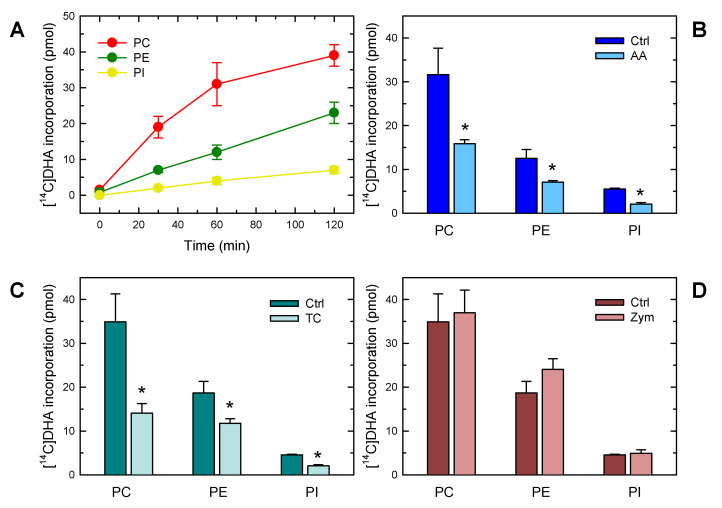
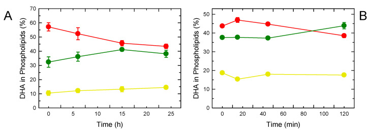
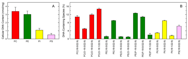
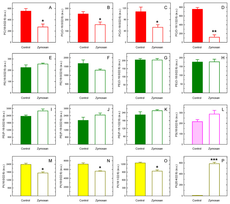
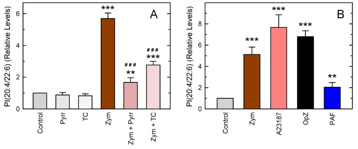
In this work, the incorporation of docosahexaenoic acid (DHA) in mouse resident peritoneal macrophages and its redistribution within the various phospholipid classes were investigated. Choline glycerophospholipids (PC) behaved as the major initial acceptors of DHA. Prolonged incubation with the fatty acid resulted in the transfer of DHA from PC to ethanolamine glycerophospholipids (PE), reflecting phospholipid remodeling. This process resulted in the cells containing similar amounts of DHA in PC and PE in the resting state. Mass spectrometry-based lipidomic analyses of phospholipid molecular species indicated a marked abundance of DHA in ether phospholipids. Stimulation of the macrophages with yeast-derived zymosan resulted in significant decreases in the levels of all DHA-containing PC and PI species; however, no PE or PS molecular species were found to decrease. In contrast, the levels of an unusual DHA-containing species, namely PI(20:4/22:6), which was barely present in resting cells, were found to markedly increase under zymosan stimulation. The levels of this phospholipid also significantly increased when the calcium-ionophore A23187 or platelet-activating factor were used instead of zymosan to stimulate the macrophages. The study of the route involved in the synthesis of PI(20:4/22:6) suggested that this species is produced through deacylation/reacylation reactions. These results define the increases in PI(20:4/22:6) as a novel lipid metabolic marker of mouse macrophage activation, and provide novel information to understand the regulation of phospholipid fatty acid turnover in activated macrophages.

Keywords: arachidonic acid; docosahexaenoic acid; inflammation; lipid signaling; membrane phospholipid; monocytes/macrophages.


Conflict of interest statement

The authors declare no conflict of interest. The funders had no role in the design of the study; in the collection, analyses, or interpretation of data; in the writing of the manuscript, or in the decision to publish the results.

Figures

Figure 1
Figure 1
Characterization of DHA incorporation into macrophage phospholipids. (A) Time-course of incorporation into different phospholipid classes. The cells were incubated with [14C]DHA, and incorporation of the fatty acid was measured in PC (red), PE (green) and PI (yellow). (B) Effect of AA on the incorporation of [14C]DHA into different phospholipid classes. The cells were exposed to [14C]DHA for 60 min in the absence (blue) or presence (light blue) of AA. (C) Effect of triacsin C (TC) on the incorporation of [14C]DHA into different phospholipid classes. The cells were exposed to [14C]DHA for 60 min in the absence (green) or presence (light green) of 3 µM triacsin C. (D) Effect of zymosan stimulation on the incorporation of [14C]DHA into different phospholipid classes. The cells were exposed to [14C]DHA for 60 min in the absence (maroon) or presence (light maroon) of 0.5 mg/mL zymosan, which acted as a cellular stimulant. The results are shown as means ± standard error of the mean (n = 3). * p < 0.05, significance of control (Ctrl) cells versus cells treated with AA, TC, or zymosan.
Figure 2
Figure 2
Phospholipid DHA remodeling. Mouse resident peritoneal macrophages (A) or RAW 264.7 macrophage-like cells (B) were pulse-labeled with [14C]DHA, washed, and incubated without label for the indicated periods of time. The radioactivity incorporated into each phospholipid class is given as a percentage of the radioactivity present in phospholipids. PC is indicated in red, PE in green, and PI in yellow. Data are shown as means ± standard error of the mean (n = 3).
Figure 3
Figure 3
Phospholipid distribution of DHA in murine peritoneal macrophages. (A) Cellular DHA content in phospholipids, as assessed by GC-MS, is shown by class. (B) Profile of major DHA-containing phospholipid molecular species, as assessed by LC-MS. Choline phospholipids (PC) are shown in red; ethanolamine phospholipids (PE) are shown in green; phosphatidylinositol (PI) is shown in yellow; phosphatidylserine (PS) is shown in pink. The data are expressed as means ± standard error (n = 3).
Figure 4
Figure 4
Changes in DHA-containing species after macrophage stimulation with zymosan. The cells were stimulated with 0.5 mg/mL zymosan for 1 h. Afterward, the content of DHA-containing PC (AD), PE (EK), PS (L), and PI (MP) molecular species was determined via LC-MS. The data are shown as mean values ± standard error (n = 3). * p < 0.05, ** p < 0.01, *** p < 0.001, significantly different from the corresponding species in control unstimulated cells. a.u., arbitrary units.
Figure 5
Figure 5
Characterization of PI(20:4/22:6) formation in mouse peritoneal macrophages. (A) Influence of pyrrophenone (1 µM, Pyrr) and triacsin C (3 µM, TC) on the levels of PI(20:4/22:6) after zymosan stimulation. (B) Effect of different stimuli on the production of PI(20:4/22:6). The cells were untreated (Ctrl) or treated with 0.5 mg/mL zymosan (Zym), 1 µM ionophore A23187, 0.5 mg/mL opsonized zymosan (OpZ), or 100 nM PAF for 1 h, as indicated. The data are shown as mean values ± standard error (n = 4). ** p < 0.01, *** p < 0.001, significantly different from control unstimulated cells. ### p < 0.001, significantly different from zymosan-stimulated cells.

Similar articles

Cited by

References

    1. Moore K.J., Tabas I. Macrophages in the pathogenesis of atherosclerosis. Cell. 2011;145:341–355. doi: 10.1016/j.cell.2011.04.005. - DOI - PMC - PubMed
    1. Murray P.J., Wynn T.A. Protective and pathogenic functions of macrophage subsets. Nat. Rev. Immunol. 2011;11:723–737. doi: 10.1038/nri3073. - DOI - PMC - PubMed
    1. Ginhoux F., Jung S. Monocytes and macrophages: Developmental pathways and tissue homeostasis. Nat. Rev. Immunol. 2014;14:392–404. doi: 10.1038/nri3671. - DOI - PubMed
    1. Viola A., Munari F., Sánchez-Rodríguez R., Scolaro T., Castegna A. The metabolic signature of macrophage responses. Front. Immunol. 2019;10:1462. doi: 10.3389/fimmu.2019.01462. - DOI - PMC - PubMed
    1. Ross E.A., Devitt A., Johnson J.R. Macrophages: The good, the bad, and the gluttony. Front. Immunol. 2021;12:708186. doi: 10.3389/fimmu.2021.708186. - DOI - PMC - PubMed

Publication types