. 2023 Nov 11;13(11):1638.
doi: 10.3390/biom13111638.

Mitochondrial Proteomes in Neural Cells: A Systematic Review

Affiliations

Mitochondrial Proteomes in Neural Cells: A Systematic Review

Aya Nusir et al. Biomolecules. .

Abstract

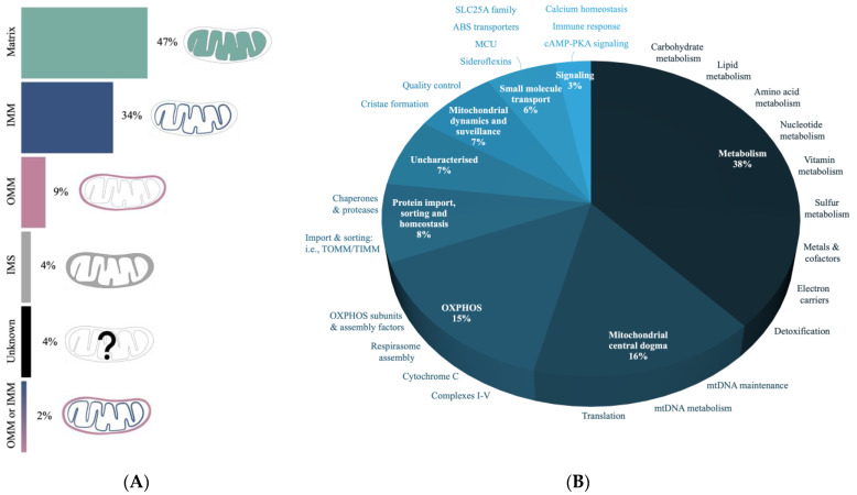
Mitochondria are ancient endosymbiotic double membrane organelles that support a wide range of eukaryotic cell functions through energy, metabolism, and cellular control. There are over 1000 known proteins that either reside within the mitochondria or are transiently associated with it. These mitochondrial proteins represent a functional subcellular protein network (mtProteome) that is encoded by mitochondrial and nuclear genomes and significantly varies between cell types and conditions. In neurons, the high metabolic demand and differential energy requirements at the synapses are met by specific modifications to the mtProteome, resulting in alterations in the expression and functional properties of the proteins involved in energy production and quality control, including fission and fusion. The composition of mtProteomes also impacts the localization of mitochondria in axons and dendrites with a growing number of neurodegenerative diseases associated with changes in mitochondrial proteins. This review summarizes the findings on the composition and properties of mtProteomes important for mitochondrial energy production, calcium and lipid signaling, and quality control in neural cells. We highlight strategies in mass spectrometry (MS) proteomic analysis of mtProteomes from cultured cells and tissue. The research into mtProteome composition and function provides opportunities in biomarker discovery and drug development for the treatment of metabolic and neurodegenerative disease.

Keywords: bioinformatics; energetics; evolution; mass spectrometry; neurodegeneration; protein quantification; synapses.


Conflict of interest statement

The authors declare no conflict of interest.

Figures

Figure 1
Figure 1
The structure of and genomic properties of mitochondria. (A) Mitochondrial compartments include the outer mitochondrial membrane (OMM); the intermembrane space (IMS); the inner mitochondrial membrane (IMM), which folds to form cristae; and a matrix that houses mitochondrial DNA (mtDNA) and 80S ribosomes (mtRibosomes). Inset: mtDNA binds core proteins that interact with peripheral proteins, mtRibosomes, and the IMM, forming a nucleoid. Core proteins include transcription factor A (TFAM), Twinkle (TWNKL), RNA polymerase (POLRMT), single-stranded DNA binding protein (mtSSB), and polymerase subunit gamma (POLG), whereas peripheral proteins include ATPase family AAA domain containing protein 3 (ATAD3) and prohibitin (PHB1 and PHB2). (B) Human mtDNA (16,569 bp) encodes 2 rRNA, 22 tRNA, and 13 proteins that form subunits of oxidative phosphorylation (OXPHOS) complexes, including NADH dehydrogenases (ND1, ND2, ND3, ND4, ND4L, ND5, and ND6), cytochrome c oxidases (COX1, COX2, and COX3), ATP synthases (ATP6 and ATP8), and cytochrome b (CyB).
Figure 2
Figure 2
Adaptive properties in mitochondrial quality control. Mitochondrial-damage associated molecular patterns (mtDAMPs) appear in inflammatory states including high concentrations of mtDNA, ATP, calcium ions (Ca2+), cytochrome c (CyC), reactive oxygen species (ROS), and cardiolipin (CL). Mitochondria react to mtDAMPs via fission, fusion, derivation of mitochondrial-derived vesicles (MDVs), and mitophagy. Fission is mediated by fission 1 (FIS1) and dynamin-related protein 1 (DRP1), forming rounder mitochondria. Fusion is regulated by mitofusin (MFN1 and MFN2) and optic atrophy 1 (OPA1), forming elongated mitochondria. (1a–b) Fission and fusion exist in a dynamic equilibrium during healthy states: hyperfission (increased FIS1 and DRP1) is associated with decreases in mitochondrial membrane potential (mtΨ) and ATP production; hyperfusion is associated with increased mtΨ and ATP. (2) Single membrane MDVs express translocase of the outer mitochondrial membrane 20 (TOMM20), carrying damaged components for lysosomal degradation. (3) Mitophagy degradation through various pathways such as Parkin-mediated mitophagy, which begins with the phosphorylation of Parkin by PTEN-induced putative kinase 1 (PINK1) and the ubiquitination of mitochondrial substrates (mtSubstates), triggering optineurin interaction with microtubule-associated protein light chain 3 (LC3) and leading to the formation of a phagophore and mitophagosome that fuses with the lysosomes.
Figure 3
Figure 3
The transport and function of synaptic mitochondria. Movement of mitochondria along the axonal cytoskeleton is mediated by kinesin and dynein microtubule motors that are complexed with Milton/TRAK proteins and Miro, a protein localized to the outer membrane of mitochondria. Kinesin traffics mitochondria from soma to axon terminals (anterogradely) and dynein back to the soma (retrogradely). As neurons develop, the rate of mitochondrial trafficking to synapses decreases, hence, synapses are dense in mitochondria and accommodate high energy demands at presynaptic terminals. Synaptic mitochondria exhibit unique properties including higher levels of ROS, fission, and lower ATP production.
Figure 4
Figure 4
Summary of mtProteome analysis. (1) Mitochondria isolated from mouse models or cultured cells. (2) Mitochondrial extraction obtained using centrifugation and/or density gradient separation. Mitochondria can also be isolated using tagged affinity purification (e.g., HA-Mito-tag). (3) Mitochondrial fraction purity can be confirmed using a Western blot detection of mitochondrial proteins (e.g., CyC). (4) Protein samples can be prepared for MS analysis using internal tagging (e.g., isotopic or isobaric) or label-free quantitative approaches. (5) MS analysis using methods such as electrospray ionization (ESI) liquid chromatography (LC) enables peptide separation and detection. (6) Protein results can be further analyzed using bioinformatic tools such as STRING network analysis.
Figure 5
Figure 5
Annotation of mtProteomes in CNS using MitoCarta3.0. (A) The submitochondrial distribution profiles of the mtProteome based on compartments: matrix, inner mitochondrial membrane (IMM), outer mitochondrial membrane (OMM), and intermembrane space (IMS). (B) Characterization of mtProteome-based functional properties. Most proteins are involved in various types of metabolism, followed by the mtDNA regulation and oxidative phosphorylation (OXPHOS).

References

    1. Margulis L. Origin of Eukaryotic Cells: Evidence and Research Implications for a Theory of the Origin and Evolution of Microbial, Plant, and Animal Cells on the Precambrian Earth. Yale University Press; New Haven, CT, USA: 1970.
    1. Gray M.W. Mitochondrial Evolution. Cold Spring Harb. Perspect. Biol. 2012;4:a011403. doi: 10.1101/cshperspect.a011403. - DOI - PMC - PubMed
    1. Wiedemann N., Pfanner N. Mitochondrial Machineries for Protein Import and Assembly. Annu. Rev. Biochem. 2017;86:685–714. doi: 10.1146/annurev-biochem-060815-014352. - DOI - PubMed
    1. Roger A.J., Muñoz-Gómez S.A., Kamikawa R. The Origin and Diversification of Mitochondria. Curr. Biol. 2017;27:R1177–R1192. doi: 10.1016/j.cub.2017.09.015. - DOI - PubMed
    1. Liu M., Spremulli L. Interaction of Mammalian Mitochondrial Ribosomes with the Inner Membrane. J. Biol. Chem. 2000;275:29400–29406. doi: 10.1074/jbc.M002173200. - DOI - PubMed

Publication types