. 2023 Nov 13;13(11):1644.
doi: 10.3390/biom13111644.

Placentas from Women with Late-Onset Preeclampsia Exhibit Increased Expression of the NLRP3 Inflammasome Machinery

Affiliations

Placentas from Women with Late-Onset Preeclampsia Exhibit Increased Expression of the NLRP3 Inflammasome Machinery

Luis M Garcia-Puente et al. Biomolecules. .

Abstract

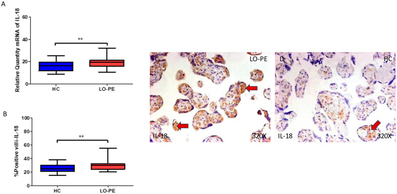
Pre-eclampsia is a harmful and potentially lethal medical condition during pregnancy clinically diagnosed by hypertension and commonly accompanied by proteinuria and multiorgan affections. According to the time of diagnosis, it is differentiated between early-onset (EO-PE) and late-onset preeclampsia (LO-PE). Despite being less dangerous and presenting distinct pathophysiological signatures, LO-PE has a greater prevalence than EO-PE, both having significant consequences on the placenta. Previous works have evidenced that exacerbated inflammation in this organ might play a potential pathogenic role in the development of pre-eclampsia, and there is some preliminary evidence that the hyperactivation of inflammasomes can be related to the altered immunoinflammatory responses observed in the placentas of these patients. However, the precise role of inflammasomes in the placentas of women with LO-PE remains to be fully understood. In this work, we have studied the gene and protein expression of the main components related to the canonical and non-canonical pathways of the inflammasome NLRP3 (NLRP3, ASC, caspase 1, caspase 5, caspase 8, interleukin 1β, and interleukin 18) in the placental tissue of women with LO-PE. Our results show a marked increase in all these components in the placentas of women who have undergone LO-PE, suggesting that NLRP3 inflammasome plays a potentially pathophysiological role in the development of this entity. Future works should aim to evaluate possible translational approaches to this dysregulation in these patients.

Keywords: NLRP3 inflammasome; caspases; interleukin 18; interleukin 1β; late-onset pre-eclampsia (LO-PE); placenta.


Conflict of interest statement

The authors declare no conflict of interest.

Figures

Figure 1
Figure 1
(A) NLRP3 mRNA expression in women with LO-PE and HC. (B) IRS scores for NLRP3 expression in the placental villi of the LO-PE and HC group. (C,D) Images showing immunostaining for NLRP3 in the placental villi of the LO-PE and HC. p < 0.001 (***). Red arrows: Tissue expression of NLRP3 was strongly evidenced in the syncytiotrophoblast layer of women with LO-PE, whereas for HC, it is more limited to the cytotrophoblast and other cells located in the inner of placental villi.
Figure 2
Figure 2
(A) ASC mRNA expression in women with LO-PE and HC. (B) IRS scores for ASC expression in the placental villi of the LO-PE and HC group. (C,D) Images showing immunostaining for ASC in the placental villi of the LO-PE and HC. p < 0.001 (***). In red arrows, it can be observed that expression of this component in the placentas of women with LO-PE is mainly found in the syncytiotrophoblast layer, whereas for HC, it is observed in cytotrophoblast and the other cells present in the inner of the placental villi.
Figure 3
Figure 3
(A) Caspase 1 mRNA expression in women with LO-PE and HC. (B) IRS scores for caspase 1 expression in the placental villi of the LO-PE and HC group. (C,D) Images showing immunostaining for caspase 1 in the placental villi of the LO-PE and HC. p < 0.01 (**). In red arrows, the expression of caspase 1 in the syncytiotrophoblast layer and also in the cytotrophoblast and other cells in the inner layer of the placental villi in women with LO-PE is highlighted, whereas for HC, it is more located in the syncytiotrophoblast layer.
Figure 4
Figure 4
(A) Caspase 5 mRNA expression in women with LO-PE and HC. (B) IRS scores for caspase 5 expression in the placental villi of the LO-PE and HC group. (C,D) Images showing immunostaining for caspase 5 in the placental villi of the LO-PE and HC. p < 0.001 (***). In red arrows, caspase 5 expression is observed throughout the placental villi of women affected by LO-PE in comparison to HC, in which this marker can be slightly observed in the cytotrophoblasts and other cells in the inner layer of the placental villi.
Figure 5
Figure 5
(A) Caspase 8 mRNA expression in women with LO-PE and HC. (B) IRS scores for caspase 8 expression in the placental villi of the LO-PE and HC group. (C,D) Images showing immunostaining for caspase 8 in the placental villi of the LO-PE and HC. p < 0.01 (**); p < 0.05 (*). Red arrows: caspase 8 expression is evident in the cytotrophoblasts and other cells located in the inner of the placental villi of women affected by LO-PE in comparison to HC, in which the expression of this marker can be differentially observed in syncytiotrophoblasts.
Figure 6
Figure 6
(A) IL-1β mRNA expression in women with LO-PE and HC. (B) IRS scores for IL-1β expression in the placental villi of the LO-PE and HC group. (C,D) Images showing immunostaining for IL-1β in the placental villi of the LO-PE and HC. p < 0.001 (***). Red arrow: IL-1β is highly expressed in the syncytiotrophoblast layer of the placentas of women affected by LO-PE in comparison to HC, in which the expression of this marker is slight and almost limited to some syncytiotrophoblasts and cytotrophoblasts.
Figure 7
Figure 7
(A) Caspase 1 mRNA expression in women with LO-PE and HC. (B) IRS scores for caspase 1 expression in the placental villi of the LO-PE and HC group. (C,D) Images showing immunostaining for caspase 1 in the placental villi of the LO-PE and HC. p < 0.01 (**). Red arrows: IL-18 expression was strongly displayed throughout the placental villi of women affected by LO-PE in comparison to HC, in which a moderate expression of this cytokine can be observed in cytotrophoblast and other cells located in the inner of the placental villi.
Figure 8
Figure 8
Graphical abstract of the results observed in our study.

Similar articles

Cited by

References

    1. Garovic V.D., White W.M., Vaughan L., Saiki M., Parashuram S., Garcia-Valencia O., Weissgerber T.L., Milic N., Weaver A., Mielke M.M. Incidence and Long-Term Outcomes of Hypertensive Disorders of Pregnancy. J. Am. Coll. Cardiol. 2020;75:2323–2334. doi: 10.1016/j.jacc.2020.03.028. - DOI - PMC - PubMed
    1. Hutcheon J.A., Lisonkova S., Joseph K. Epidemiology of Pre-Eclampsia and the Other Hypertensive Disorders of Pregnancy. Best Pract. Res. Clin. Obstet. Gynaecol. 2011;25:391–403. doi: 10.1016/j.bpobgyn.2011.01.006. - DOI - PubMed
    1. Voto L.S., Zeitune M.G. Perinatology. Springer; Cham, Switzerland: 2022. Preeclampsia; pp. 707–746. - DOI
    1. Filipek A., Jurewicz E. Preeclampsia—A Disease of Pregnant Women. Postep. Biochem. 2018;64:229–232. doi: 10.18388/pb.2018_146. - DOI - PubMed
    1. Ives C.W., Sinkey R., Rajapreyar I., Tita A.T.N., Oparil S. Preeclampsia-Pathophysiology and Clinical Presentations: JACC State-of-the-Art Review. J. Am. Coll. Cardiol. 2020;76:1690–1702. doi: 10.1016/j.jacc.2020.08.014. - DOI - PubMed

Publication types

Substances

LinkOut - more resources