. 2023 Nov 16;13(11):1655.
doi: 10.3390/biom13111655.

DPH1 Gene Mutations Identify a Candidate SAM Pocket in Radical Enzyme Dph1•Dph2 for Diphthamide Synthesis on EF2

Affiliations

DPH1 Gene Mutations Identify a Candidate SAM Pocket in Radical Enzyme Dph1•Dph2 for Diphthamide Synthesis on EF2

Koray Ütkür et al. Biomolecules. .

Abstract

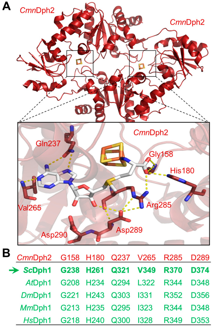
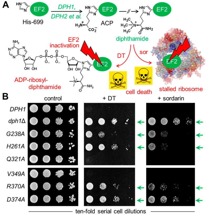
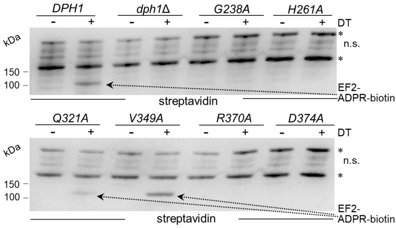
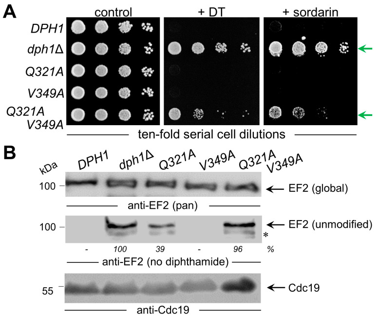
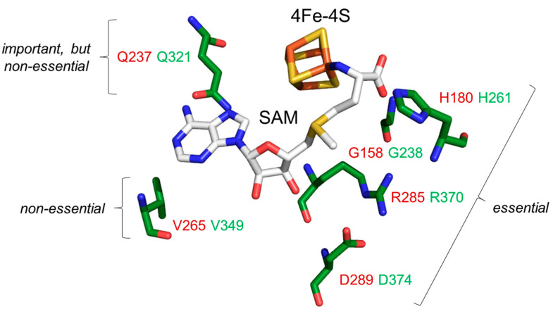
In eukaryotes, the Dph1•Dph2 dimer is a non-canonical radical SAM enzyme. Using iron-sulfur (FeS) clusters, it cleaves the cosubstrate S-adenosyl-methionine (SAM) to form a 3-amino-3-carboxy-propyl (ACP) radical for the synthesis of diphthamide. The latter decorates a histidine residue on elongation factor 2 (EF2) conserved from archaea to yeast and humans and is important for accurate mRNA translation and protein synthesis. Guided by evidence from archaeal orthologues, we searched for a putative SAM-binding pocket in Dph1•Dph2 from Saccharomyces cerevisiae. We predict an SAM-binding pocket near the FeS cluster domain that is conserved across eukaryotes in Dph1 but not Dph2. Site-directed DPH1 mutagenesis and functional characterization through assay diagnostics for the loss of diphthamide reveal that the SAM pocket is essential for synthesis of the décor on EF2 in vivo. Further evidence from structural modeling suggests particularly critical residues close to the methionine moiety of SAM. Presumably, they facilitate a geometry specific for SAM cleavage and ACP radical formation that distinguishes Dph1•Dph2 from classical radical SAM enzymes, which generate canonical 5'-deoxyadenosyl (dAdo) radicals.

Keywords: ADP ribosylation; Dph1•Dph2; EF2 diphthamide modification; SAM; Saccharomyces cerevisiae; diphtheria toxin; radical SAM enzymes.


Conflict of interest statement

K.M. and U.B. are employed by and members of Roche Pharma Research & Early Development (pRED) and are co-inventors on patent applications that cover assays to detect the presence or absence of diphthamide. Roche is interested in targeted therapies and diagnostics. All other authors declare no conflict of interest.

Figures

Figure 1
Figure 1
The SAM-binding pocket in archaeal Dph2 subunits of ACP synthase dimers from Candidatus methanoperedens nitroreducens (CmnDph2•Dph2) was conserved in the Dph1 subunit of eukaryal counterparts (Dph1•Dph2). (A) Crystal structure of CmnDph2•Dph2 homodimer in complex with SAM (PDB: 6BXN) revealed specific SAM-interacting residues (orange dotted lines represent polar contacts): Gly-158, His-180, Gln-237, Val-265, Arg-285, Asp2-89 and Asp-290 [14]. (B) Excerpt of the amino acid sequence alignment between CmnDph2 (in red) and Dph1 subunits from model eukaryotes (in green): S. cerevisiae (Sc), A. thaliana (At), D. melanogaster (Dm), M. musculus (Mm) and H. sapiens (Hs). The table sums up eukaryal Dph1 residues that were conserved or similar to the SAM pocket from CmnDph2. Residues identical between CmnDph2 and ScDph1 are labeled in bold, with the arrow highlighting the amino acid positions chosen for alanine-specific substitution mutagenesis of yeast gene DPH1. For full alignment details, see Figures S1 and S2.
Figure 2
Figure 2
Site-directed DPH1 mutagenesis uncovered a candidate SAM pocket in yeast Dph1•Dph2 relevant to diphthamide synthesis on EF2. (A) Simplified scheme showing diphthamide synthesis initiated by the DPH1 and DPH2 gene products and other factors (et al.) to modify His-699 on yeast EF2 with ACP. Subsequent enzymatic steps that completed diphthamide are not shown in detail for simplicity. Diphthamide is pathologically relevant; it can be hijacked by diphtheria toxin (DT) for inhibitory ADP ribosylation of EF2 and induction of cell death (skull and crossbones) or complexed by antifungal sordarin (sor) to irreversibly stall elongating ribosomes. (B) Cell growth assays in response to DT and sordarin to phenotypically diagnose diphthamide synthesis on EF2. As indicated, yeast tester strains comprised wild-type (DPH1) and null mutant (dph1∆) controls as well as the candidate SAM pocket mutants (G238A, H261A, Q321A V349A, R370A and D374A). Ten-fold serial cell dilutions were cultivated for 2–3 days at 30 °C without DT or sordarin (left panel: control) under conditions of endogenous DT fragment A production from a galactose-inducible expression plasmid (middle panel: + DT) [19] or in the presence of 12.5 µg/mL antifungal (right panel: +sordarin) sufficient to inhibit the wild-type (DPH1) control. Green arrows indicate DT and sordarin resistance.
Figure 3
Figure 3
Diphthamide-dependent ADP ribosylation of EF2 by DT in vitro. Total protein extracts obtained from yeast strains with the indicated genetic backgrounds were treated with (+) or without (−) DT (200 ng) in the presence of 5 µM biotin-NAD at 25 °C for 1 h. Detection of the biotin moiety transferred with ADP-ribose to EF2 by DT involved Western blots with a streptavidin peroxidase conjugate. The reaction product (EF2-ADPR-biotin) with a molecular weight of ~100 kDa is denoted by dotted arrows. Unspecific (n.s.) bands marked with an asterisk likely represent endogenously biotinylated yeast proteins. Note that V349A produces wild-type-like EF2-ADPR-biotin signals diagnostic for EF2 diphthamide modification, while significantly weaker signals from Q321A suggest reduced (but not entirely abolished) Dph1 activity. Original images can be found in Supplementary Materials (Figure S8).
Figure 4
Figure 4
Characterization of single Q321A and V391A mutants alone and in tandem. (A) Phenotypic spot assays diagnostic for diphthamide modification defects. As indicated, the tester strains comprised wild-type (DPH1) and null-mutant (dph1∆) controls as well as single (Q321A or V391A) and double (Q321A V391A) mutants. Ten-fold serial cell dilutions were cultivated and grown under conditions essentially described in Figure 2B legend. DT and sordarin resistance traits are indicated (green arrows). (B) Analysis of the EF2 modification state using anti-EF2(pan) antibodies for global EF2 recognition and anti-eEF2(no diphthamide) antibodies to specifically detect unmodified EF2 [37]. Quantifications of unmodified EF2 signals relative to dph1∆ are given as percentage (%) values. For Western blots, protein extracts were obtained from tester strains with the genetic backgrounds as indicated in (A). EF2 degradation products are marked with an asterisk. Detection of Cdc19 with anti-Cdc19 antibodies is shown in parallel Western blots. Note that the combination of the two single Q321A and V391A mutations in the double mutant is phenotypically additive (A) and causes diphthamide modification defects (B) comparable to the null mutant (dph1∆). Original images can be found in Supplementary Materials (Figure S9).
Figure 5
Figure 5
Functional distinctions of individual SAM pocket residues identified in subunit Dph1 from the yeast Dph1•Dph2 dimer. An AlphaFold2-based structural model [35,36] of Dph1 was aligned to the structure of CmnDph2 in complex with SAM (PDB: 6BXN). Protein structures of CmnDph2 are hidden, while the 4Fe-4S and SAM cofactors remain shown as sticks. Dph1 amino acids (green) are part of a SAM pocket (Gly-238, His-261, Gln-321, Val-349, Arg-370 and Asp-374 conserved to CmnDph2 residues (red) with Gly-158, His-180, Gln-237, Val-265, Arg-285 and Asp-289, respectively (see also Figure 1A)). Residues in proximity to the SAM methionine moiety are essential (Gly-238, His-261, Arg-370 and Asp-374), while amino acids close to the adenine are important but nonessential (Gln-321) or dispensable and nonessential (Val-349).

Similar articles

Cited by

References

    1. Frey P.A., Hegeman A.D., Ruzicka F.J. The radical SAM superfamily. Crit. Rev. Biochem. Mol. Biol. 2008;43:63–88. doi: 10.1080/10409230701829169. - DOI - PubMed
    1. Landgraf B.J., McCarthy E.L., Booker S.J. Radical S-adenosylmethionine enzymes in human health and disease. Annu. Rev. Biochem. 2016;85:485–514. doi: 10.1146/annurev-biochem-060713-035504. - DOI - PubMed
    1. Broderick J.B., Duffus B.R., Duschene K.S., Shepard E.M. Radical S-adenosylmethionine enzymes. Chem. Rev. 2014;114:4229–4317. doi: 10.1021/cr4004709. - DOI - PMC - PubMed
    1. Yokoyama K., Lilla E.A. C-C bond forming radical SAM enzymes involved in the construction of carbon skeletons of cofactors and natural products. Nat. Prod. Rep. 2018;35:660–694. doi: 10.1039/C8NP00006A. - DOI - PMC - PubMed
    1. Hoffman B.M., Broderick W.E., Broderick J.B. Mechanism of radical initiation in the radical SAM enzyme superfamily. Annu. Rev. Biochem. 2023;92:333–349. doi: 10.1146/annurev-biochem-052621-090638. - DOI - PMC - PubMed

Publication types

MeSH terms

LinkOut - more resources