. 2023 Nov 17;13(11):1659.
doi: 10.3390/biom13111659.

Dithiothreitol Affects the Fertilization Response in Immature and Maturing Starfish Oocytes

Affiliations

Dithiothreitol Affects the Fertilization Response in Immature and Maturing Starfish Oocytes

Nunzia Limatola et al. Biomolecules. .

Abstract

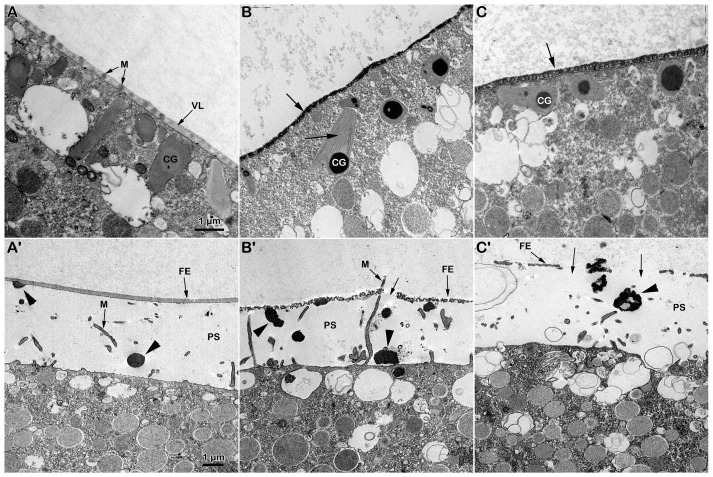
Immature starfish oocytes isolated from the ovary are susceptible to polyspermy due to the structural organization of the vitelline layer covering the oocyte plasma membrane, as well as the distribution and biochemical properties of the actin cytoskeleton of the oocyte cortex. After the resumption of the meiotic cycle of the oocyte triggered by the hormone 1-methyladenine, the maturing oocyte reaches fertilizable conditions to be stimulated by only one sperm with a normal Ca2+ response and cortical reaction. This cytoplasmic ripening of the oocyte, resulting in normal fertilization and development, is due to the remodeling of the cortical actin cytoskeleton and germinal vesicle breakdown (GVBD). Since disulfide-reducing agents such as dithiothreitol (DTT) are known to induce the maturation and GVBD of oocytes in many species of starfish, we analyzed the pattern of the fertilization response displayed by Astropecten aranciacus oocytes pre-exposed to DTT with or without 1-MA stimulation. Short treatment of A. aranciacus immature oocytes with DTT reduced the rate of polyspermic fertilization and altered the sperm-induced Ca2+ response by changing the morphology of microvilli, cortical granules, and biochemical properties of the cortical F-actin. At variance with 1-MA, the DTT treatment of immature starfish oocytes for 70 min did not induce GVBD. On the other hand, the DTT treatment caused an alteration in microvilli morphology and a drastic depolymerization of the cortical F-actin, which impaired the sperm-induced Ca2+ response at fertilization and the subsequent embryonic development.

Keywords: DTT; actin; calcium; disulfide-reducing agents; fertilization; oocyte maturation; polyspermy; starfish.


Conflict of interest statement

The authors declare no conflict of interest.

Figures

Figure 1
Figure 1
Brief exposure to DTT before insemination reduces the extent of polyspermy in immature (GV-stage) oocytes of A. aranciacus. (A) Insemination of GV-stage oocytes suspended in NSW induces the formation of multiple fertilization cones (arrowheads) due to polyspermic fertilization. Note the localized separation of the fertilization envelope (arrow) in some restricted surface areas. (B) DTT treatment (10 min) reduces the number of fertilization cones (arrowhead) as fewer sperm activate and penetrate the oocytes. Oocyte-incorporated sperm (arrowheads) were visualized via the fluorescent dye binding to DNA (CE) and were counted to be presented in the histograms (F). Epifluorescence photomicrographs of representative oocytes inseminated in NSW with (D) or without DTT exposure (C) were compared with the ones inseminated after DTT exposure (10 min) and washed with NSW (E). * Tukey’s post hoc test, p < 0.01.
Figure 2
Figure 2
Effect of DTT on the surface of A. aranciacus immature oocytes before and after insemination. (A) Ultrastructure of a control GV-stage oocyte in NSW visualized with transmission electron microscopy (TEM) at lower and higher magnification (inset). The vitelline layer (VL) appears as a homogenous layer in which microvilli (M) are orderly embedded. (B) A brief DTT treatment (10 min) of the oocytes induces changes in the VL structure and microvillar morphology, which is more evident in the regions where cytoplasm bulges next to the microvilli exhibiting interdigitation profile (inset). (C) The TEM micrograph of the immature oocytes inseminated in NSW depicts a fertilization cone (arrow) formed beneath partially elevating FE due to CG exocytosis (arrowhead) and microvilli (M) elongation. (D) Insemination of DTT-treated oocytes evidences partial destruction of the VL (arrows), which now forms the fertilization envelope (FE), and more frequent occurrence of the electron-dense content within the CGs being released in the perivitelline space (PS) (arrowheads).
Figure 3
Figure 3
DTT-induced changes in the cortical F-actin distribution and dynamic before and after insemination of starfish immature oocytes. Live A. aranciacus GV-stage oocytes injected with Alexa 568-phalloidin were treated with DTT and inseminated to monitor the real-time changes in the F-actin with confocal laser scanning microscopy. Network of F-actin distribution in control oocytes in NSW before (A) and after insemination: 5 min (A′) and 20 min (A″). The overlay in (A′) with images of fluorescently labeled sperm head (blue) shows the formation of multiple fertilization cones (arrow) with polymerization of actin fibers to incorporate the sperm (A″, arrowhead). (B) DTT treatment (10 mM, 10 min) induces alteration in the outermost cortical F-actin structure (note the partially detaching of actin filaments from the GV nuclear envelope indicated by an arrow), which compromises the subsequent formation of the actin fibers (arrowhead) incorporating sperm 5 min (B′) and sometimes even 20 min after insemination (B″). This tendency is evident in DTT-treated oocytes inseminated after washing in NSW (C). The less defined cortical polymerization of actin filaments (arrowhead) at 5 min (C′) and 20 min (C″) may explain why the oocytes are penetrated by fewer sperm, as shown in Figure 1.
Figure 4
Figure 4
DTT exposure does not induce GVBD in immature A. aranciacus oocytes and alters their cortical reaction upon insemination. (A) A GV-stage oocyte stimulated with the natural maturing hormone 1-MA underwent GVBD by 70 min incubation, displaying the total and equidistant elevation of the fertilization envelope (FE) 5 min upon insemination. (B) A GV-stage oocyte treated for 70 min with DTT failed to undergo GVBD and intermixing of the nucleoplasm with the cytoplasm. Five minutes after insemination, these treated oocytes did not display a clear sign of FE elevation under the light microscope. (C) DTT (10 min) added after the incubation of immature oocytes with 1-MA for 60 min was sufficient to compromise the exocytosis of the CGs and the subsequent elevation of the fertilization envelope (FE), despite the apparently normal proceedings of GVBD. The number of DNA-stained sperm in the zygotes was assessed 10 min after insemination by counting the fluorescent signals corresponding to the sperm head (arrowheads) in oocytes stimulated for 70 min with 1-MA (D), oocytes treated for 70 min with DTT (E), oocytes stimulated for 60 min with 1-MA and then treated with DTT for 10 min (F). The results are presented in histograms (G). Predominantly, only one sperm entered (monospermic) in all the described experimental conditions.
Figure 5
Figure 5
DTT treatment interferes with F-actin dynamics in maturing oocytes. A. aranciacus immature oocytes pre-injected with Alexa 568-phalloidin were stimulated for 70 min with 1-MA, and the changes in F-actin distribution were monitored before (A) and after insemination with confocal laser scanning microscopy: 5 min (A′); 20 min (A″). Note that actin filaments oriented perpendicularly to the plasma membrane of the unfertilized mature egg. The overlay image in (A′) indicates that sperm addition promotes the elevation of the fertilization envelope (FE) and cortical F-actin remodeling along the fertilization cone to capture the sperm about 5 min after insemination (blue spot and arrow) and more profoundly in the cytoplasm 20 min later (A″, blue spot and arrow). (B) Treatment of an immature oocyte for 70 min with DTT does not induce GVBD but reorganizes the cortical actin cytoskeleton in a way that is distinct from what 1-MA does. As the cortical actin cytoskeleton is more depolymerized in these eggs, the subsequent insemination does not produce the fertilization cone formation by 5 min (B′). Only 20 min after sperm addition is it possible to detect actin fibers in the cytoplasm with an altered structural organization (B″, arrow). (C) The cortical F-actin remodeling in A. aranciacus oocytes stimulated for 60 min with 1-MA to induce GVBD and then exposed to DTT for 10 min. The effect of the short external application of DTT is manifested in the impairment of the cortical actin dynamics following fertilization in the presence of DTT, leading to the formation of a morphologically altered fertilization cone and F-actin structure to incorporate the sperm 5 min and 20 min after insemination (blue spots and arrows in (C′) and (C″), respectively). (D) The cortical actin distribution in an oocyte washed with NSW after being incubated for 60 min with 1-MA and then with DTT for 10 min. Insemination in NSW still affects the cortical actin dynamics to form the fertilization cone and to bring in the sperm (arrows) by 5 min (D′) and 20 min (D″) after insemination.
Figure 6
Figure 6
Ultrastructural analyses of DTT-treated immature oocytes before and after insemination. (A) TEM micrograph of an A. aranciacus GV-stage oocyte stimulated with 1-MA for 70 min. The microvilli (M) are ensheathed by the vitelline layer (VL). Beneath that, cortical granules (CGs) are positioned perpendicularly to the mature egg surface. (A′) Five minutes after insemination, the electron-dense rounded content of the CGs (arrowhead) is now extruded into the perivitelline space (PS) by exocytosis. Note the thick layer of the fertilization envelope (FE). (B) Treatment of immature oocytes with DTT for 70 min heavily alters the structure of the VL, to the extent that it prevents the visualization of the embedded microvilli (arrow). The internal structure of the CGs in these oocytes exhibits a more electron-dense content filamentous bodies (arrow). (B′) Five minutes after insemination, the elevated fertilization envelope (FE) shows partial disruption (arrow). The effect of DTT on the CGs is also evidenced by the altered morphology of their electron-dense content (arrowheads) exocytosed in the perivitelline space (PS). (C) The surface of an immature oocyte treated with 1-MA for 60 min and for an additional 10 min by DTT. Microvilli (arrow), cortical granules (CG). (C′) The short DTT treatment (10 min after stimulation with 1-MA for 60 min) was sufficient to alter the structure of the VL and CGs (arrowhead), as indicated by the rupture of the FE (arrows) in zygotes examined 5 min after insemination.
Figure 7
Figure 7
DTT preincubation alters the Ca2+ responses in starfish oocytes inseminated at the GV-stage. (A) The instantaneous increases in Ca2+ levels in an immature A. aranciacus oocyte after insemination in NSW. The Ca2+ signals detected upon sperm addition are the initiations of multiple Ca2+ waves (Ca2+ spots, arrowheads) due to stimulation by numerous sperm. Then, the cortical Ca2+ increase (the cortical flash, arrow) is triggered, occurring simultaneously at the periphery of the oocyte cortex. The multiple Ca2+ waves converge and run together to propagate to the opposite pole. (B) Ca2+ responses in an immature oocyte inseminated after 10 min of pretreatment with DTT. Ca2+ spot (arrowhead) and cortical flash (arrow) (C) Ca2+ responses in the same DTT-pretreated oocytes that were inseminated after washing in NSW. Ca2+ spot (arrowhead) and cortical flash (arrow). (D) Quantified Ca2+ trajectories represented in relative fluorescence unit (RFU) as defined in Section 2. The moment of the first detected Ca2+ signal was taken as t = 0, and the time is expressed in the minute and second format (mm: ss).
Figure 8
Figure 8
Alteration in the Ca2+ response at fertilization of immature oocytes induced to undergo maturation with DTT treatment. (A) The pseudocolor images of the Ca2+ response at fertilization of a control A. aranciacus GV-stage oocyte stimulated for 70 min with 1-MA. Sperm binding triggers the release of Ca2+ simultaneously at the periphery of the naturally matured egg (cortical flash, arrow), which is then followed by a Ca2+ wave (CW) that propagates from the sperm–egg fusion site to the opposite pole. (B) Treatment of immature oocytes with DTT for 70 min inhibits the sperm-induced CF and reduces the peak amplitude of the CW. The arrowhead indicates the initiation of the sperm-induced CW. (C) A short treatment with DTT (10 min) after stimulation of immature oocytes with 1-MA for 60 min (to induce GVBD) is sufficient to alter the Ca2+ response at fertilization. Cortical flash (arrow). (D) The washing out of DTT before insemination of eggs treated as in C still affects the Ca2+ response at fertilization. The arrowhead indicates the initiation of the sperm-induced CW. The pseudocolor fluorescent images and the graphs in (E) (inset) evidence the CF and CW being lower than the control in amplitude. (E) Quantified Ca2+ trajectories are represented in relative fluorescence unit (RFU).

Similar articles

Cited by

References

    1. Kanatani H. Maturation-Inducing Substance in Starfishes. Int. Rev. Cytol. 1973;35:253–298. doi: 10.1016/S0074-7696(08)60356-3. - DOI - PubMed
    1. Meijer L., Guerrier P. Maturation and Fertilization in Starfish Oocytes. Int. Rev. Cytol. 1984;86:129–196. doi: 10.1016/s0074-7696(08)60179-5. - DOI - PubMed
    1. Kishimoto T. MPF-based meiotic cell cycle control: Half a century of lessons from starfish oocytes. Proc. Jpn. Acad. Ser. B. 2018;94:180–203. doi: 10.2183/pjab.94.013. - DOI - PMC - PubMed
    1. Chiba K. Oocyte Maturation in Starfish. Cells. 2020;9:476. doi: 10.3390/cells9020476. - DOI - PMC - PubMed
    1. Dale B., de Santis A., Hoshi M. Membrane response to 1-methyladenine requires the presence of the nucleus. Nature. 1979;282:89–90. doi: 10.1038/282089a0. - DOI - PubMed

Publication types

LinkOut - more resources