Extracellular Mechanical Stimuli Alters the Metastatic Progression of Prostate Cancer Cells within 3D Tissue Matrix
- PMID: 38002395
- PMCID: PMC10669840
- DOI: 10.3390/bioengineering10111271
Extracellular Mechanical Stimuli Alters the Metastatic Progression of Prostate Cancer Cells within 3D Tissue Matrix
Abstract
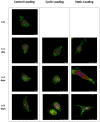
This study aimed to understand extracellular mechanical stimuli's effect on prostate cancer cells' metastatic progression within a three-dimensional (3D) bone-like microenvironment. In this study, a mechanical loading platform, EQUicycler, has been employed to create physiologically relevant static and cyclic mechanical stimuli to a prostate cancer cell (PC-3)-embedded 3D tissue matrix. Three mechanical stimuli conditions were applied: control (no loading), cyclic (1% strain at 1 Hz), and static mechanical stimuli (1% strain). The changes in prostate cancer cells' cytoskeletal reorganization, polarity (elongation index), proliferation, expression level of N-Cadherin (metastasis-associated gene), and migratory potential within the 3D collagen structures were assessed upon mechanical stimuli. The results have shown that static mechanical stimuli increased the metastasis progression factors, including cell elongation (p < 0.001), cellular F-actin accumulation (p < 0.001), actin polymerization (p < 0.001), N-Cadherin gene expression, and invasion capacity of PC-3 cells within a bone-like microenvironment compared to its cyclic and control loading counterparts. This study established a novel system for studying metastatic cancer cells within bone and enables the creation of biomimetic in vitro models for cancer research and mechanobiology.
Keywords: EQUicycler; actin; bone; cancer; cyclic; cytoskeleton; elongation; extracellular; invasion; loading; mechanical; metastasis; prostate; static; stimuli.
Conflict of interest statement
The authors declare no conflict of interest.
Figures


Similar articles
-
Design and Validation of Equiaxial Mechanical Strain Platform, EQUicycler, for 3D Tissue Engineered Constructs.Biomed Res Int. 2017;2017:3609703. doi: 10.1155/2017/3609703. Epub 2017 Jan 12. Biomed Res Int. 2017. PMID: 28168197 Free PMC article.
-
Mechanobiological evaluation of prostate cancer metastasis to bone using an in vitro prostate cancer testbed.J Biomech. 2021 Jan 4;114:110142. doi: 10.1016/j.jbiomech.2020.110142. Epub 2020 Nov 21. J Biomech. 2021. PMID: 33290947 Free PMC article.
-
Perfusion bioreactor enabled fluid-derived shear stress conditions for novel bone metastatic prostate cancer testbed.Biofabrication. 2021 Apr 2;13(3). doi: 10.1088/1758-5090/abd9d6. Biofabrication. 2021. PMID: 33418550
-
The matrix environmental and cell mechanical properties regulate cell migration and contribute to the invasive phenotype of cancer cells.Rep Prog Phys. 2019 Jun;82(6):064602. doi: 10.1088/1361-6633/ab1628. Epub 2019 Apr 4. Rep Prog Phys. 2019. PMID: 30947151 Review.
-
Molecular insights into prostate cancer progression: the missing link of tumor microenvironment.J Urol. 2005 Jan;173(1):10-20. doi: 10.1097/01.ju.0000141582.15218.10. J Urol. 2005. PMID: 15592017 Review.
Cited by
-
Mechanical Loading of Osteocytes via Oscillatory Fluid Flow Regulates Early-Stage PC-3 Prostate Cancer Metastasis to Bone.Adv Biol (Weinh). 2025 Apr;9(4):e2400824. doi: 10.1002/adbi.202400824. Epub 2025 Feb 19. Adv Biol (Weinh). 2025. PMID: 39969425 Free PMC article.
-
Nanostructured Biomaterials in 3D Tumor Tissue Engineering Scaffolds: Regenerative Medicine and Immunotherapies.Int J Mol Sci. 2024 May 16;25(10):5414. doi: 10.3390/ijms25105414. Int J Mol Sci. 2024. PMID: 38791452 Free PMC article. Review.
-
Environmental Exposure to Bisphenol A Enhances Invasiveness in Papillary Thyroid Cancer.Int J Mol Sci. 2025 Jan 19;26(2):814. doi: 10.3390/ijms26020814. Int J Mol Sci. 2025. PMID: 39859529 Free PMC article.
References
-
- Caley M.P., King H., Shah N., Wang K., Rodriguez-Teja M., Gronau J.H., Waxman J., Sturge J. Tumor-associated Endo180 requires stromal-derived LOX to promote metastatic prostate cancer cell migration on human ECM surfaces. Clin. Exp. Metastasis. 2016;33:151–165. doi: 10.1007/s10585-015-9765-7. - DOI - PMC - PubMed
Grants and funding
LinkOut - more resources
Full Text Sources
Research Materials

