

Quantitative Evaluation of Caries and Calculus with Ultrahigh-Resolution Optical Coherence Tomography
- PMID: 38002442
- PMCID: PMC10669567
- DOI: 10.3390/bioengineering10111317
Quantitative Evaluation of Caries and Calculus with Ultrahigh-Resolution Optical Coherence Tomography
Abstract
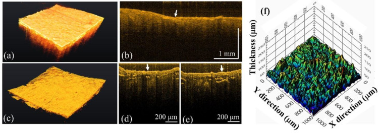
Dental caries on the crown's surface is caused by the interaction of bacteria and carbohydrates, which then gradually alter the tooth's structure. In addition, calculus is the root of periodontal disease. Optical coherence tomography (OCT) has been considered to be a promising tool for identifying dental caries; however, diagnosing dental caries in the early stage still remains challenging. In this study, we proposed an ultrahigh-resolution OCT (UHR-OCT) system with axial and transverse resolutions of 2.6 and 1.8 μm for differentiating the early-stage dental caries and calculus. The same teeth were also scanned by a conventional spectral-domain OCT (SD-OCT) system with an axial resolution of 7 μm. The results indicated that early-stage carious structures such as small cavities can be observed using UHR-OCT; however, the SD-OCT system with a lower resolution had difficulty identifying it. Moreover, the estimated surface roughness and the scattering coefficient of enamel were proposed for quantitatively differentiating the different stages of caries. Furthermore, the thickness of the calculus can be estimated from the UHR-OCT results. The results have demonstrated that UHR-OCT can detect caries and calculus in their early stages, showing that the proposed method for the quantitative evaluation of caries and calculus is potentially promising.
Keywords: calculus; caries; optical coherence tomography; roughness; scattering.
Conflict of interest statement
The authors declare no conflict of interest.
Figures


Similar articles
-
Visualization and tissue classification of human breast cancer images using ultrahigh-resolution OCT.Lasers Surg Med. 2017 Mar;49(3):258-269. doi: 10.1002/lsm.22654. Epub 2017 Mar 6. Lasers Surg Med. 2017. PMID: 28264146 Free PMC article.
-
Evaluation of calculus imaging on root surfaces by spectral-domain optical coherence tomography.Photodiagnosis Photodyn Ther. 2019 Mar;25:275-279. doi: 10.1016/j.pdpdt.2019.01.014. Epub 2019 Jan 14. Photodiagnosis Photodyn Ther. 2019. PMID: 30648636
-
Design and Simulation of an Ultrahigh-resolution Spectral-domain Optical Coherence Tomography.J Med Signals Sens. 2025 Apr 19;15:12. doi: 10.4103/jmss.jmss_36_24. eCollection 2025. J Med Signals Sens. 2025. PMID: 40351778 Free PMC article.
-
Ultra high-resolution anterior segment optical coherence tomography in the diagnosis and management of ocular surface squamous neoplasia.Ocul Surf. 2014 Jan;12(1):46-58. doi: 10.1016/j.jtos.2013.11.001. Epub 2013 Nov 9. Ocul Surf. 2014. PMID: 24439046 Free PMC article. Review.
-
Evaluation of dental caries, tooth crack, and age-related changes in tooth structure using optical coherence tomography.Jpn Dent Sci Rev. 2020 Nov;56(1):109-118. doi: 10.1016/j.jdsr.2020.08.001. Epub 2020 Oct 2. Jpn Dent Sci Rev. 2020. PMID: 33033549 Free PMC article. Review.
Cited by
-
Enhancing Oral Cancer Detection: A Systematic Review of the Diagnostic Accuracy and Future Integration of Optical Coherence Tomography with Artificial Intelligence.J Clin Med. 2024 Sep 29;13(19):5822. doi: 10.3390/jcm13195822. J Clin Med. 2024. PMID: 39407882 Free PMC article. Review.
References
-
- Franco E., Saunders C.P., Roberts G.J., Suwanprasit A. Dental disease, caries related microflora and salivary IgA of children with severe congenital cardiac disease: An epidemiological and oral microbial survey. Pediatr. Dent. 1996;18:228–235. - PubMed
-
- Pitts N.B., Zero D.T., Marsh P.D., Ekstrand K., Weintraub J.A., Ramos-Gomez F., Tagami J., Twetman S., Tsakos G., Ismail A. Dental caries. Nat. Rev. Dis. Primers. 2017;3:17030. - PubMed
-
- Kinane D.F., Stathopoulou P.G., Papapanou P.N. Periodontal diseases. Nat. Rev. Dis. Primers. 2017;3:17038. - PubMed
Grants and funding
LinkOut - more resources
Full Text Sources
