. 2023 Nov 18;10(11):1330.
doi: 10.3390/bioengineering10111330.

Functional Load Capacity of Teeth with Reduced Periodontal Support: A Finite Element Analysis

Affiliations

Functional Load Capacity of Teeth with Reduced Periodontal Support: A Finite Element Analysis

Marco Dederichs et al. Bioengineering (Basel). .

Abstract

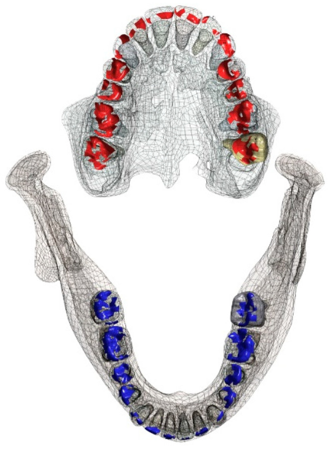
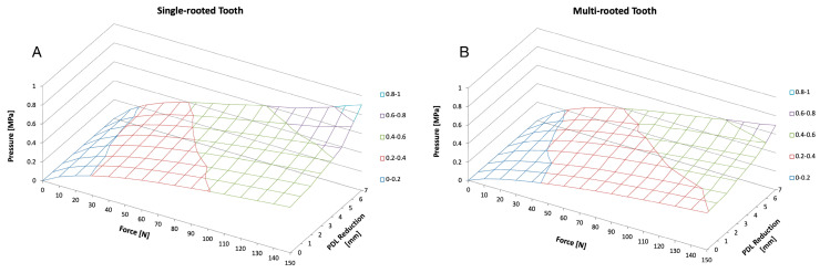
The purpose of this study was to investigate the functional load capacity of the periodontal ligament (PDL) in a full arch maxilla and mandible model using a numerical simulation. The goal was to determine the functional load pattern in multi- and single-rooted teeth with full and reduced periodontal support. CBCT data were used to create 3D models of a maxilla and mandible. The DICOM dataset was used to create a CAD model. For a precise description of the surfaces of each structure (enamel, dentin, cementum, pulp, PDL, gingiva, bone), each tooth was segmented separately, and the biomechanical characteristics were considered. Finite Element Analysis (FEA) software computed the biomechanical behavior of the stepwise increased force of 700 N in the cranial and 350 N in the ventral direction of the muscle approach of the masseter muscle. The periodontal attachment (cementum-PDL-bone contact) was subsequently reduced in 1 mm increments, and the simulation was repeated. Quantitative (pressure, tension, and deformation) and qualitative (color-coded images) data were recorded and descriptively analyzed. The teeth with the highest load capacities were the upper and lower molars (0.4-0.6 MPa), followed by the premolars (0.4-0.5 MPa) and canines (0.3-0.4 MPa) when vertically loaded. Qualitative data showed that the areas with the highest stress in the PDL were single-rooted teeth in the cervical and apical area and molars in the cervical and apical area in addition to the furcation roof. In both single- and multi-rooted teeth, the gradual reduction in bone levels caused an increase in the load on the remaining PDL. Cervical and apical areas, as well as the furcation roof, are the zones with the highest functional stress. The greater the bone loss, the higher the mechanical load on the residual periodontal supporting structures.

Keywords: attachment loss; bone loss; finite element analysis; mandible; maxilla; periodontium; teeth.


Conflict of interest statement

The authors declare no conflict of interest.

Figures

Figure 1
Figure 1
From the DICOM format to Finite Element Mesh. CBCT data (A) were transformed into the CAD file format as a segmented 3D model of the dento–alveolar complex (B) followed by the creation of surface mesh (C).
Figure 2
Figure 2
CAD-formatted 3D model (A) including posterior single- and multi-rooted teeth for = reduced periodontium analysis. The FEA model (B) shows that the forces were applied from the maxillary teeth and the maxilla (1, highlighted in red) toward the teeth and bone (blue) in the mandible (2).
Figure 3
Figure 3
CAD-formatted 3D model with indications of loading and boundary conditions. The maxilla (A) was set as fixated. The red arrow at the lower left first molar illustrates the direction in which the force was applied with maximal 700 N in the cranial and 350 N in the ventral direction (B) towards the maxilla. The blue axis (C) connecting the two condyles represents the axis of rotation. The red area reflects the insertion of the masseter muscle.
Figure 4
Figure 4
Areas of initial occlusal contact. In red the contacts on the maxillary teeth and in blue the contact area of the teeth in the mandible.
Figure 5
Figure 5
Pressure distribution under functional loading on teeth with full periodontal support. Areas of stress are colored yellow and red. The pressure distribution appears to be most prominent in the cervical area of the molars, premolars, and canine teeth (circle in the magnified area). In molars, the furcation roof (circled in the magnification) shows the most stressed area (see also Figure 6).
Figure 6
Figure 6
Distribution of forces under an axial load (Pressure stress—positive values, tensile stress—negative values). Under a simulated axial load, the pressure in the periodontium of the individual teeth is shown in the upper row diagrams (A). The upper first molars, and the second and first lower molars thereby carry the most force, followed by the premolars, and the canines (B). The anteriors are only minimally impacted by axial load. Teeth are numbered according to the Universal Numbering System.
Figure 7
Figure 7
Highlighted illustration of the bony housing of a single-rooted and a multi-rooted tooth during functional load. The area of the interradicular bone appears to be a highly stressed region.
Figure 8
Figure 8
Illustration of changes in force distribution when reducing the periodontal support. In fully periodontally supported teeth, the highest stress under loading conditions was found at the cervical area, the apical area, and in molars in the furcation area. When the periodontal support was reduced (shown here for 60% attachment), the remaining periodontally supported root structure carried the load, and the stress zones were found to be more evenly distributed along the root, with a highlight at the apices of the teeth.
Figure 9
Figure 9
Graphical illustration of the correlations between increasing force and a reduction in PDL on the pressure development of a single (A) and a multi-rooted (B) tooth (as shown in Figure 7). The illustration depicts a single-rooted tooth (left hand) and a multi-rooted tooth (right hand) as shown in Figure 8. In general, pressure on PDL increases with an increasing force as well as with an increasing PDL reduction.

References

    1. Gilbert G., Shelton B., Chavers L., Bradford E.H. Predicting tooth loss during a population based study: Role of attachment level in the presence of other dental conditions. J. Periodontol. 2002;73:1427–1463. doi: 10.1902/jop.2002.73.12.1427. - DOI - PubMed
    1. Mühlemann H. Tooth mobility: A review of clinical aspects and research findings. J. Periodontol. 1967;38:686–708. doi: 10.1902/jop.1967.38.6_part2.686. - DOI - PubMed
    1. Goellner M., Berthold C., Holst S., Petschelt A., Wichmann M., Schmitt J. Influence of attachmen and bone loss on the mobility of incisors and canine teeth. Acta Odontol. Scand. 2013;71:656–663. doi: 10.3109/00016357.2012.711488. - DOI - PubMed
    1. Ng S., Leung W. Oral health-related quaity of life and periodontal status. Community Dent. Oral Epidemiol. 2006;34:114–122. doi: 10.1111/j.1600-0528.2006.00267.x. - DOI - PubMed
    1. Luchian I., Martu M.-A., Tatarciuc M., Scutariu M.M., Ioanid N., Pasarin L., Kappenberg-Nitescu D.C., Sioustis I.-A., Solomon S.M. Using fem to assess the effect of orthodontic forces on affected periodontium. Appl. Sci. 2021;11:7183. doi: 10.3390/app11167183. - DOI

LinkOut - more resources