

Assessing Machine Learning Models for Predicting Age with Intracranial Vessel Tortuosity and Thickness Information
- PMID: 38002472
- PMCID: PMC10669197
- DOI: 10.3390/brainsci13111512
Assessing Machine Learning Models for Predicting Age with Intracranial Vessel Tortuosity and Thickness Information
Abstract
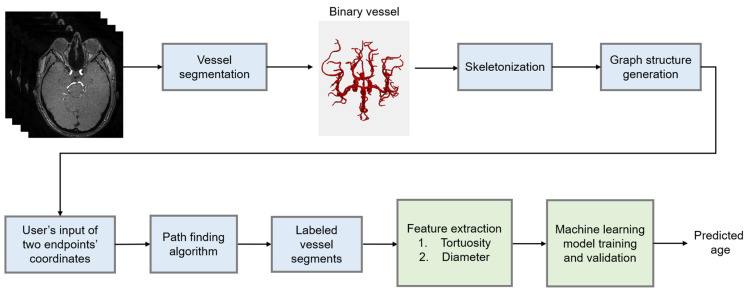
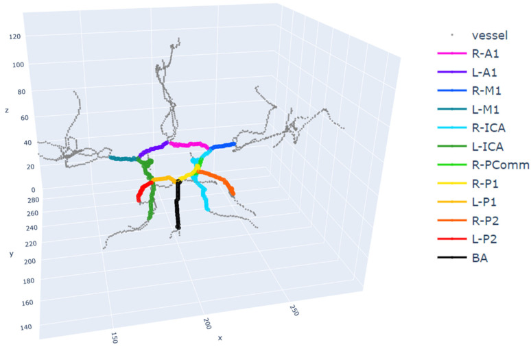
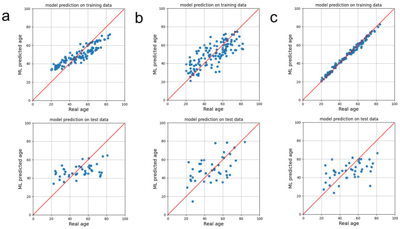
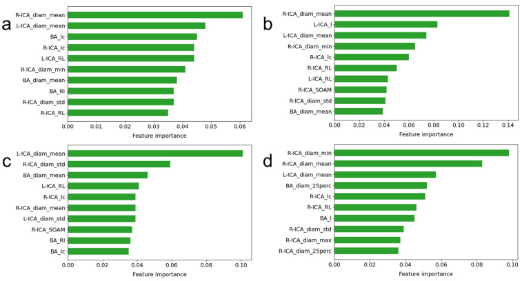
This study aimed to develop and validate machine learning (ML) models that predict age using intracranial vessels' tortuosity and diameter features derived from magnetic resonance angiography (MRA) data. A total of 171 subjects' three-dimensional (3D) time-of-flight MRA image data were considered for analysis. After annotations of two endpoints in each arterial segment, tortuosity features such as the sum of the angle metrics, triangular index, relative length, and product of the angle distance, as well as the vessels' diameter features, were extracted and used to train and validate the ML models for age prediction. Features extracted from the right and left internal carotid arteries (ICA) and basilar arteries were considered as the inputs to train and validate six ML regression models with a four-fold cross validation. The random forest regression model resulted in the lowest root mean square error of 14.9 years and the highest average coefficient of determination of 0.186. The linear regression model showed the lowest average mean absolute percentage error (MAPE) and the highest average Pearson correlation coefficient (0.532). The mean diameter of the right ICA vessel segment was the most important feature contributing to prediction of age in two out of the four regression models considered. An ML of tortuosity descriptors and diameter features extracted from MRA data showed a modest correlation between real age and ML-predicted age. Further studies are warranted for the assessment of the model's age predictions in patients with intracranial vessel diseases.
Keywords: age prediction; feature extraction; intracranial artery; machine learning; magnetic resonance angiography; medical image analysis.
Conflict of interest statement
The authors declare no conflict of interest.
Figures
Similar articles
-
Quantification of morphometry and intensity features of intracranial arteries from 3D TOF MRA using the intracranial artery feature extraction (iCafe): A reproducibility study.Magn Reson Imaging. 2019 Apr;57:293-302. doi: 10.1016/j.mri.2018.12.007. Epub 2018 Dec 20. Magn Reson Imaging. 2019. PMID: 30580079 Free PMC article.
-
Four Severity Levels for Grading the Tortuosity of a Retinal Fundus Image.J Imaging. 2022 Sep 22;8(10):258. doi: 10.3390/jimaging8100258. J Imaging. 2022. PMID: 36286352 Free PMC article.
-
Machine learning assessment of myocardial ischemia using angiography: Development and retrospective validation.PLoS Med. 2018 Nov 13;15(11):e1002693. doi: 10.1371/journal.pmed.1002693. eCollection 2018 Nov. PLoS Med. 2018. PMID: 30422987 Free PMC article.
-
Development of a quantitative intracranial vascular features extraction tool on 3D MRA using semiautomated open-curve active contour vessel tracing.Magn Reson Med. 2018 Jun;79(6):3229-3238. doi: 10.1002/mrm.26961. Epub 2017 Oct 17. Magn Reson Med. 2018. PMID: 29044753 Free PMC article.
-
MR angiography of the spine and spinal cord.Top Magn Reson Imaging. 2003 Dec;14(6):444-60. doi: 10.1097/00002142-200312000-00003. Top Magn Reson Imaging. 2003. PMID: 14872165 Review.
Cited by
-
Quantitative assessment of the oral microvasculature using optical coherence tomography angiography.Front Bioeng Biotechnol. 2024 Sep 20;12:1464562. doi: 10.3389/fbioe.2024.1464562. eCollection 2024. Front Bioeng Biotechnol. 2024. PMID: 39372434 Free PMC article.
-
Brain age gap estimation using attention-based ResNet method for Alzheimer's disease detection.Brain Inform. 2024 Jun 4;11(1):16. doi: 10.1186/s40708-024-00230-1. Brain Inform. 2024. PMID: 38833039 Free PMC article.
References
-
- Guillén M.F. 2030: How Today’s Biggest Trends Will Collide and Reshape the Future of Everything. St. Martin’s Press; New York, NY, USA: 2020.
-
- Hepp T., Blum D., Armanious K., Scholkopf B., Stern D., Yang B., Gatidis S. Uncertainty estimation and explainability in deep learning-based age estimation of the human brain: Results from the German National Cohort MRI study. Comput. Med. Imaging Graph. 2021;92:101967. doi: 10.1016/j.compmedimag.2021.101967. - DOI - PubMed
Grants and funding
LinkOut - more resources
Full Text Sources
Miscellaneous
