

Neoadjuvant Chemotherapy with Laser Interstitial Thermal Therapy in Central Nervous System Neuroblastoma: Illustrative Case and Literature Review
- PMID: 38002476
- PMCID: PMC10669297
- DOI: 10.3390/brainsci13111515
Neoadjuvant Chemotherapy with Laser Interstitial Thermal Therapy in Central Nervous System Neuroblastoma: Illustrative Case and Literature Review
Abstract
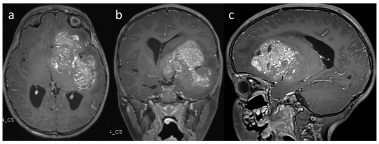
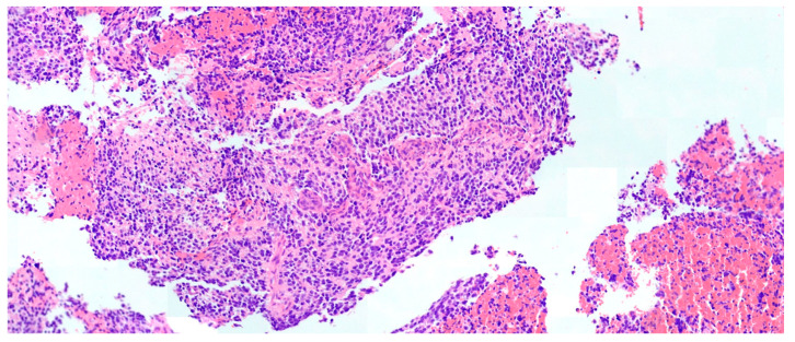
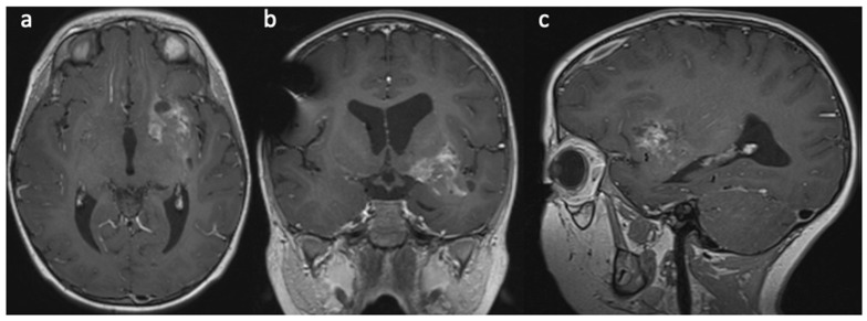
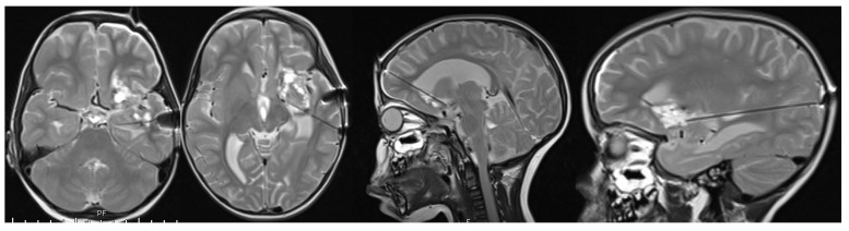
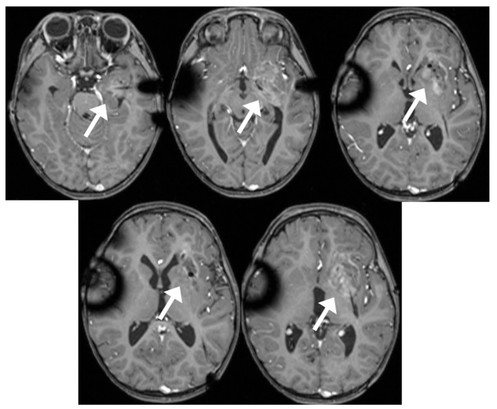
Primitive neuroectodermal tumors of the central nervous system, or CNS neuroblastoma, are rare neoplasms in children. Recently, methylation profiling enabled the discovery of four distinct entities of these tumors. The current treatment paradigm involves surgical resection followed by chemotherapy and radiation. However, upfront surgical resection carries high surgical morbidity in this patient population due to their young age, tumor vascularity, and often deep location in the brain. We report a case of CNS neuroblastoma that can be successfully treated with neoadjuvant chemotherapy followed by minimally invasive laser interstitial thermal therapy and radiation. The patient has complete treatment with no evidence of recurrence at one year follow-up. This case illustrates a potential paradigm shift in the treatment of these rare tumors can be treated using minimally invasive surgical approach to achieve a favorable outcome.
Keywords: CNS neuroblastoma; LITT; laser interstitial thermal therapy.
Conflict of interest statement
The authors declare no conflict of interest.
Figures

Similar articles
-
Laser interstitial thermal therapy followed by minimal-access transsulcal resection for the treatment of large and difficult to access brain tumors.Neurosurg Focus. 2016 Oct;41(4):E14. doi: 10.3171/2016.8.FOCUS16233. Neurosurg Focus. 2016. PMID: 27690658
-
Role of Laser Interstitial Thermal Therapy in the Management of Primary and Metastatic Brain Tumors.Curr Treat Options Oncol. 2021 Oct 23;22(12):108. doi: 10.1007/s11864-021-00912-6. Curr Treat Options Oncol. 2021. PMID: 34687357 Review.
-
Laser interstitial thermal therapy as a radiation-sparing approach for central nervous system tumors in children with cancer predisposition syndromes: report of a child with Li-Fraumeni syndrome. Illustrative case.J Neurosurg Case Lessons. 2024 Feb 5;7(6):CASE23595. doi: 10.3171/CASE23595. Print 2024 Feb 5. J Neurosurg Case Lessons. 2024. PMID: 38315990 Free PMC article.
-
Primitive neuroectodermal tumors of the brainstem in children treated according to the HIT trials: clinical findings of a rare disease.J Neurosurg Pediatr. 2015 Mar;15(3):227-35. doi: 10.3171/2014.9.PEDS14213. Epub 2015 Jan 2. J Neurosurg Pediatr. 2015. PMID: 25555122
-
Laser Interstitial Thermal Therapy in the treatment of brain metastases and radiation necrosis.Cancer Lett. 2020 Oct 1;489:9-18. doi: 10.1016/j.canlet.2020.05.014. Epub 2020 Jun 3. Cancer Lett. 2020. PMID: 32504657 Review.
Cited by
-
Editorial for Brain Sciences Special Issue "Advances in Restorative Neurotherapeutic Technologies".Brain Sci. 2025 Mar 5;15(3):273. doi: 10.3390/brainsci15030273. Brain Sci. 2025. PMID: 40149794 Free PMC article.
-
MR-guided laser interstitial thermal therapy in the treatment of brain tumors and epilepsy.Acta Neurochir (Wien). 2024 Aug 21;166(1):344. doi: 10.1007/s00701-024-06238-0. Acta Neurochir (Wien). 2024. PMID: 39167226 Review.
References
-
- Furuta T., Moritsubo M., Muta H., Koga M., Komaki S., Nakamura H., Morioka M., Ohshima K., Sugita Y. Central nervous system neuroblastic tumor with FOXR2 activation presenting both neuronal and glial differentiation: A case report. Brain Tumor Pathol. 2020;37:100–104. doi: 10.1007/s10014-020-00370-2. - DOI - PubMed
-
- Von Hoff K., Haberler C., Schmitt-Hoffner F., Schepke E., De Rojas T., Jacobs S., Zapotocky M., Sumerauer D., Perek-Polnik M., Dufour C., et al. Therapeutic implications of improved molecular diagnostics for rare CNS embryonal tumor entities: Results of an international, retrospective study. Neuro-Oncology. 2021;23:1597–1611. doi: 10.1093/neuonc/noab136. - DOI - PMC - PubMed
-
- Korshunov A., Okonechnikov K., Schmitt-Hoffner F., Ryzhova M., Sahm F., Stichel D., Schrimpf D., Reuss D.E., Sievers P., Suwala A.K., et al. Molecular analysis of pediatric CNS-PNET revealed nosologic heterogeneity and potent diagnostic markers for CNS neuroblastoma with FOXR2-activation. Acta Neuropathol. Commun. 2021;9:20. doi: 10.1186/s40478-021-01118-5. - DOI - PMC - PubMed
Publication types
Grants and funding
LinkOut - more resources
Full Text Sources
